
Anatomy of eye acc to khurrna
anatomy of the eye diagram layers fibrous uveal nervous coat
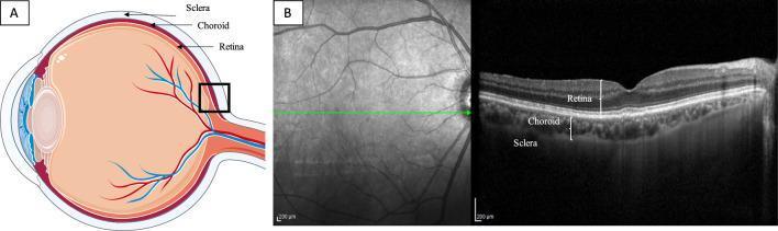
This composite educational graphic illustrates human ocular anatomy and diagnostic imaging. Section A presents a sagittal schematic diagram of the eye, highlighting the three posterior layers: the retina (inner), the choroid (middle vascular layer), and the sclera (outer fibrous layer). A black box indicates the posterior pole region being analyzed. Section B features a Spectral Domain Optical Coherence Tomography (SD-OCT) scan centered on the fovea. The left side of panel B shows a grayscale infrared fundus image with a green horizontal line indicating the scan acquisition path. The right side displays the corresponding cross-sectional OCT B-scan, which provides high-resolution visualization of the retinal architecture, the underlying choroidal layer with its characteristic vascular luminal and stromal spaces, and the interface with the sclera. Labels on the B-scan confirm the stratified arrangement of the retina, choroid, and sclera, making this a foundational resource for understanding posterior segment imaging and choroidal thickness assessment.
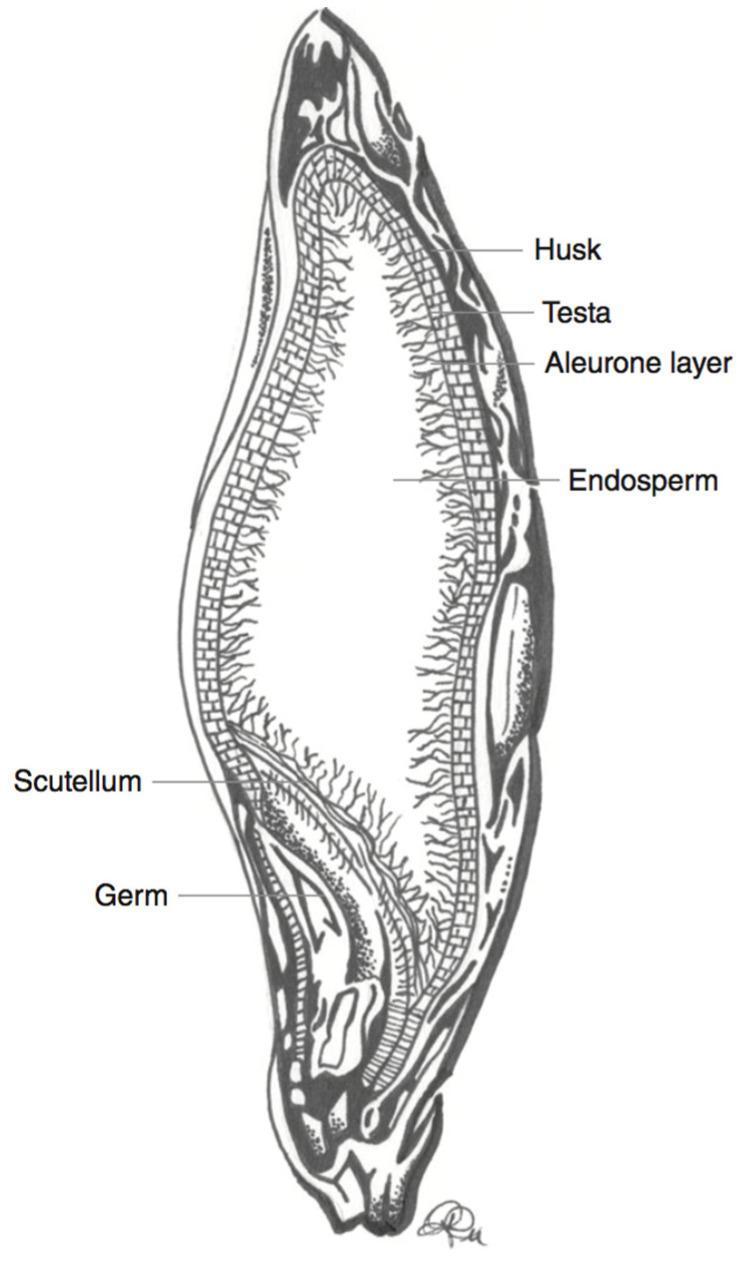
This medical-botanical schematic illustration depicts a longitudinal cross-section of a barley kernel (Hordeum vulgare), serving as a model for cereal grain anatomy relevant to human nutrition and dietetics. The diagram identifies key structural components involved in nutrient distribution, specifically the localization of anthocyanins and fiber. The anatomical layers are labeled from the periphery to the interior: the outermost fibrous Husk, the thin protective Testa, and the Aleurone layer composed of structured, brick-like cells. The largest central region is the Endosperm, characterized by a grainy, fibrous texture. At the base of the kernel, the embryo-related structures, the Scutellum and the Germ, are clearly demarcated. This diagram is educationally significant for understanding the localization of bioactive compounds like phenolic pigments, which are primarily concentrated in the outer layers (pericarp and aleurone) and are often lost during commercial milling processes.
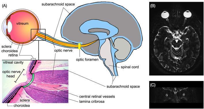
This composite educational graphic details the neuroanatomical continuity between the eye and the central nervous system. Panel A features a sagittal pathophysiology diagram illustrating the connection from the eyeball to the brain via the optic nerve. It highlights the layers of the eye (sclera, choroid, retina), the vitreum, and the optic foramen. A magnified histological inset shows the optic nerve head, lamina cribrosa, and central retinal vessels. Crucially, the diagram labels the subarachnoid space (SAS), demonstrating its continuity from the intracranial compartment to the orbital optic nerve sheath. Panel B provides clinical correlation with an axial T2-weighted MRI scan, where high-intensity (bright) signal represents cerebrospinal fluid (CSF) within the optic nerve SAS, contrasting against the dark optic nerves extending from the posterior globes. Panel C displays a coronal MRI slice further demonstrating the bilateral symmetry of the optic nerve sheaths. This material is designed to teach the physiological link between intracranial pressure and ocular health, relevant to specialties like ophthalmology, neurology, and radiology.
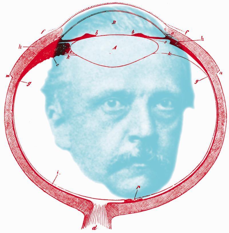
This educational illustration is an anatomical diagram of the human eye in sagittal cross-section, overlaid with a portrait of Hermann von Helmholtz. The diagram, rendered in red linework, depicts essential ophthalmic structures including the cornea (labeled B), the crystalline lens (labeled A), and the ciliary body (b). Various alphabetical annotations (c, d, e, f, g, h, i, k, m, n) indicate specific histological and anatomical regions, such as the sclera, retina, choroid layers, and the optic nerve exit (d). The lens is shown in its anatomical position behind the anterior chamber and iris. The image serves as a conceptual tribute to physiological optics, bridging the anatomy of vision with the historical figure responsible for major advancements in the field, such as the invention of the ophthalmoscope and the primary treatise on physiological optics. The spatial arrangement emphasizes the internal ocular environment, with the retinal lining and posterior segment framing the central portrait.
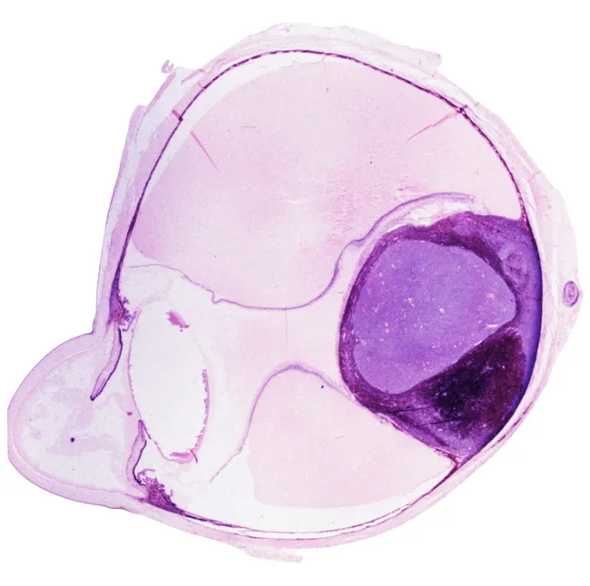
Brightfield light microscopy of a hematoxylin and eosin stained sagittal cross‑section through an ocular globe. The slide displays the layered architecture of the eye, with a prominent circular structure representing the crystalline lens encased by a thin capsule and adjacent zonular fibers. Surrounding tissues reveal the scleral white of the globe and the immediately subjacent uveal tissues, including choroid and pigmented retinal pigment epithelium, transitioning to the multi‑layered neural retina at the inner surface. Nuclei appear dark purple on hematoxylin, while cytoplasm and extracellular matrix stain pink to pale pink, creating distinct optic nerve/retinal nerve fiber layer boundaries peripherally and centrally. The vitreous body appears relatively acellular and eosinophilic, separating the lens from the retina. The optic axis and ciliary body regions may be suggested at the periphery by tubular structures and vascular patterns. Overall, this image demonstrates normal ocular histomorphology suitable for educational reference, anatomical atlas documentation, and pattern recognition training in ophthalmic pathology. Potential clinical applications include teaching ocular anatomy (globe, lens, retina, sclera, choroid), diagnosing cataract or retinal disorders in histological specimens, and serving as a baseline for comparative studies in ocular development and disease. This representation emphasizes laminar organization, including anterior corneal–limbal regions not visible here, posterior retina layers such as ganglion cell layer and inner nuclear layer, and lens substructures—capsule, cortex, and nucleus—facilitating recognition tasks for students and clinicians. High-quality reference for ophthalmology education.
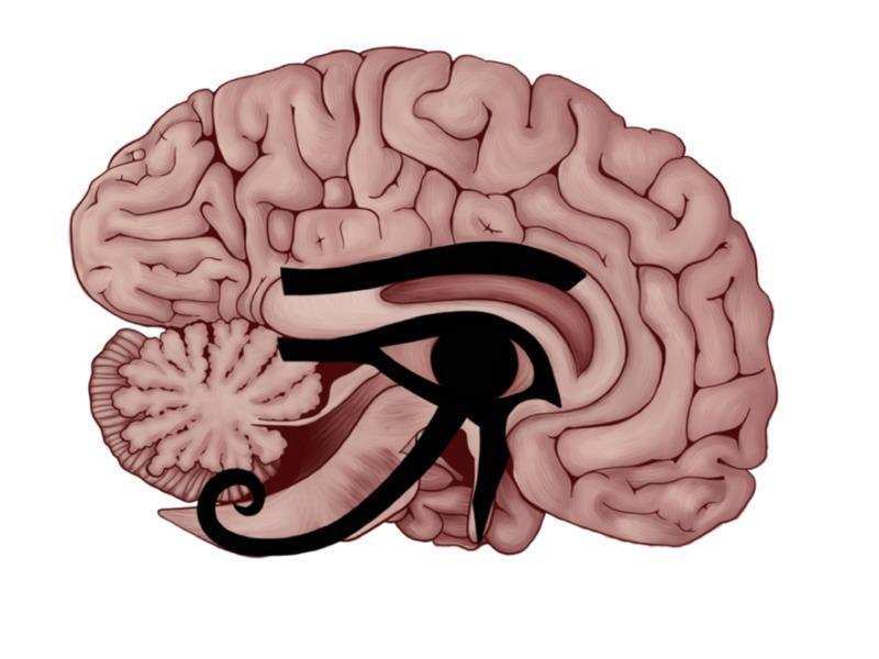
This medical illustration presents an anatomical diagram of a human brain in a mid-sagittal section, featuring the superimposition of the Eye of Horus over key neuroanatomical structures. The underlying anatomy displays the cerebrum with its cortical gyri and sulci, the corpus callosum, the cerebellum showing the arbor vitae pattern, the brainstem, and the diencephalic region. The Eye of Horus is strategically aligned to illustrate perceived correlations between its geometric components and functional brain anatomy: the eyebrow corresponds to the corpus callosum; the pupil is centered over the thalamus and interthalamic adhesion (massa intermedia); the anterior triangular component aligns with the olfactory trigone; and the descending extensions correspond to the pathways for hearing (Brodmann areas 41/42), taste, and the somatosensory tract. The image serves as a comparative conceptual diagram linking ancient symbolic art with functional neuroanatomy, highlighting the central nervous system's sensory processing centers and commissural fibers.
cross section of eye cornea lens retina choroid sclera diagram
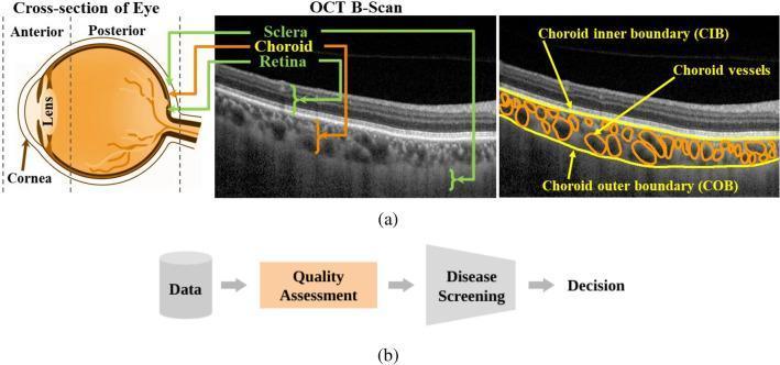
This educational graphic illustrates the anatomical and diagnostic visualization of the posterior segment of the human eye. Section (a) contains three panels: a sagittal cross-sectional diagram of the eye showing the anterior (cornea, lens) and posterior segments; a sample Optical Coherence Tomography (OCT) B-scan identifying the retina (innermost), choroid (middle), and sclera (outermost); and an annotated OCT B-scan specifically delineating the choroid. The annotated scan highlights the choroidal inner boundary (CIB), the choroidal outer boundary (COB) at the choroid-sclera interface, and individual choroidal vessels within the vascular stroma. Section (b) presents a diagnostic flowchart for clinical decision-making, emphasizing 'Quality Assessment' as a mandatory intermediary step between data acquisition and disease screening. This material is designed for ophthalmology education, focusing on retinal imaging, choroidal biomarker quantification (such as thickness and vascularity index), and the importance of image quality in automated diagnostic pipelines for conditions like central serous chorioretinopathy and age-related macular degeneration.
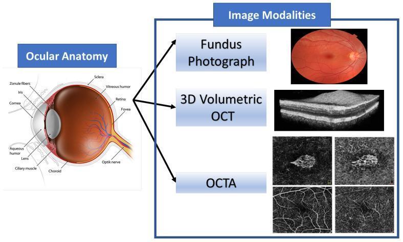
This educational graphic combines an anatomical diagram of the human eye with corresponding ophthalmic imaging modalities. The 'Ocular Anatomy' section features a detailed sagittal cross-section identifying key structures: the cornea, aqueous humor, iris, pupil, biconvex lens, zonule fibers, and ciliary muscle in the anterior segment; and the vitreous humor, retina, fovea, choroid, sclera, and optic nerve in the posterior segment. The 'Image Modalities' section illustrates diagnostic tools used to visualize these structures. A color Fundus Photograph provides a surface view of the retina, optic disc, and vasculature. A 3D Volumetric Optical Coherence Tomography (OCT) scan displays a high-resolution, grayscale cross-sectional view of retinal layers, highlighting tissue stratification. Finally, Optical Coherence Tomography Angiography (OCTA) panels show non-invasive, depth-resolved images of the retinal and choroidal microvasculature. This composite serves as a reference for correlating anatomical landmarks with clinical diagnostic imaging used in the management of retinal diseases such as Age-related Macular Degeneration (AMD).
This educational illustration is an anatomical diagram of the human eye in sagittal cross-section, overlaid with a portrait of Hermann von Helmholtz. The diagram, rendered in red linework, depicts essential ophthalmic structures including the cornea (labeled B), the crystalline lens (labeled A), and the ciliary body (b). Various alphabetical annotations (c, d, e, f, g, h, i, k, m, n) indicate specific histological and anatomical regions, such as the sclera, retina, choroid layers, and the optic nerve exit (d). The lens is shown in its anatomical position behind the anterior chamber and iris. The image serves as a conceptual tribute to physiological optics, bridging the anatomy of vision with the historical figure responsible for major advancements in the field, such as the invention of the ophthalmoscope and the primary treatise on physiological optics. The spatial arrangement emphasizes the internal ocular environment, with the retinal lining and posterior segment framing the central portrait.
Anatomy of the Eye (as per Khurrana's Comprehensive Ophthalmology)
Note: Khurrana's Comprehensive Ophthalmology is not directly available in our textbook library. The following is presented according to Khurrana's well-known organizational framework, supplemented by Kanski's Clinical Ophthalmology and Ganong's Review of Medical Physiology available in our library.
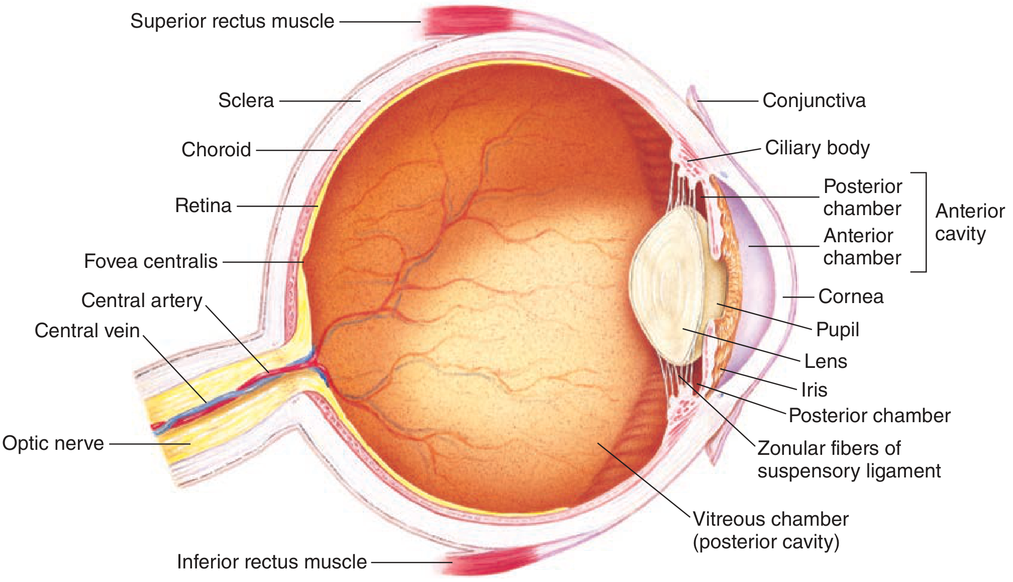
Overview
- Fibrous coat (outer)
- Uveal coat / Vascular coat (middle)
- Nervous coat / Retina (inner)
1. Fibrous Coat (Outer Coat)
A. Sclera (Posterior 5/6)
- Tough, opaque, white fibrous tissue made of dense collagen bundles
- Provides structural rigidity and protects intraocular contents
- Perforated posteriorly by the lamina cribrosa (where optic nerve fibres pass through)
- Externally, is continuous with the dural sheath of the optic nerve
- Anteriorly, is covered by the conjunctiva (bulbar part)
- Blood supply: poor (from episcleral vessels)
B. Cornea (Anterior 1/6)
- Transparent, avascular, highly innervated structure
- Refractive power: +43 diopters (most powerful refractive surface of the eye)
- Horizontal diameter: ~11.7 mm; vertical diameter: ~10.6 mm
- Radius of curvature: ~7.8 mm anteriorly
| Layer | Key Features |
|---|---|
| 1. Epithelium | Stratified squamous non-keratinized, 5-6 cell layers; regenerates from limbal stem cells |
| 2. Bowman's layer | Acellular condensation of anterior stroma; does NOT regenerate if damaged |
| 3. Stroma | Thickest layer (90% of corneal thickness); regular collagen lamellae (orthogonal arrangement = transparency) |
| 4. Descemet's membrane | Basement membrane of endothelium; thickens with age; regenerates |
| 5. Endothelium | Single layer of hexagonal cells; maintains corneal dehydration (deturgescence); does NOT regenerate in adults |
2. Uveal Coat (Middle / Vascular Coat)
A. Iris (Anteriormost)
- Colored, opaque diaphragm with central aperture = pupil
- Two muscles:
- Sphincter pupillae - circular fibers, constricts pupil (miosis) - parasympathetic (CN III)
- Dilator pupillae - radial fibers, dilates pupil (mydriasis) - sympathetic (superior cervical ganglion)
- Two layers of epithelium on posterior surface (both pigmented)
- Blood supply from major arterial circle of iris (MACI) formed by long posterior ciliary arteries (LPCAs)
B. Ciliary Body (Middle)
- Triangular in cross-section, located between iris and choroid
- Two parts:
- Pars plana (flat part, posterior) - 3.5-4 mm wide; surgical access site (pars plana vitrectomy)
- Pars plicata (corrugated part, anterior) - bears ~70 ciliary processes
- Functions:
- Aqueous humour production by ciliary processes (via active secretion and ultrafiltration)
- Accommodation via ciliary muscle (Helmholtz theory): circular/longitudinal smooth muscle fibers
- Ciliary zonules (suspensory ligaments of lens) attach from ciliary body to lens equator
- Blood supply: anterior ciliary arteries + LPCAs
C. Choroid (Posterior 2/3)
- Highly vascular, brown/black pigmented layer between sclera and retina
- Extends from ora serrata anteriorly to optic nerve posteriorly
- Layers (outside to inside):
- Suprachoroid (potential space)
- Large vessel layer (Haller's layer)
- Medium vessel layer (Sattler's layer)
- Choriocapillaris - fenestrated capillaries nourishing outer retina
- Bruch's membrane - innermost layer (5 sub-layers), separates choroid from RPE
- Blood supply: short posterior ciliary arteries (SPCAs)
- Function: Nourishes outer retina (RPE + photoreceptors); acts as a heat sink
3. Nervous Coat (Inner Coat) - Retina
Extent
- Anteriorly ends at ora serrata
- Posteriorly: macula lutea is the area of central vision (~5.5 mm temporal to optic disc)
- Fovea centralis is the central depression of the macula - area of highest visual acuity (only cones, no rods)
- Foveal avascular zone (FAZ): ~0.5 mm diameter, no vessels
- Optic disc (blind spot): 1.5 mm diameter, where optic nerve exits; contains no photoreceptors
Ten Layers of Retina (from outside to inside)
| Layer | Content |
|---|---|
| 1. RPE (Retinal Pigment Epithelium) | Single layer of pigmented cells; phagocytoses shed outer segments; forms outer blood-retinal barrier |
| 2. Photoreceptor layer | Rods (120 million, peripheral, scotopic/dim light) and Cones (6 million, central, photopic/color) |
| 3. External limiting membrane | Zonula adherens junctions between Müller cells and photoreceptors |
| 4. Outer nuclear layer | Nuclei of photoreceptors |
| 5. Outer plexiform layer | Synapses between photoreceptors and bipolar/horizontal cells |
| 6. Inner nuclear layer | Nuclei of bipolar, amacrine, Müller, and horizontal cells |
| 7. Inner plexiform layer | Synapses between bipolar, amacrine, and ganglion cells |
| 8. Ganglion cell layer | Nuclei of ganglion cells |
| 9. Nerve fiber layer | Axons of ganglion cells (become optic nerve) |
| 10. Internal limiting membrane | Basement membrane of Müller cells; inner boundary |
- Inner retina (layers 1-6 from inside): central retinal artery (branch of ophthalmic artery)
- Outer retina (rods/cones, RPE): choriocapillaris (via diffusion through Bruch's membrane)
- Inner blood-retinal barrier: tight junctions of retinal capillary endothelium
- Outer blood-retinal barrier: tight junctions of RPE cells
4. Contents of the Eyeball
Aqueous Humour
- Clear, protein-free fluid
- Produced by ciliary processes (active secretion ~70%, ultrafiltration ~20%, diffusion ~10%)
- Fills anterior chamber (between cornea and iris) and posterior chamber (between iris, ciliary body, and lens)
- Drained via trabecular meshwork → Schlemm's canal → episcleral veins (conventional route)
- Minor route: uveoscleral (through ciliary body)
- Normal IOP: 10-21 mmHg
- Obstructed drainage → raised IOP → glaucoma
Lens
- Transparent, biconvex, avascular, acellular (no nerves or blood vessels)
- Refractive power: +18 diopters (at rest); increases with accommodation
- Parts: anterior capsule > epithelium > cortex > nucleus > posterior capsule
- Held by zonular fibers (zonules of Zinn) attached to ciliary body
- Accommodation: ciliary muscle contracts → zonules relax → lens becomes more convex → increased power (near vision)
- Equatorial lens cells proliferate throughout life → lens grows and becomes less flexible → presbyopia
Vitreous Humour
- Transparent gel, 99% water + collagen fibrils + hyaluronic acid
- Fills the vitreous chamber (posterior cavity) = 4/5 of globe volume (~4 mL)
- Maintains globe shape; supports retina; allows light transmission
- Attachments: vitreous base (ora serrata), around optic disc, at macula, around retinal vessels
- Cloquet's canal: remnant of hyaloid artery running through vitreous
5. Chambers of the Eye
| Chamber | Location | Contents |
|---|---|---|
| Anterior chamber | Between cornea and iris | Aqueous humour |
| Posterior chamber | Between iris, zonule, and lens | Aqueous humour |
| Vitreous chamber | Between lens and retina | Vitreous humour |
6. Accessory Structures
Eyelids
- Protect the eye; distribute tear film via blinking
- Layers (outer to inner): Skin → Subcutaneous tissue (no fat) → Orbicularis oculi muscle → Orbital septum → Tarsal plate → Palpebral conjunctiva
- Tarsal plates: dense fibrous tissue containing Meibomian glands (sebaceous glands, ~25 in upper, ~20 in lower lid)
- Meibomian glands secrete lipid layer of tear film
- Glands of Zeis: sebaceous glands of eyelashes
- Glands of Moll: modified sweat glands
Conjunctiva
- Thin mucous membrane covering anterior sclera (bulbar conjunctiva) and inner eyelids (palpebral conjunctiva)
- Goblet cells secrete mucin (innermost layer of tear film)
- Rich in lymphoid tissue (CALT - conjunctiva-associated lymphoid tissue)
Lacrimal Apparatus
- Lacrimal gland (superolateral orbit) → secretes aqueous layer of tears
- Tears flow across cornea and drain via:
- Puncta → Canaliculi (vertical 2 mm then horizontal 8 mm) → common canaliculus → Lacrimal sac → Nasolacrimal duct → inferior meatus of nose
- Valve of Rosenmüller (prevents reflux into canaliculi)
- Valve of Hasner (at nasolacrimal duct opening)
- Tear film has 3 layers:
- Lipid (outer) - from Meibomian glands
- Aqueous (middle) - from lacrimal gland
- Mucin (inner) - from goblet cells
7. Extraocular Muscles
| Muscle | Primary Action | Nerve Supply |
|---|---|---|
| Medial rectus | Adduction | CN III (oculomotor) |
| Lateral rectus | Abduction | CN VI (abducens) |
| Superior rectus | Elevation, intorsion, adduction | CN III |
| Inferior rectus | Depression, extorsion, adduction | CN III |
| Superior oblique | Intorsion, depression, abduction | CN IV (trochlear) |
| Inferior oblique | Extorsion, elevation, abduction | CN III |
- All muscles arise from the annulus of Zinn (common tendinous ring at orbital apex) EXCEPT inferior oblique (from anteromedial orbital floor)
- All muscles insert into sclera anterior to equator
- Fick's axes: X (horizontal), Y (sagittal/anteroposterior), Z (vertical) - axes of rotation
8. Blood Supply of the Eye
| Structure | Artery |
|---|---|
| Retina (inner) | Central retinal artery |
| Choroid | Short posterior ciliary arteries |
| Iris, ciliary body | Long posterior + anterior ciliary arteries |
| Optic nerve head | Short posterior ciliary arteries + circle of Zinn-Haller |
| Extraocular muscles | Anterior ciliary arteries (from muscular branches of ophthalmic artery) |
9. Nerve Supply of the Eye
| Structure | Nerve |
|---|---|
| Corneal sensation | Nasociliary branch of V1 (long ciliary nerves) |
| Pupil constriction | Parasympathetic from EW nucleus via CN III → ciliary ganglion → short ciliary nerves |
| Pupil dilatation | Sympathetic from superior cervical ganglion → long ciliary nerves |
| Retina → optic nerve | CN II (optic nerve, actually a CNS tract) |
| Accommodation | Parasympathetic (CN III → ciliary ganglion → short ciliary nerves → ciliary muscle) |