Appendicular skeleton all bones with image leveling
Reading File
Finding Sources
Finding Sources
Reading File
Reading File
Loading Image
Loading Image
Finding Sources
Finding Sources
Reading File
Finding Sources
Finding Sources
Reading File
Loading Image
Loading Image
Reading File
Finding Sources
Finding Sources
Reading File
Reading File
Loading Image
Loading Image
Loading Image
Finding Sources
Reading File
I now have all the key content needed to write a comprehensive, image-rich answer. Let me compose the full response.
Appendicular Skeleton - Complete Overview with Images
The appendicular skeleton consists of 126 bones in the upper and lower limbs (extremities). These are divided into two girdles and their associated free limb bones.
- Gray's Anatomy for Students; Imaging Anatomy Vol. 3, Bones, Joints, Vessels and Nerves
Overview: Axial vs. Appendicular
The skeleton divides into:
- Axial skeleton (80 bones) - skull, vertebral column, ribs, sternum
- Appendicular skeleton (126 bones) - all four limbs
UPPER LIMB (64 bones total - 32 per side)
1. Pectoral (Shoulder) Girdle - 4 bones (2 per side)
| Bone | Count | Notes |
|---|---|---|
| Clavicle | 2 | "Collarbone"; only bony connection between upper limb and axial skeleton |
| Scapula | 2 | Triangular flat bone; includes glenoid cavity, acromion, coracoid process, spine of scapula |
2. Arm (Brachium) - 2 bones (1 per side)
| Bone | Count | Key Landmarks |
|---|---|---|
| Humerus | 2 | Head, anatomic neck, surgical neck, greater & lesser tubercles, intertubercular groove, deltoid tuberosity, radial groove, capitulum, trochlea, medial & lateral epicondyles, olecranon fossa |
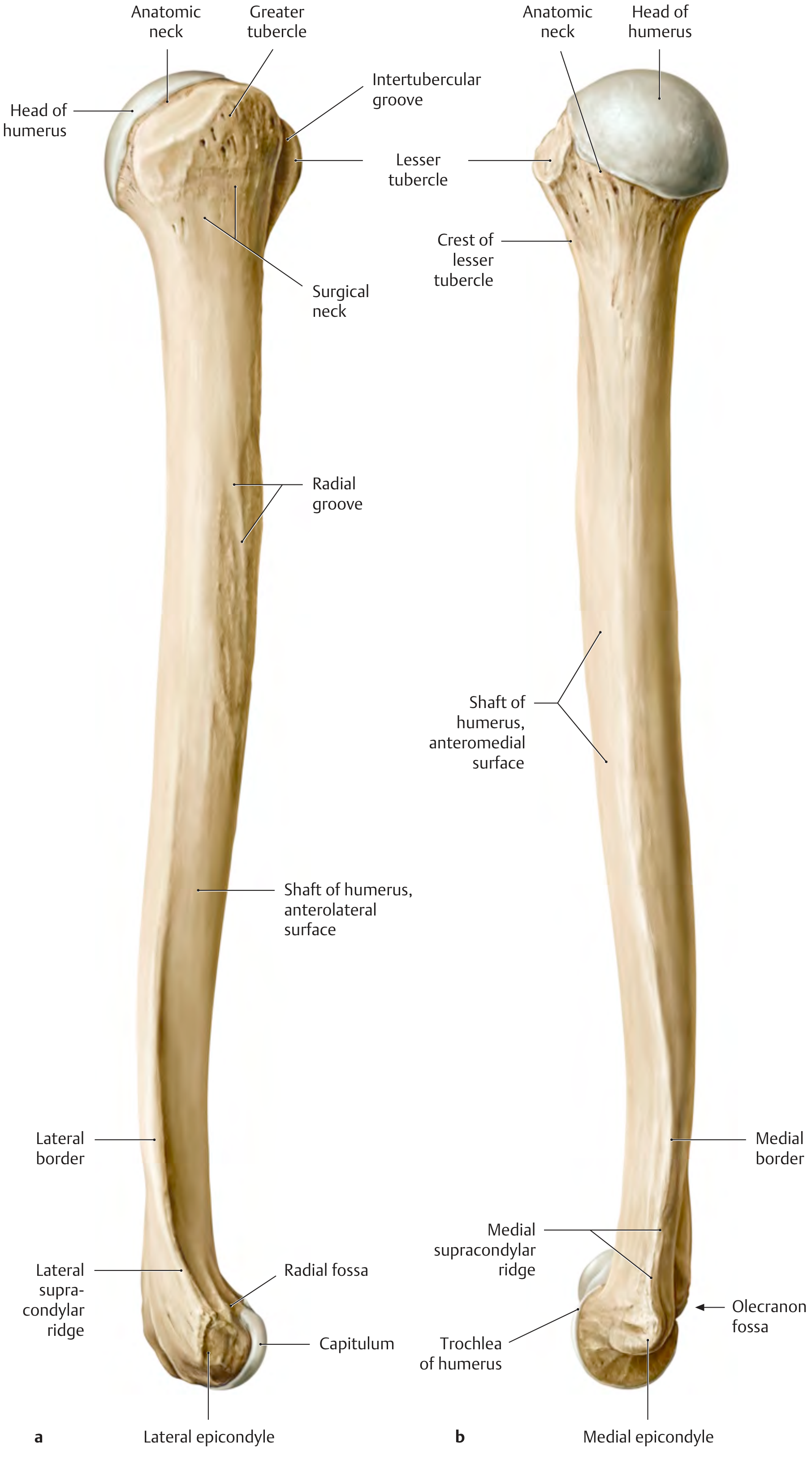
THIEME Atlas of Anatomy - The right humerus, anterior (a) and posterior (b) views
3. Forearm (Antebrachium) - 4 bones (2 per side)
| Bone | Count | Key Landmarks |
|---|---|---|
| Radius | 2 | Head (articular fovea, articular circumference), neck, radial tuberosity, shaft, styloid process of radius, dorsal tubercle (Lister's tubercle) |
| Ulna | 2 | Olecranon, trochlear notch, coronoid process, radial notch, ulnar tuberosity, shaft, styloid process of ulna, head of ulna |
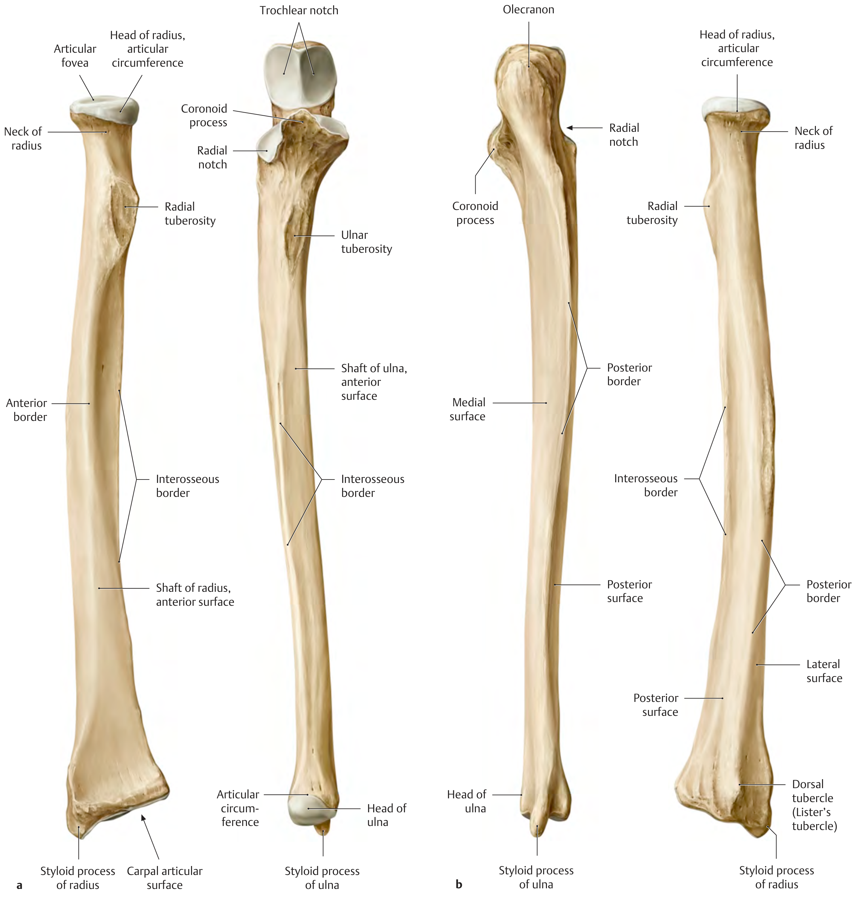
THIEME Atlas of Anatomy - Right radius and ulna, anterior and posterior views
4. Wrist (Carpals) - 16 bones (8 per hand)
Arranged in two rows of 4 bones each:
| Row | Bones (lateral to medial) | Mnemonic |
|---|---|---|
| Proximal row | Scaphoid, Lunate, Triquetrum, Pisiform | She Looks Too Pretty |
| Distal row | Trapezium, Trapezoid, Capitate, Hamate | To Touch Carla's Hand |
Functional column organization:
- Scaphoid (radial) column - Scaphoid, Trapezium, Trapezoid
- Lunate (central) column - Lunate, Capitate
- Triquetral (ulnar) column - Triquetrum, Hamate (Pisiform = sesamoid in FCU tendon)
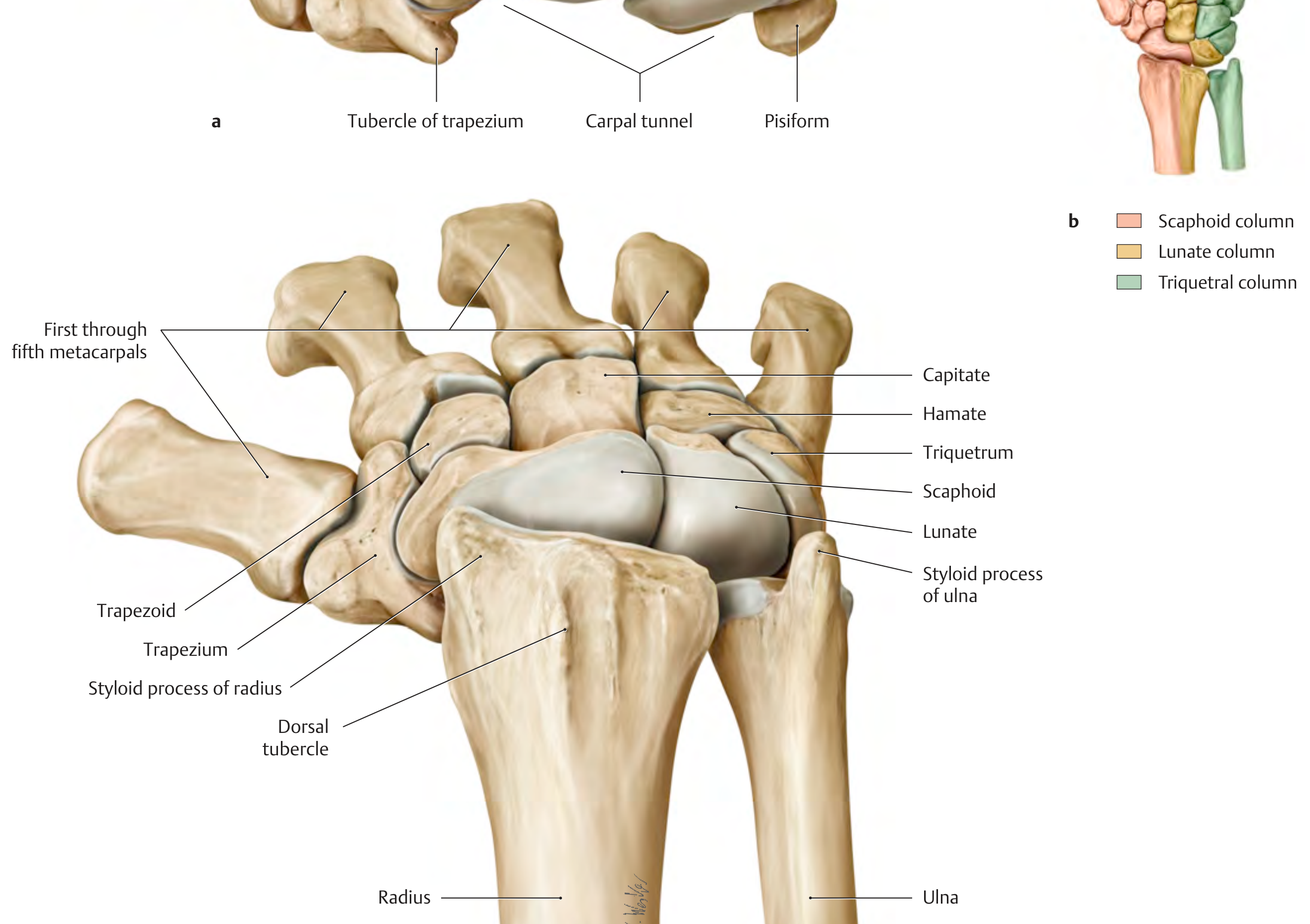
THIEME Atlas of Anatomy - Carpal bones showing scaphoid, lunate, triquetrum, trapezoid, trapezium, capitate, and hamate
5. Hand Bones - 38 bones (19 per hand)
| Segment | Bones | Count per hand |
|---|---|---|
| Metacarpals | Metacarpals I-V | 5 |
| Phalanges of thumb (digit 1) | Proximal + Distal | 2 |
| Phalanges of fingers (digits 2-5) | Proximal + Middle + Distal (x4) | 12 |
| Total per hand | 19 |
Thumb has 2 phalanges; fingers have 3 phalanges each.
LOWER LIMB (62 bones total - 31 per side)
1. Pelvic (Hip) Girdle - 2 bones
| Bone | Count | Notes |
|---|---|---|
| Hip bone (Os coxae) | 2 | Each fuses from 3 parts - ilium, ischium, pubis - fusing at the triradiate cartilage (complete fusion by ~25 years). Together they form the pelvis |
The three components of the hip bone:
- Ilium - largest, forms the iliac crest, ASIS, AIIS, greater sciatic notch
- Ischium - forms the ischial tuberosity ("sitting bone"), ischial spine
- Pubis - forms the pubic symphysis anteriorly, pubic tubercle
2. Thigh - 2 bones (1 per side)
| Bone | Count | Key Landmarks |
|---|---|---|
| Femur | 2 | Longest bone in body. Head (articulates with acetabulum), neck (neck-shaft angle ~122°, anteversion ~10-15°), greater & lesser trochanters, intertrochanteric line (anterior) & crest (posterior), shaft with linea aspera, medial & lateral condyles, patellar surface |
| Patella | 2 | Largest sesamoid bone; embedded in quadriceps tendon; protects the knee joint |
3. Leg (Crus) - 4 bones (2 per side)
| Bone | Count | Key Landmarks |
|---|---|---|
| Tibia | 2 | Medial & lateral condyles, intercondylar eminence, tibial plateau, tibial tuberosity (patellar ligament attachment), anterior border ("shin"), medial malleolus |
| Fibula | 2 | Head, neck, shaft, lateral malleolus (forms lateral wall of ankle mortise) |
4. Ankle (Tarsals) - 14 bones (7 per foot)
| Bone | Notes |
|---|---|
| Talus | Articulates with tibia/fibula above (ankle mortise); transmits entire body weight to foot |
| Calcaneus | Largest tarsal bone; forms the heel; calcaneal tuberosity for Achilles tendon |
| Navicular | Medial side; articulates with head of talus |
| Cuboid | Lateral side; articulates with calcaneus |
| Medial cuneiform | |
| Intermediate cuneiform | |
| Lateral cuneiform |
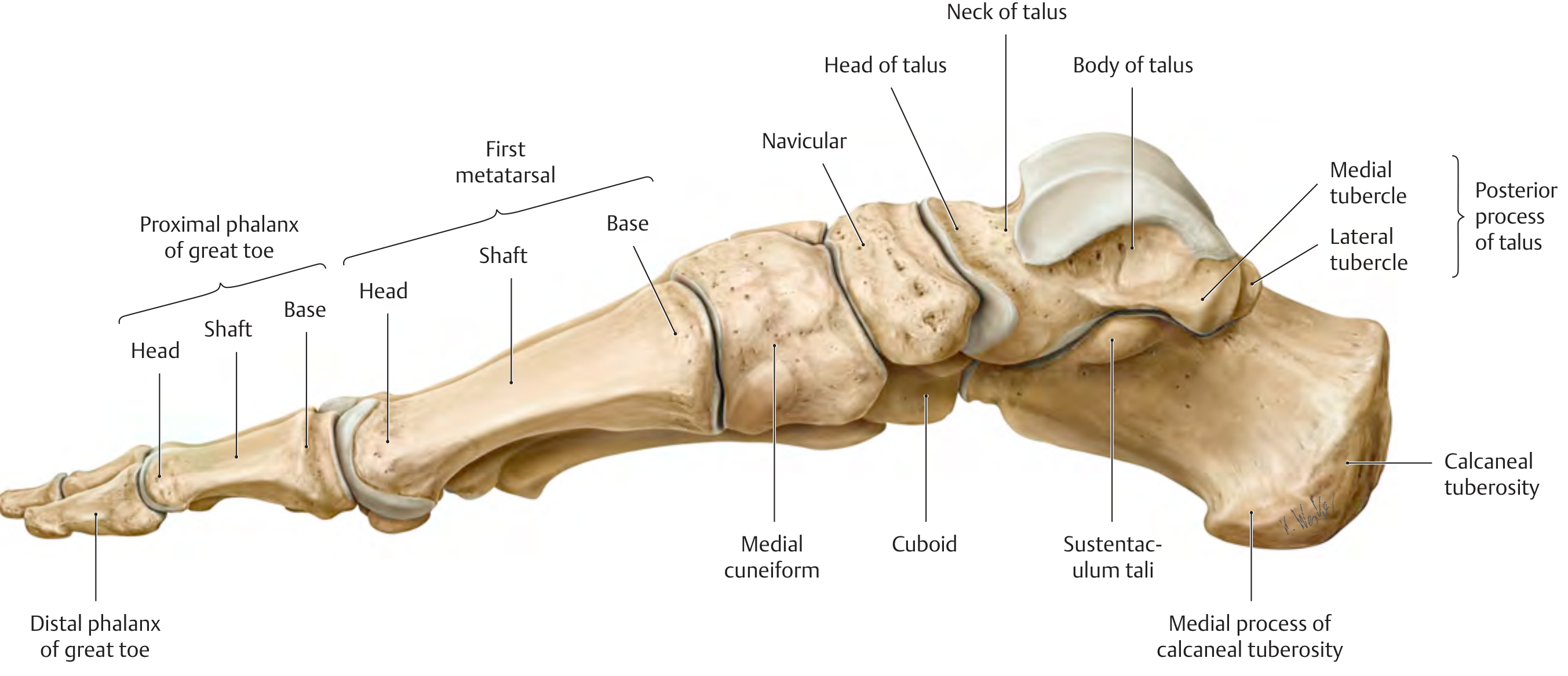
THIEME Atlas of Anatomy - Right foot, medial view showing all tarsal bones, metatarsals, and phalanges
5. Foot Bones - 38 bones (19 per foot)
| Segment | Bones | Count per foot |
|---|---|---|
| Metatarsals | Metatarsals I-V | 5 |
| Phalanges of great toe (digit 1) | Proximal + Distal | 2 |
| Phalanges of toes (digits 2-5) | Proximal + Middle + Distal (x4) | 12 |
| Total per foot | 19 |
Great toe has 2 phalanges; lateral 4 toes have 3 phalanges each (same pattern as hand).
Complete Count Summary
| Region | Bones per side | Total (both sides) |
|---|---|---|
| Pectoral girdle (clavicle + scapula) | 2 | 4 |
| Arm - humerus | 1 | 2 |
| Forearm - radius + ulna | 2 | 4 |
| Carpals (wrist) | 8 | 16 |
| Metacarpals | 5 | 10 |
| Phalanges (hand) | 14 | 28 |
| Upper limb total | 32 | 64 |
| Pelvic girdle (hip bone) | 1 | 2 |
| Thigh - femur + patella | 2 | 4 |
| Leg - tibia + fibula | 2 | 4 |
| Tarsals (ankle) | 7 | 14 |
| Metatarsals | 5 | 10 |
| Phalanges (foot) | 14 | 28 |
| Lower limb total | 31 | 62 |
| APPENDICULAR TOTAL | 63 | 126 |
Bone Classification in the Appendicular Skeleton
| Type | Examples |
|---|---|
| Long bones | Humerus, radius, ulna, femur, tibia, fibula, metacarpals, metatarsals, phalanges |
| Short bones | Carpals (wrist), tarsals (ankle) |
| Flat bones | Scapula |
| Irregular bones | Hip bone (os coxae) |
| Sesamoid bones | Patella (largest sesamoid), pisiform |
Sources: Gray's Anatomy for Students; THIEME Atlas of General Anatomy and Musculoskeletal System; Imaging Anatomy Vol. 3 - Bones, Joints, Vessels and Nerves; Miller's Review of Orthopaedics 9th Ed.
Facial bones all with images leveling
Finding Sources
Finding Sources
Reading File
Finding Sources
Reading File
Loading Image
Reading File
Finding Sources
Reading File
Finding Sources
Loading Image
Finding Sources
Finding Sources
Finding Sources
Reading File
Reading File
Loading Image
Reading File
Reading File
Reading File
This is a shared conversation. Sign in to Orris to start your own chat.