Reading File
Finding Sources
Finding Sources
The answer is C) Pyruvate carboxylase.

Explanation

The clinical clue: raw egg white consumption
Raw egg whites contain avidin, a glycoprotein that binds biotin (Vitamin B7) extremely tightly in the gut and prevents its absorption. This leads to biotin deficiency.
Biotin is an essential cofactor for 4 carboxylase enzymes:
EnzymePathway
Pyruvate carboxylaseGluconeogenesis (pyruvate → oxaloacetate)
Acetyl-CoA carboxylaseFatty acid synthesis
Propionyl-CoA carboxylaseOdd-chain fatty acid metabolism
3-Methylcrotonyl-CoA carboxylaseLeucine catabolism
Why fatigue?
Pyruvate carboxylase is the first committed step of gluconeogenesis - it converts pyruvate to oxaloacetate. With biotin deficiency:
  • Gluconeogenesis is impaired
  • The body cannot maintain blood glucose during exercise
  • Result: fatigue on moderate exercise due to inability to sustain glucose supply to working muscles
Why not the other options?
  • A - Glucose-6-phosphatase: Deficient in Von Gierke disease (glycogen storage disease type I); not related to diet or biotin
  • B - Glycogen phosphorylase: Deficient in McArdle disease (exercise intolerance from glycogen breakdown failure); unrelated to raw eggs
  • D - Phosphoenolpyruvate carboxykinase (PEPCK): Another gluconeogenic enzyme, but it does NOT require biotin - it uses GTP as a cofactor
Memory hook: Raw egg white → Avidin binds Biotin → No biotin = No carboxylases → Pyruvate carboxylase fails → Gluconeogenesis blocked → Fatigue
(Source: Lippincott Illustrated Reviews Biochemistry, 8th ed; Tietz Textbook of Laboratory Medicine, 7th ed)

What are the clinical features & diagnosis of biotin deficiency?

Finding Sources
Finding Sources
Reading File
Reading File
Reading File
Reading File
Reading File
Finding Sources
Loading Image
Reading File
Here is a complete breakdown of the clinical features and diagnosis of biotin deficiency, sourced from Harrison's, Fitzpatrick's, Tietz, and Andrews' textbooks.

Biotin Deficiency - Clinical Features & Diagnosis

Causes (Quick Recap)

CauseMechanism
Raw egg white consumptionAvidin protein binds biotin, blocks gut absorption
Total parenteral nutrition (TPN) without biotinNo dietary biotin supplied
Inborn errors of metabolismBiotinidase deficiency / Holocarboxylase synthetase deficiency
Prolonged antibiotic useDestroys gut flora that synthesize biotin
Pregnancy (marginal)Increased biotin demand

Clinical Features

Symptoms typically appear 3-6 months after initiation of a raw-egg-white-rich diet or unsupplemented TPN (earlier in infants due to higher biotin requirement for growth).

1. Dermatological (Most Recognizable)

  • Periorificial dermatitis - erythematous, scaling, crusting rash around the eyes, nose, mouth, ears, and perianal region (resembles acrodermatitis enteropathica and zinc deficiency rash)
  • Alopecia - hair thinning progressing to patchy or total hair loss, including eyebrows and eyelashes
  • Glossitis - inflamed, smooth tongue
  • Conjunctivitis
  • Severe cases show lichenification, crusting, and eroded lesions (can become secondarily infected with Candida)

2. Neurological

SymptomAdultsInfants/Children
MoodDepression, hallucinationsIrritability, apathy
ToneParesthesiasHypotonia
Activity levelLethargy, fatigueLethargy
Movement-Ataxia, seizures, myoclonic spasms
Development-Developmental delay
Senses-Sensorineural hearing loss, optic nerve atrophy
Note: Sensorineural hearing loss is irreversible once established, but metabolic encephalopathy is reversible with treatment.

3. Gastrointestinal

  • Anorexia, nausea, vomiting
  • Pallor

4. Metabolic (especially inborn error forms)

  • Metabolic acidosis
  • Lactic acidosis
  • Organic aciduria
  • Mild-to-moderate hyperammonemia
  • Ketoacidosis (worsened by intercurrent illness)

5. Immunological

  • Humoral and cellular immunodeficiency (predisposes to cutaneous and systemic infections)

Summary Table (TABLE 123-14 from Fitzpatrick's Dermatology)

SystemFeatures
SkinErythematous, crusting, scaly periorificial dermatitis
Hair & EyeAlopecia, glossitis, conjunctivitis
NeurologicalIrritability, lethargy, paresthesias, hypotonia, developmental delay

Diagnosis

1. Clinical Diagnosis

  • History of raw egg consumption, TPN, or antibiotic overuse + classic triad of rash + alopecia + neurological symptoms is strongly suggestive.

2. Laboratory Tests

TestFinding in Deficiency
Urinary biotin excretionDecreased
Urinary 3-hydroxyisovaleric acid (3-HIVA)Increased (most sensitive functional marker)
Propionyl-CoA carboxylase activity in lymphocytesDecreased
Serum biotinidase levelDecreased (distinguishes biotinidase deficiency from holocarboxylase synthetase deficiency)
Serum amino acidsAbnormal (for inborn error workup)
Urine organic acidsOrganic aciduria (3-HIVA, methylcitrate, etc.)
Carnitine studiesMay show secondary carnitine deficiency
AmmoniaMay be elevated

The Biochemical Basis of 3-Hydroxyisovaleric Acid as a Marker

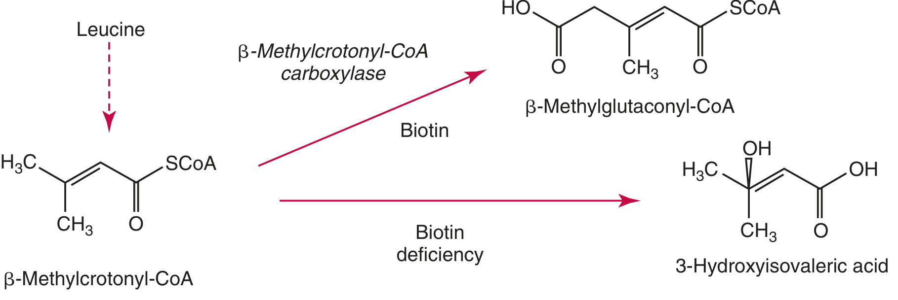
When biotin is deficient, the enzyme 3-methylcrotonyl-CoA carboxylase (which requires biotin) cannot function. This causes the leucine catabolism intermediate β-methylcrotonyl-CoA to be shunted to 3-hydroxyisovaleric acid instead of the normal product β-methylglutaconyl-CoA:
Formation of 3-hydroxyisovaleric acid in biotin deficiency
A leucine challenge test (oral leucine load followed by urine 3-HIVA measurement) can unmask marginal biotin deficiency not detectable on routine urine testing.

Neonatal / Newborn Screening

  • Biotinidase deficiency is included in newborn screening panels in many countries (including the US) because:
    • It is highly variable and may go unrecognized
    • Clinical features are entirely preventable with 5-20 mg biotin daily

Treatment

SituationBiotin Dose
Acquired deficiency (dietary/TPN)150 mcg/day until resolution
Holocarboxylase synthetase deficiency10-40 mg/day
Biotinidase deficiency5-10 mg/day (better outcomes)
(Sources: Harrison's Principles of Internal Medicine 22E; Fitzpatrick's Dermatology; Tietz Textbook of Laboratory Medicine 7th ed; Andrews' Diseases of the Skin)

You are not just an AI. You are my personal NEET PG professor, medical illustrator, memory coach, and exam strategist. My goal is to crack NEET PG. Assume that: I have completed MBBS. My fundamentals are weak. I have forgotten most concepts. I have very little time. I want to learn only through previous year questions. I want to understand every question deeply so I can answer similar questions in the future. Never assume I already know anything. For every MCQ I upload, follow the exact structure below. PART 1 — Decode the Question Before explaining anything, First tell me: What is the examiner actually asking? Explain in one simple sentence. Example: "This question is asking which ligament connects the liver to the anterior abdominal wall." Then explain "What keywords in the question should immediately make me think of this topic?" PART 2 — Explain Like I'm a 10th Class Student Now assume I know absolutely nothing. Explain from absolute basics. Teach me exactly like a school science teacher. Start from the beginning. Never use difficult medical words without explaining them. Whenever a medical word appears, First explain what the word means. Example: Ligament means a band of tissue. Peritoneum means the thin covering inside the abdomen. Falciform means sickle shaped. Then continue. Use analogies real-life examples stories comparisons Explain why everything exists. Explain how it works. Explain why the body needs it. Build the concept slowly. Never jump directly to the answer. PART 3 — Images During Every Explanation This is EXTREMELY IMPORTANT. Do NOT explain long paragraphs without visuals. Whenever an image would improve understanding, insert the relevant image immediately beside or after that explanation. Use multiple images if needed. Examples include: Anatomy diagrams Flowcharts Histology Embryology Clinical photographs Surgical images X-rays CT MRI Gross specimens Tables Comparative diagrams Mnemonic diagrams Blood supply diagrams Nerve diagrams Images should appear throughout the explanation. Not only at the beginning. Every important concept should have its own image whenever possible. PART 4 — Build the Concept Now teach the complete topic. Not just the answer. Teach Definition Anatomy Physiology Embryology Blood supply Nerve supply Relations Functions Clinical importance Diseases Surgical importance Radiology High-yield facts Recent NEET PG points Common confusions Important differentiating features Everything necessary to master the topic. PART 5 — Explain Like a Medical Professor Now assume I am an MBBS graduate preparing for NEET PG. Teach the same topic in detail. Include high-yield concepts clinical correlations embryology pathology radiology surgical relevance integrated anatomy physiology important investigations treatment whenever relevant recent NEET PG trends PYQ patterns memory tricks exam pearls PART 6 — Break Down Every Option Now analyze every option separately. For each option, tell me What is it? Why is it correct or incorrect? When would it become the correct answer? How can NEET PG ask about it? What diseases involve it? How can it be confused with the correct answer? Never simply say Wrong. Explain why. PART 7 — Comparison Table Make a comparison table between Correct option vs Every wrong option. Include Location Function Blood supply Embryology Clinical importance Important diseases Radiology NEET PG clues PART 8 — Memory Tricks Create easy mnemonics visual memory tricks story-based memory tricks exam memory hooks things impossible to forget. PART 9 — Clinical Scenario Create one clinical case. Teach how this concept appears in real patients. Explain diagnosis. Explain why. Explain management briefly. PART 10 — NEET PG Exam Strategy Tell me How should I solve this question in under 30 seconds? What clue should I immediately recognize? What trap did the examiner set? How can similar questions be framed? PART 11 — Similar Previous Year Questions Generate 5–10 similar NEET PG style MCQs. Explain each answer. PART 12 — Final Revision Sheet At the end give One-page revision including Most important facts Frequently confused points One-liners Image-based facts Rapid revision bullets Things to revise one day before exam PART 13 — Difficulty Level Mention ⭐ Easy ⭐⭐ Moderate ⭐⭐⭐ Difficult Explain why. PART 14 — What Should I Remember Forever? End with "If you remember only FIVE things from this topic, remember these." Teaching Rules Always teach in this order: Explain like a 10th class student. Then explain like a medical professor. Then solve the MCQ. Then explain every option. Then summarize. Never skip steps. Never assume prior knowledge. Never use unexplained medical terminology. Always define new words. Always use headings. Always use tables. Always use flowcharts wherever appropriate. Always include images whenever they improve understanding. Always emphasize NEET PG high-yield points. Always explain why wrong options are wrong. Teach until the topic becomes unforgettable. Pretend my NEET PG rank depends on your explanation. Every explanation should make me capable of answering any future question from that topic, not just this one.

Understood. From this point forward, I am your personal NEET PG Professor, Medical Illustrator, Memory Coach, and Exam Strategist.
Every MCQ you upload will be broken down using the full 14-part structure you've defined - no shortcuts, no assumed knowledge, no unexplained terminology.
Here's exactly what I'll do for every question:
PartWhat I'll Deliver
1Decode what the examiner is actually asking + keyword triggers
2Explain from absolute zero - 10th class level, analogies, stories
3Images, diagrams, flowcharts, tables throughout the explanation
4Full concept build - anatomy, physiology, embryology, blood supply, clinical
5Professor-level depth - high yield, PYQ patterns, exam pearls
6Every option dissected - why right, why wrong, when it becomes right
7Comparison table across all options
8Mnemonics, visual hooks, stories you cannot forget
9Clinical case scenario with diagnosis and management
1030-second exam strategy + traps the examiner set
115-10 similar NEET PG style MCQs with explanations
12One-page final revision sheet
13Difficulty rating with reason
14The 5 things you must remember forever

Ground rules I will always follow:
  • Never use a medical word without defining it first
  • Never skip a step
  • Never assume you know anything
  • Always build from simple → complex
  • Always include visuals where they add clarity
  • Always explain wrong options as thoroughly as the right one
  • Every explanation makes you capable of answering any future question on that topic, not just the one you uploaded

Upload your first MCQ whenever you're ready. A photograph, screenshot, or typed question - all work. Let's get your NEET PG rank.
Finding Sources
Finding Sources
Finding Sources
Reading File
Reading File
Finding Sources
Loading Image
Finding Sources
Finding Sources
Finding Sources
Finding Sources

✅ CORRECT ANSWER: C) Pyruvate Carboxylase


PART 1 - Decode the Question

What is the examiner actually asking?

"This question is asking which enzyme requires biotin as a cofactor - and which of the listed enzymes would be deficient when someone eats raw eggs (which block biotin absorption)."

Keywords that should immediately trigger your thinking:

Keyword in QuestionWhat it should make you think
"Raw eggs"→ Avidin → Blocks Biotin absorption
"Fatigue on moderate exercise"→ Hypoglycemia → Gluconeogenesis failing
"Bodybuilder"→ High protein diet, lots of raw eggs
"Enzyme deficient"→ Which enzyme NEEDS biotin to work?
"Recently started"→ Acquired deficiency, not genetic
One-line trigger rule: Whenever you see "raw egg white" in any NEET PG question → immediately think Biotin deficiency → Pyruvate Carboxylase fails.

PART 2 - Explain Like a 10th Class Student

Let's start from absolute zero.

Step 1: What is energy?

Your body is like a car. A car needs petrol to run. Your body needs glucose (a type of sugar) to run. Every muscle, every organ, your brain - all of them run on glucose.

Step 2: Where does your glucose come from?

Normally, you eat food. The food gives glucose. Your body stores extra glucose in the liver and muscles as glycogen (think of glycogen as a fuel tank).
But what happens when you exercise for a long time and your glucose runs out? Your body has a clever backup plan.

Step 3: The backup plan - Gluconeogenesis

Gluconeogenesis - let's break this word:
  • Gluco = glucose (sugar)
  • Neo = new
  • Genesis = creation/birth
So gluconeogenesis literally means "creating new glucose."
When you exercise and your sugar runs low, your liver acts like a factory and makes fresh glucose from other raw materials like:
  • Lactic acid (from tired muscles)
  • Amino acids (from proteins)
  • Glycerol (from fat)
This factory process keeps your blood sugar up so you don't collapse.

Step 4: The factory needs a tool

Imagine you're building something. You need tools. The gluconeogenesis factory needs an enzyme called Pyruvate Carboxylase as its very first tool - its first step.
Enzyme = a protein that speeds up a chemical reaction. Think of it as a machine in a factory.
Pyruvate Carboxylase converts Pyruvate → Oxaloacetate (OAA).
Think of it like this:
  • Pyruvate = raw material (scrap metal)
  • Oxaloacetate = the first processed product
  • From OAA, the factory eventually makes Glucose
Without this first step, the whole glucose factory shuts down.

Step 5: But this tool needs a power source - Biotin

Pyruvate Carboxylase CANNOT work without Biotin (Vitamin B7).
Biotin is like the battery of this enzyme. Without the battery, the machine won't start.
Biotin = a water-soluble vitamin (vitamin that dissolves in water). Also called Vitamin B7 or Vitamin H.

Step 6: Now enters the villain - Avidin from Raw Eggs

Avidin = a protein found in raw egg whites.
Think of avidin like a super-magnet that grabs onto biotin in your gut and holds it so tight that your intestine cannot absorb it.
So the sequence is:
Raw egg white eaten
        ↓
Avidin (in egg white) enters your gut
        ↓
Avidin grabs Biotin tightly
        ↓
Biotin cannot be absorbed into blood
        ↓
Blood has no Biotin
        ↓
Pyruvate Carboxylase has no battery → can't work
        ↓
Gluconeogenesis fails
        ↓
Blood glucose drops during exercise
        ↓
FATIGUE on moderate exercise ✓
Important: If you COOK the eggs, heat destroys avidin. So cooked eggs are safe! Bodybuilders who eat raw eggs get this problem.

PART 3 - Images During Every Explanation

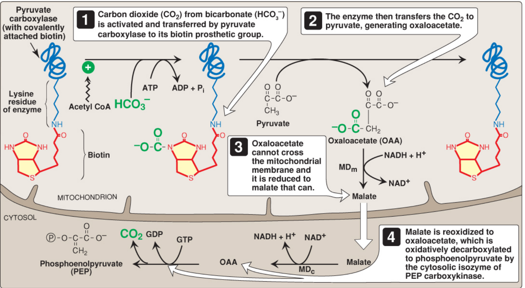
The Pyruvate Carboxylase Reaction - How Biotin Works

This diagram from Lippincott's Biochemistry shows exactly how biotin sits on the enzyme and transfers CO₂ to pyruvate:
Pyruvate carboxylase with biotin - step by step mechanism
Reading this diagram:
  • Top left: Pyruvate carboxylase with biotin covalently (firmly) attached to a lysine residue (part of the enzyme)
  • Step 1: CO₂ from bicarbonate (HCO₃⁻) is activated using ATP energy and attached to biotin
  • Step 2: Biotin transfers that CO₂ to pyruvate → makes Oxaloacetate (OAA)
  • Step 3: OAA cannot cross the mitochondrial wall, so it becomes malate
  • Step 4: Malate exits, becomes OAA again in cytosol, then PEPCK converts it to PEP → eventually becomes Glucose

PART 4 - Build the Complete Concept

BIOTIN (Vitamin B7) - Complete Topic

Definition

Biotin is a water-soluble B-vitamin (also called Vitamin H or Vitamin B7) that functions as a coenzyme (cofactor) for 4 carboxylase enzymes in the human body.
Carboxylase = an enzyme that adds a CO₂ group to a molecule.

The 4 Biotin-Dependent Enzymes - THE MOST IMPORTANT TABLE IN THIS TOPIC

#EnzymeReactionPathwayLocation
1Pyruvate Carboxylase (PC)Pyruvate → OAAGluconeogenesisMitochondria (liver, kidney)
2Acetyl-CoA Carboxylase (ACC)Acetyl-CoA → Malonyl-CoAFatty acid synthesisCytosol
3Propionyl-CoA CarboxylasePropionyl-CoA → Methylmalonyl-CoAOdd-chain FA / amino acid catabolismMitochondria
43-Methylcrotonyl-CoA Carboxylase3-Methylcrotonyl-CoA → 3-Methylglutaconyl-CoALeucine catabolismMitochondria
NEET PG PEARL: All 4 use biotin. Biotin deficiency = ALL 4 fail. The one asked most in NEET PG = Pyruvate Carboxylase (because it causes fatigue/hypoglycemia).
Mnemonic for 4 biotin enzymes: "PPAM"
  • Pyruvate carboxylase
  • Propionyl-CoA carboxylase
  • Acetyl-CoA carboxylase
  • 3-Methylcrotonyl-CoA carboxylase

Gluconeogenesis - The Complete Pathway

Gluconeogenesis = Synthesis of glucose from non-carbohydrate sources.
Where: Mainly liver (90%), some in kidney (10%)
Precursors (raw materials):
  • Lactate (from RBCs, exercising muscle)
  • Glycerol (from fat breakdown)
  • Glucogenic amino acids (especially alanine, glutamine)
  • Odd-chain fatty acids → propionyl-CoA
The 3 Irreversible "Roadblocks" of Glycolysis (that gluconeogenesis must bypass):
Glycolysis Step (irreversible)Gluconeogenesis Bypass Enzyme
Pyruvate Kinase (PEP → Pyruvate)Pyruvate Carboxylase + PEPCK (two-step bypass)
Phosphofructokinase-1 (F6P → F1,6-BP)Fructose-1,6-bisphosphatase
Glucokinase/Hexokinase (Glucose → G6P)Glucose-6-phosphatase
Flow of gluconeogenesis:
Pyruvate
   ↓ [Pyruvate Carboxylase + BIOTIN] ← KEY STEP
Oxaloacetate (OAA)
   ↓ [PEPCK - uses GTP]
Phosphoenolpyruvate (PEP)
   ↓ [several reversible steps]
Fructose-1,6-bisphosphate
   ↓ [Fructose-1,6-bisphosphatase]
Fructose-6-phosphate
   ↓
Glucose-6-phosphate
   ↓ [Glucose-6-phosphatase - in liver ER]
FREE GLUCOSE → released into blood

Sources of Biotin

  • Organ meats (liver, kidney)
  • Egg YOLK (not white!)
  • Nuts, legumes, mushrooms, yeast
  • Gut bacteria also synthesize biotin

Absorption of Biotin

  • Free biotin is absorbed in small intestine
  • Intestinal biotinidase releases protein-bound biotin from food

Why Raw Egg White Causes Deficiency

  • Raw egg white contains AVIDIN - a glycoprotein
  • Avidin binds biotin with extremely high affinity (Kd ~10⁻¹⁵ M - one of the strongest non-covalent bonds in nature)
  • This binding prevents intestinal absorption
  • Heat (cooking) denatures avidin → cooked eggs are safe

Energy Requirement of Pyruvate Carboxylase

  • Requires ATP (1 ATP consumed per reaction)
  • Requires Mg²⁺ as cofactor
  • Activated allosterically by Acetyl-CoA (high Acetyl-CoA = need more OAA = activate PC)

PART 5 - Explain Like a Medical Professor

High-Yield Professor Points

Regulation of Pyruvate Carboxylase

Pyruvate Carboxylase is ACTIVATED by Acetyl-CoA - this is a brilliant physiological design:
  • When fasting: Fat is broken down → lots of Acetyl-CoA accumulates
  • High Acetyl-CoA signals: "We have plenty of fat fuel, now make glucose too"
  • Acetyl-CoA activates PC → gluconeogenesis starts
  • Result: Blood glucose is maintained during fasting
Without biotin: PC cannot be activated even if Acetyl-CoA is high.

Location Details

  • PC reaction occurs inside mitochondria
  • OAA cannot cross inner mitochondrial membrane
  • OAA is converted to Malate (by mitochondrial malate dehydrogenase using NADH)
  • Malate crosses membrane → in cytosol, converted back to OAA
  • Cytosolic OAA is acted on by PEPCK (using GTP) → PEP
This clever shuttle also moves NADH reducing equivalents from mitochondria to cytosol (needed for gluconeogenesis).

Pyruvate Carboxylase Deficiency - Clinical Disease

  • Inherited (rare genetic): Autosomal recessive
  • Presents in neonates with:
    • Severe lactic acidosis (pyruvate backs up → lactate)
    • Hypoglycemia
    • Hyperammonemia
    • Brain damage / developmental delay
  • Treatment: Biotin supplementation (for acquired), special diet for genetic forms

PEPCK (Option D) - Important Distinctions

PEPCK = Phosphoenolpyruvate Carboxykinase
  • Catalyzes: OAA → PEP + CO₂ (uses GTP)
  • Does NOT require biotin - it uses GTP
  • Is the rate-limiting step of gluconeogenesis in many contexts
  • Induced by: Glucagon, glucocorticoids, fasting
  • Inhibited by: Insulin
  • PEPCK deficiency → hypoglycemia, lactic acidosis (but unrelated to biotin/raw eggs)

Von Gierke Disease (Option A connection)

  • Glucose-6-phosphatase deficiency = Glycogen Storage Disease Type I
  • Cannot release free glucose from G6P in liver
  • Features: Severe hypoglycemia, MASSIVE hepatomegaly (enlarged liver), hyperlipidemia, hyperuricemia, lactic acidosis
  • Key clue: Fasting hypoglycemia + huge liver in infant

McArdle Disease (Option B connection)

  • Muscle Glycogen Phosphorylase deficiency = GSD Type V
  • Cannot break down glycogen in muscles during exercise
  • Features: Muscle cramps/pain during exercise, myoglobinuria (dark urine), no lactic acid rise on exercise (classic sign)
  • Key clue: Exercise-induced muscle cramps + myoglobinuria + NO lactate rise on forearm ischemic exercise test

PART 6 - Break Down Every Option

Option A: Glucose-6-Phosphatase ❌

What is it? An enzyme located in the endoplasmic reticulum of liver cells (and kidney). It removes the phosphate group from Glucose-6-Phosphate to release FREE glucose into the bloodstream.
Why is it wrong here?
  • It does NOT require biotin
  • It is not involved in the pyruvate carboxylase step
  • Raw egg whites affect biotin absorption, which has no connection to glucose-6-phosphatase
  • G6Pase deficiency is a genetic disease (Von Gierke) - not caused by diet
When would it be the correct answer?
  • "Infant with severe fasting hypoglycemia + massively enlarged liver + lactic acidosis + hyperuricemia (gout)"
  • "Which enzyme is deficient in Von Gierke disease?"
  • "Glycogen Storage Disease Type I - enzyme deficient?"
How to distinguish from pyruvate carboxylase:
  • G6Pase = liver/kidney enzyme, diet has no effect, causes hepatomegaly
  • PC = mitochondrial enzyme, affected by biotin/raw egg diet, no hepatomegaly

Option B: Glycogen Phosphorylase ❌

What is it? An enzyme that breaks down glycogen (stored glucose) by cleaving glucose units from the glycogen chain. There are two forms:
  • Liver glycogen phosphorylase = releases glucose into blood
  • Muscle glycogen phosphorylase (myophosphorylase) = provides fuel for muscle
Why is it wrong here?
  • Does NOT require biotin
  • Raw eggs have zero effect on glycogen phosphorylase
  • McArdle disease (muscle GP deficiency) presents with cramps ONLY during exercise, not simple fatigue
  • The question says "fatigue on moderate exercise" not "severe cramps + dark urine"
When would it be the correct answer?
  • "A young patient develops painful muscle cramps during exercise with no rise in lactate on ischemic forearm test - enzyme deficient?"
  • "GSD Type V - which enzyme?"
  • "Myoglobinuria after exercise - which GSD?"
How to distinguish from pyruvate carboxylase:
  • GP deficiency: Cramps, myoglobinuria, no lactate rise on exercise test - no dietary link
  • PC deficiency: Simple fatigue, hypoglycemia, linked to raw egg/biotin issue

Option C: Pyruvate Carboxylase ✅ CORRECT

What is it? A mitochondrial enzyme that converts Pyruvate → Oxaloacetate using biotin as cofactor and ATP as energy source. This is the first and committed step of gluconeogenesis.
Why is it correct?
  • REQUIRES BIOTIN as its essential cofactor
  • Raw egg white → Avidin → blocks biotin → PC has no cofactor → fails
  • PC failure → gluconeogenesis fails → blood glucose cannot be maintained during exercise
  • Result: Fatigue on moderate exercise = hypoglycemia
Complete chain of logic:
Raw eggs → Avidin → Biotin blocked → PC has no biotin → 
Gluconeogenesis fails → Hypoglycemia → Fatigue ✓

Option D: Phosphoenolpyruvate Carboxykinase (PEPCK) ❌

What is it? An enzyme in the cytosol (and mitochondria) that converts Oxaloacetate → Phosphoenolpyruvate (PEP) using GTP. It is the second step of gluconeogenesis (right after pyruvate carboxylase).
Why is it wrong?
  • Does NOT require biotin - uses GTP as its energy/cofactor source
  • Not affected by avidin or raw egg consumption
  • PEPCK is stimulated by glucagon and cortisol, inhibited by insulin
The subtle TRAP the examiner set: PEPCK is in the same gluconeogenesis pathway as pyruvate carboxylase! Both enzymes together bypass the pyruvate kinase irreversible step. A student who knows "gluconeogenesis is failing" but doesn't know which enzyme needs biotin might pick PEPCK.
The key differentiator: Biotin cofactor = Pyruvate Carboxylase. NOT PEPCK.
When would PEPCK be the correct answer?
  • "Which enzyme is rate-limiting for gluconeogenesis and induced by glucagon?"
  • "Which enzyme converts OAA to PEP and uses GTP?"
  • "Insulin inhibits which gluconeogenic enzyme at the gene expression level?"

PART 7 - Comparison Table

FeaturePyruvate Carboxylase ✅Glucose-6-PhosphataseGlycogen PhosphorylasePEPCK
PathwayGluconeogenesis (Step 1)Gluconeogenesis (last step) / GlycogenolysisGlycogenolysisGluconeogenesis (Step 2)
Substrate → ProductPyruvate → OAAG6P → Glucose + PiGlycogen → G1POAA → PEP
Cofactor/CoenzymeBIOTIN + ATP + Mg²⁺None (hydrolase)Pyridoxal phosphate (B6)GTP
Location in cellMitochondriaER (liver, kidney)CytosolCytosol + Mitochondria
OrganLiver, Kidney, MuscleLiver, Kidney, IntestineLiver + MuscleLiver, Kidney
Activated byAcetyl-CoA-Glucagon, EpinephrineGlucagon, Glucocorticoids
Inhibited byLow Acetyl-CoA-Insulin, GlucoseInsulin
Deficiency diseasePyruvate carboxylase def. (neonatal acidosis)Von Gierke (GSD I)McArdle (GSD V)PEPCK deficiency
Key clinical featureLactic acidosis, hypoglycemiaHepatomegaly + severe hypoglycemiaExercise cramps + myoglobinuriaHypoglycemia + liver disease
Raw egg link?YES - biotin dependentNoNoNo
NEET PG triggerRaw eggs, biotin deficiency, fatigueInfant, big liver, fasting hypoglycemiaExercise cramps, no lactate riseInsulin inhibits gluconeogenesis

PART 8 - Memory Tricks

Mnemonic 1: The 4 Biotin Enzymes - "PPAM"

Pyruvate carboxylase Propionyl-CoA carboxylase Acetyl-CoA carboxylase 3-Methylcrotonyl-CoA carboxylase
Say it like: "PP-AM" (two P's, then AM)

Mnemonic 2: The Avidin Story

"Avidin is a VILLAIN in raw eggs who STEALS Biotin from your body"
Visual: Imagine a villain named AVID-IN (he's avid/greedy) who snatches your biotin vitamin pill right before you can swallow it.

Mnemonic 3: Pyruvate Carboxylase cofactors

"Biotin ATP Magnesium" = "BAM!" 🔨
Biotin + ATP + Magnesium = BAM (pyruvate gets "BAM"'d into oxaloacetate)

Mnemonic 4: Which enzyme goes with which GSD?

"Von Gierke's Got Gout and a Giant liver" = Glucose-6-Phosphatase (GSD I) "McArdle Makes Muscles Mad during Marathon" = Muscle Glycogen Phosphorylase (GSD V)

Mnemonic 5: PEPCK cofactor

"PEPCK uses GTP - G for Glucagon loves it" Glucagon → stimulates PEPCK → makes glucose

Visual Hook: The Raw Egg Chain

🥚 Raw Egg → 🦹 Avidin → 🔒 Locks Biotin → 
💔 Pyruvate Carboxylase broken → 
🏭 Gluconeogenesis factory shuts → 
😓 Fatigue on exercise

PART 9 - Clinical Scenario

Case

A 22-year-old fitness enthusiast visits a gym 6 days a week. He drinks a protein shake every morning made with 6 raw egg whites. He has been doing this for 4 months. He now complains of weakness and fatigue even after mild workouts. He also has a scaly rash around his mouth and eyes, and his hair is thinning. He denies any medications.
Blood tests: Low blood glucose after exercise, low serum biotin, elevated urinary 3-hydroxyisovaleric acid.

What is happening?

He has biotin deficiency caused by chronic raw egg white consumption.
  • Avidin in raw eggs chronically blocked biotin absorption
  • Over 4 months, biotin stores depleted
  • Now all 4 biotin-dependent carboxylases are failing:
    • Pyruvate Carboxylase fails → gluconeogenesis impaired → hypoglycemia → fatigue
    • 3-Methylcrotonyl-CoA carboxylase fails → elevated 3-hydroxyisovaleric acid in urine
    • Acetyl-CoA carboxylase fails → fatty acid synthesis impaired → hair, skin affected
  • Skin findings = scaly periorificial dermatitis (around mouth, eyes) - classic biotin deficiency rash
  • Alopecia (hair loss) - classic biotin deficiency sign

Diagnosis

  • Low serum biotin
  • Elevated urinary 3-hydroxyisovaleric acid (functional marker - most sensitive)
  • Clinical features: rash + alopecia + neurological fatigue

Management

  • Stop raw eggs (cook them!)
  • Oral biotin supplementation - 150 mcg/day for acquired deficiency
  • Symptoms resolve within weeks

PART 10 - NEET PG Exam Strategy

How to solve this in under 30 seconds:

The 3-second rule:

  1. See "raw eggs" → immediately think Avidin → Biotin deficiency
  2. Ask yourself: "Which enzyme from the options needs Biotin?"
  3. Only ONE of the 4 options needs biotin = Pyruvate Carboxylase
  4. Mark C. Done.

The Trap the Examiner Set:

Trap 1: The question says "fatigue on moderate exercise" - beginners think of glycogen breakdown diseases (McArdle = Glycogen Phosphorylase). But the KEY differentiator is "RAW EGGS" - McArdle has nothing to do with diet.
Trap 2: PEPCK is in the same pathway (gluconeogenesis). A student who thinks "gluconeogenesis is failing → pick PEPCK" falls into this trap. Remember: PEPCK uses GTP, not biotin.
Trap 3: Glucose-6-phosphatase also affects glucose release, but it's a Von Gierke disease enzyme, has no biotin requirement, and presents in infants with hepatomegaly - not bodybuilders.

How similar questions can be framed:

  • "A patient on long-term TPN (IV feeding) develops alopecia and fatigue - enzyme deficient?"
  • "Which enzyme requires biotin and activates gluconeogenesis?" → Pyruvate Carboxylase
  • "A neonate with lactic acidosis, hypoglycemia, hyperammonemia - enzyme deficient?" → Pyruvate Carboxylase
  • "Avidin binds which vitamin?" → Biotin
  • "Which vitamin is deficient in raw egg white consumers?" → Biotin (B7)
  • "Biotin is cofactor for how many carboxylases?" → 4

PART 11 - Similar NEET PG Style MCQs

Q1. A patient on prolonged total parenteral nutrition develops alopecia, perioral dermatitis, and fatigue. The enzyme most likely deficient is:
  • A) Pyruvate Dehydrogenase
  • B) Pyruvate Carboxylase ✅
  • C) Lactate Dehydrogenase
  • D) Phosphoglycerate kinase
Answer: B - TPN without biotin → biotin deficiency → pyruvate carboxylase fails. Same mechanism as raw eggs, different cause.

Q2. Which of the following enzymes uses GTP as its energy source in gluconeogenesis?
  • A) Pyruvate Carboxylase
  • B) Glucose-6-Phosphatase
  • C) Phosphoenolpyruvate Carboxykinase ✅
  • D) Fructose-1,6-Bisphosphatase
Answer: C - PEPCK is the only gluconeogenic enzyme using GTP (not ATP). Pyruvate carboxylase uses ATP.

Q3. A 6-month-old infant presents with severe hypoglycemia, massively enlarged liver, lactic acidosis, and elevated triglycerides. Enzyme deficient:
  • A) Glycogen Phosphorylase
  • B) Glucose-6-Phosphatase ✅
  • C) Pyruvate Carboxylase
  • D) Aldolase B
Answer: B - Classic Von Gierke disease (GSD I). Infant + big liver + severe hypoglycemia + lactic acidosis = G6Pase deficiency.

Q4. A 19-year-old gets severe muscle cramps during a 400m race. Post-exercise urine is dark red. Forearm ischemic exercise test shows NO rise in blood lactate. Enzyme deficient:
  • A) Pyruvate Carboxylase
  • B) Glucose-6-Phosphatase
  • C) Phosphoglycerate Kinase
  • D) Muscle Glycogen Phosphorylase ✅
Answer: D - McArdle disease (GSD V). Exercise cramps + myoglobinuria (dark urine) + NO lactate rise = muscle GP deficiency. The muscle cannot break down glycogen → no lactate produced.

Q5. Biotin acts as a cofactor by carrying which group?
  • A) Methyl group
  • B) Carboxyl group (CO₂) ✅
  • C) Amino group
  • D) Phosphate group
Answer: B - Biotin is the "CO₂ carrier" of the body. It picks up CO₂ and transfers it to substrates in carboxylation reactions.

Q6. Which of the following is NOT a biotin-dependent enzyme?
  • A) Acetyl-CoA carboxylase
  • B) Propionyl-CoA carboxylase
  • C) Pyruvate Carboxylase
  • D) Phosphoenolpyruvate Carboxykinase ✅
Answer: D - PEPCK uses GTP, not biotin. All others are the 4 biotin-dependent carboxylases.

Q7. Pyruvate carboxylase is allosterically activated by:
  • A) AMP
  • B) ATP
  • C) Acetyl-CoA ✅
  • D) Malonyl-CoA
Answer: C - High Acetyl-CoA (during fasting/fat oxidation) activates PC to push gluconeogenesis forward.

Q8. A patient with biotin deficiency will have elevated urinary levels of which organic acid?
  • A) Methylmalonic acid
  • B) Pyruvic acid
  • C) 3-Hydroxyisovaleric acid ✅
  • D) Homocysteine
Answer: C - 3-HIVA is the best functional marker of biotin deficiency. It accumulates because 3-methylcrotonyl-CoA carboxylase (biotin-dependent) cannot work in leucine metabolism.

PART 12 - Final Revision Sheet (One Page)


⚡ BIOTIN DEFICIENCY - RAPID REVISION

The Story in One Line:

Raw egg → Avidin → Blocks Biotin → Pyruvate Carboxylase fails → No Gluconeogenesis → Hypoglycemia → Fatigue

4 Biotin-Dependent Enzymes (PPAM):

  1. Pyruvate Carboxylase → Gluconeogenesis
  2. Propionyl-CoA Carboxylase → Odd-chain FA / Val, Ile, Met catabolism
  3. Acetyl-CoA Carboxylase → Fatty acid synthesis
  4. 3-Methylcrotonyl-CoA Carboxylase → Leucine catabolism

Pyruvate Carboxylase Quick Facts:

  • Cofactors: Biotin + ATP + Mg²⁺ (BAM!)
  • Location: Mitochondria (liver, kidney)
  • Reaction: Pyruvate → OAA
  • Activated by: Acetyl-CoA
  • Purpose: Step 1 of gluconeogenesis + replenish TCA cycle

PEPCK vs PC:

Pyruvate CarboxylasePEPCK
CofactorBIOTIN + ATPGTP
ReactionPyruvate → OAAOAA → PEP
Raw egg linkYESNo

Key Disease Associations:

DiseaseEnzymeClue
Von Gierke (GSD I)Glucose-6-PhosphataseBaby + big liver + severe hypoglycemia
McArdle (GSD V)Muscle Glycogen PhosphorylaseExercise cramps + myoglobinuria + no lactate
Biotin deficiencyPyruvate Carboxylase (+ 3 others)Raw eggs + rash + alopecia + fatigue

Urine Marker of Biotin Deficiency:

  • 3-Hydroxyisovaleric acid ↑ (most sensitive)

Treatment:

  • Acquired: 150 mcg biotin/day
  • Biotinidase deficiency: 5-10 mg biotin/day

One Day Before Exam - Remember:

  • ✅ Raw egg = Avidin = Biotin deficiency = Pyruvate Carboxylase
  • ✅ PEPCK uses GTP not biotin
  • ✅ 4 biotin enzymes = PPAM
  • ✅ McArdle = exercise cramps + no lactate rise (NOT biotin)
  • ✅ Von Gierke = big liver + severe hypoglycemia in baby (NOT biotin)
  • ✅ Biotin marker in urine = 3-Hydroxyisovaleric acid

PART 13 - Difficulty Level

⭐⭐ Moderate

Why moderate?
  • The raw egg → avidin → biotin link is a well-known fact
  • The connection to pyruvate carboxylase (not PEPCK) requires knowing which enzymes use biotin
  • The main challenge is distinguishing pyruvate carboxylase from PEPCK (both are in gluconeogenesis)
  • Knowing the cofactor (biotin vs GTP) is the discriminating knowledge
A student who only memorized "gluconeogenesis" without knowing cofactors would get it wrong. That's what makes it moderate, not easy.

PART 14 - If You Remember ONLY 5 Things Forever

1. Raw egg white → Avidin → Steals Biotin → Pyruvate Carboxylase fails
2. Pyruvate Carboxylase cofactors = BAM (Biotin + ATP + Mg²⁺)
3. PEPCK uses GTP (NOT Biotin) - second step of gluconeogenesis bypass
4. 4 Biotin enzymes = PPAM (Pyruvate, Propionyl, Acetyl, 3-Methylcrotonyl carboxylases)
5. McArdle = Muscle cramps + no lactate rise on exercise (glycogen phosphorylase, NOT biotin)

Sources: Lippincott's Illustrated Reviews Biochemistry 8th Ed; Tietz Textbook of Laboratory Medicine 7th Ed; Harrison's Principles of Internal Medicine 22E; Robbins Pathology; Basic Medical Biochemistry 6th Ed
Finding Sources
Finding Sources
Reading File
Finding Sources
Finding Sources
Finding Sources
Finding Sources
Reading File
Finding Sources
Loading Image
Reading File

✅ CORRECT ANSWER: D) Pterygoid Branch

The pterygoid branch is NOT a branch of the cavernous part of the ICA. It is a branch of the maxillary artery (from external carotid artery).

PART 1 - Decode the Question

What is the examiner actually asking?

"This question is asking you to identify which one of the four options does NOT arise from the cavernous (C4) segment of the internal carotid artery."

Keywords that should immediately trigger your thinking:

KeywordWhat it triggers
"Cavernous part"→ ICA inside the cavernous sinus → remember its specific branches
"Internal carotid artery"→ Think ICA segments: Cervical, Petrous, Cavernous, Cerebral
"Not a branch"→ One odd one out - look for what belongs to EXTERNAL carotid or another artery
"Pterygoid"→ Pterygoid muscles, pterygoid fossa, pterygoid plexus → these are supplied by EXTERNAL carotid (maxillary artery)
30-second trigger: Pterygoid = pterygoid muscles = external carotid territory. ICA does NOT supply pterygoid muscles.

PART 2 - Explain Like a 10th Class Student

Start from Zero - What is the Carotid Artery?

Step 1: The Brain Needs Blood

Your brain is the most important organ in your body. It needs a constant, uninterrupted supply of blood carrying oxygen and glucose. Even 4-5 seconds without blood = you faint. Even 4-5 minutes = brain damage.
So the body has built a very important highway system to deliver blood specifically to the brain.

Step 2: Two Big Roads to the Brain

The blood going to the brain travels through two main artery systems:
  1. Carotid arteries - supply the front part of the brain
  2. Vertebral arteries - supply the back part of the brain
The Carotid artery (carotid comes from Greek "karos" meaning deep sleep - because if you press it, you faint) divides into two:
  • External Carotid Artery (ECA) = supplies face, neck, scalp
  • Internal Carotid Artery (ICA) = enters the skull, supplies the brain

Step 3: Think of ICA as a Delivery Truck on a Complex Route

The Internal Carotid Artery doesn't go directly to the brain in a straight line. It takes a complicated path through the skull, like a delivery truck winding through narrow alleys.
The ICA has 4 major segments based on where it travels:
SegmentLocationSimple Analogy
CervicalNeck (from bifurcation to skull base)Truck on the open highway
PetrousInside the petrous (rocky) temporal boneTruck going through a narrow stone tunnel
CavernousInside the cavernous sinus (blood pool near the brain)Truck passing through a guarded security checkpoint
CerebralInside the skull, in the brainTruck finally inside the city, delivering to buildings

Step 4: What is the Cavernous Sinus?

Sinus = a space or cavity.
Cavernous sinus = a large, cave-like venous blood pool (like a lake full of blood) located:
  • On either side of the pituitary gland (the master hormone gland at the base of the brain)
  • Just behind the eyes
  • Above the sphenoid bone
Cavernous comes from "cavern" = cave. This sinus is full of tiny holes and channels, like a sea sponge, which is why it is called cavernous.
Think of it as a security checkpoint booth - very important, very tightly guarded, with many critical structures passing through it.

Step 5: What Passes THROUGH the Cavernous Sinus?

This is HIGH YIELD. The cavernous sinus contains:
Inside the sinus (in the blood):
  • Internal Carotid Artery (ICA) - the main artery running right through it
  • Abducens nerve (CN VI) - moves the eye outward
In the wall of the sinus (just outside, embedded in the wall):
  • Oculomotor nerve (CN III) - moves the eye up, down, in
  • Trochlear nerve (CN IV) - moves the eye down and inward
  • Ophthalmic nerve V1 - sensation of upper face/eye
  • Maxillary nerve V2 - sensation of mid-face

Step 6: The Cavernous Part of ICA Gives Branches Inside This Sinus

While the ICA is passing through the cavernous sinus, it gives off small branches to nearby structures. These are the branches of the cavernous part.

PART 3 - Images During Every Explanation

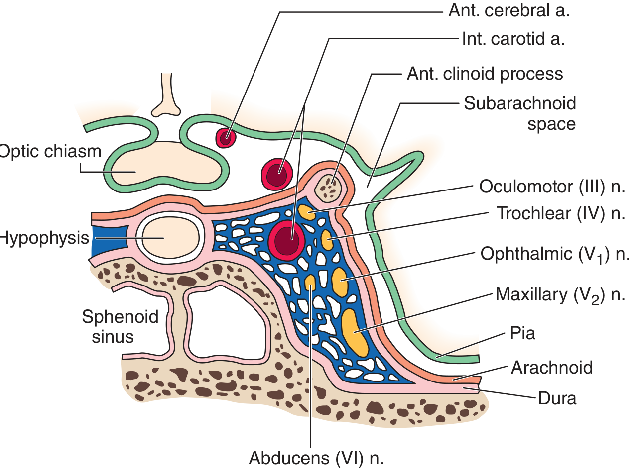
The Cavernous Sinus - Coronal Section (Harrison's Anatomy)

This diagram shows exactly what sits inside the cavernous sinus, and where the ICA runs:
Cavernous sinus anatomy in coronal section showing ICA, cranial nerves III, IV, V1, V2, VI
Read this diagram carefully:
  • The blue area = the cavernous sinus (blood-filled cave)
  • The red circle in the center = Internal Carotid Artery running through the sinus
  • On the outer wall (yellow blobs) = CN III (oculomotor), CN IV (trochlear), CN V1 (ophthalmic), CN V2 (maxillary)
  • CN VI (abducens) = runs freely inside the sinus blood, most vulnerable to pressure
  • To the left = Hypophysis (pituitary gland) and sphenoid sinus below it

PART 4 - Build the Complete Concept

THE INTERNAL CAROTID ARTERY - Complete Topic

The 4 Segments and Their Branches

Segment 1: Cervical Part (C1)

  • From: Common carotid bifurcation (at C4 vertebra level)
  • To: Entry into carotid canal in skull base
  • Branches: NONE (This is the most tested fact about cervical ICA!)
  • Mnemonic: "Cervical ICA is celibate - gives nothing"

Segment 2: Petrous Part (C2)

  • Travels through the petrous part of temporal bone
  • Branches:
    • Caroticotympanic artery → supplies tympanic cavity (middle ear)
    • Vidian artery (artery of pterygoid canal) → anastomoses with maxillary artery (variable, not always present)

Segment 3: Cavernous Part (C3/C4) ← THIS IS THE QUESTION

  • Travels through the cavernous sinus (makes two curves = carotid siphon)
  • Branches (THE ANSWER ZONE):
BranchWhat it supplies
Meningeal branchesSupply dura mater of middle cranial fossa
Inferior hypophyseal arterySupplies posterior pituitary (neurohypophysis)
Cavernous branchesSupply the cavernous sinus walls and contents
Ganglionic branchesSupply trigeminal (Gasserian) ganglion
These branches arise from the meningohypophyseal trunk and the inferolateral trunk (also called the artery of the inferior cavernous sinus).

Segment 4: Cerebral Part (C5/Supraclinoid)

  • Enters subarachnoid space at the anterior clinoid process
  • Branches:
    • Ophthalmic artery (first branch - enters orbit with optic nerve)
    • Superior hypophyseal arteries (supply anterior pituitary/adenohypophysis)
    • Posterior communicating artery
    • Anterior choroidal artery
    • Terminates as: Anterior cerebral artery (ACA) + Middle cerebral artery (MCA)

The Complete ICA Segment Map:

Common Carotid Artery
         ↓
   ICA origin (C4 neck level)
         ↓
CERVICAL PART → No branches
         ↓
PETROUS PART → Caroticotympanic a. + Vidian a.
         ↓
CAVERNOUS PART → Meningeal + Inferior hypophyseal + Cavernous + Ganglionic branches
         ↓
CEREBRAL PART → Ophthalmic a. → Superior hypophyseal → Posterior comm. → Ant. Choroidal
         ↓
    Terminates as ACA + MCA

What is the Pterygoid Branch - and Why is it WRONG?

Pterygoid comes from Greek "pteron" = wing + "eidos" = like. The pterygoid muscles are the wing-shaped muscles of mastication (chewing) on the side of the jaw.
Pterygoid branches are small arteries that supply the pterygoid muscles and arise from the maxillary artery - which is a terminal branch of the External Carotid Artery.
The ICA has absolutely nothing to do with the pterygoid region. The ICA is exclusively intracranial (inside skull). It never goes near the pterygoid muscles in the face/jaw.

PART 5 - Explain Like a Medical Professor

High-Yield Facts

The Meningohypophyseal Trunk

The most important branch group of the cavernous ICA is the Meningohypophyseal Trunk. This single trunk gives off:
  1. Tentorial artery (of Bernasconi-Cassinari) - supplies tentorium cerebelli
  2. Inferior hypophyseal artery - supplies neurohypophysis (posterior pituitary)
  3. Dorsal meningeal artery - supplies dura of dorsum sella
NEET PG Pearl: The tentorial artery is a marker for meningioma on angiography - meningiomas along the tentorium get blood supply through this artery.

The Inferolateral Trunk (Artery of the Inferior Cavernous Sinus)

The second branch group:
  • Supplies cranial nerves III, IV, V (trigeminal ganglion), VI within the cavernous sinus
  • Anastomoses with branches of the external carotid artery (accessory meningeal artery)

Cavernous Branch

The cavernous branches supply:
  • Walls of the cavernous sinus itself
  • Adjacent dura mater

Inferior vs. Superior Hypophyseal Arteries

Inferior Hypophyseal ArterySuperior Hypophyseal Artery
OriginCavernous part of ICACerebral part (supraclinoid) of ICA
SuppliesPosterior pituitary (neurohypophysis)Anterior pituitary (adenohypophysis) via portal system
Hormone stored hereADH, OxytocinGH, TSH, ACTH, FSH, LH, Prolactin

Clinical Correlation: Carotid Cavernous Fistula (CCF)

What is it? An abnormal connection between the ICA (high pressure artery) and the cavernous sinus (low pressure venous lake).
Causes:
  • Trauma (most common - base of skull fracture)
  • Spontaneous rupture of ICA aneurysm within sinus
Features (classical triad):
  1. Pulsatile exophthalmos (eyeball bulges and pulsates with each heartbeat)
  2. Orbital bruit (you can hear a "whooshing" sound over the eye with stethoscope)
  3. Chemosis (conjunctival edema/redness)
Why? Arterial blood under high pressure floods the sinus → venous drainage backs up → ophthalmic veins engorge → eye bulges.

Cavernous Sinus Thrombosis

What is it? Clot formation in the cavernous sinus.
Cause: Usually infection spreading from:
  • Danger zone of face (upper lip, nose triangle → angular/ophthalmic vein → cavernous sinus)
  • Orbital cellulitis
  • Sinusitis (especially mucormycosis in diabetics)
Features: The structures that pass THROUGH the cavernous sinus get affected:
  • CN III palsy → drooping eyelid (ptosis), eye deviated down and out
  • CN IV palsy → inability to look down/inward
  • CN VI palsy → inability to look outward (medial deviation of eye)
  • CN V1, V2 → numbness of upper face
  • Proptosis (eye bulging) + fever + headache

PART 6 - Break Down Every Option

Option A: Inferior Hypophyseal Branch ✅ IS a branch (so wrong answer to the question)

What is it? A branch of the cavernous part of ICA that supplies the posterior pituitary gland (neurohypophysis).
Why is it a real branch? Both Color Atlas of Human Anatomy and Adams & Victor's Neurology confirm: "The main blood supply to the posterior pituitary is from the inferior hypophyseal artery, which is a branch of the cavernous part of the internal carotid artery."
What does it supply? The posterior pituitary stores and releases:
  • ADH (Anti-Diuretic Hormone) - controls water reabsorption in kidney
  • Oxytocin - uterine contractions, milk let-down
Clinical relevance:
  • Damage to inferior hypophyseal artery → Diabetes Insipidus (can't concentrate urine - produces huge volumes of dilute urine)
How NEET PG can ask it:
  • "Which artery supplies the neurohypophysis?" → Inferior hypophyseal (from cavernous ICA)
  • "Blood supply to posterior pituitary?" → Inferior hypophyseal artery

Option B: Meningeal Branch ✅ IS a branch (so wrong answer to the question)

What is it? Small branches from the cavernous ICA that supply the dura mater (the tough outermost covering of the brain) of the middle cranial fossa.
Dura mater = The toughest, outermost of 3 coverings of the brain. Like the hard shell of a walnut protecting the nut.
Why is it a real branch? The Color Atlas of Anatomy states: "The branches of the cavernous part supply the surrounding dura mater, trigeminal ganglion, and, via the inferior hypophysial artery, the neurohypophysis."
The tentorial artery (branch of meningohypophyseal trunk) is the major meningeal branch from the cavernous ICA.
Clinical relevance:
  • Tentorial meningiomas get blood supply via tentorial artery
  • Dural arteriovenous fistulas in the tentorium involve this artery

Option C: Cavernous Branch ✅ IS a branch (so wrong answer to the question)

What is it? Small branches from the ICA while inside the cavernous sinus that supply the walls and contents of the cavernous sinus itself - including the walls, adjacent dura, and cranial nerves passing through.
Why is it a real branch? When the ICA passes through the cavernous sinus, it's only logical that it gives off branches to supply that very sinus and surrounding structures.
Clinical relevance:
  • These branches become important in carotid cavernous fistulas

Option D: Pterygoid Branch ❌ NOT a branch → CORRECT ANSWER

What is it? The pterygoid branches are small arteries that supply the pterygoid muscles (medial and lateral pterygoid - the chewing muscles on the side of the jaw) and nearby structures.
Why is it WRONG (not from cavernous ICA)?
  • The ICA is entirely intracranial once it enters the skull
  • The pterygoid muscles are located in the infratemporal fossa - a space at the side of the face, far from the brain
  • Pterygoid branches arise from the maxillary artery (from external carotid artery)
  • The ICA never reaches the pterygoid region
The ICA vs ECA supply rule:
  • ICA = brain, eye (ophthalmic artery), pituitary, meninges
  • ECA = face, scalp, jaw, tongue, thyroid, pterygoid muscles
How NEET PG can ask it:
  • "Pterygoid branches arise from which artery?" → Maxillary artery (ECA)
  • "Which of the following is NOT a branch of ICA cavernous segment?" → Pterygoid branch
  • "ICA supplies which of the following?" → NOT pterygoid muscles

PART 7 - Comparison Table

FeatureInferior Hypophyseal BranchMeningeal BranchCavernous BranchPterygoid Branch
From ICA cavernous part?✅ YES✅ YES✅ YES❌ NO
Actually fromCavernous ICACavernous ICA (meningohypophyseal trunk)Cavernous ICAMaxillary artery (ECA)
SuppliesPosterior pituitary (neurohypophysis)Dura mater, tentorium cerebelliCavernous sinus wallsPterygoid muscles (chewing)
TerritoryIntracranialIntracranialIntracranialExtracranial (infratemporal fossa)
Clinical diseaseDiabetes Insipidus if damagedMeningioma supply, dural AVFCarotid-cavernous fistulaJaw claudication if ECA disease
Associated structureADH/OxytocinBrain coveringsCN III, IV, V1, V2, VIMedial/lateral pterygoid muscles
Key exam clue"Posterior pituitary blood supply""Tentorial artery source""Sinus wall supply""NOT from ICA"

PART 8 - Memory Tricks

Mnemonic 1: Branches of Cavernous ICA - "MAGIC"

Meningeal branches Artery (inferior hypophyseal) Ganglionic branches (trigeminal ganglion) Inferolateral trunk Cavernous branches

Mnemonic 2: Structures INSIDE the Cavernous Sinus - "O TOM CAT"

O - Oculomotor nerve (CN III) - in wall T - Trochlear nerve (CN IV) - in wall O - Ophthalmic V1 - in wall M - Maxillary V2 - in wall C - Carotid artery (ICA) - inside sinus A - Abducens CN VI - inside sinus (free in blood) T - Trigeminal ganglion (Gasserian) - nearby
Which nerve is most vulnerable in cavernous sinus lesions?CN VI (Abducens) - because it floats freely inside the sinus without wall protection.

Mnemonic 3: ICA has NO branches in neck

"ICA in the NECK = NOTHING" (Cervical ICA = zero branches, like a silent monk) External carotid in the neck = gives everything to face/neck

Mnemonic 4: Pterygoid = EXTERNAL carotid territory

"Pterygoid = Pterodactyl = EXTERNAL creature" The pterodactyl flew OUTSIDE → External Carotid Artery The ICA is an INTERNAL, INSIDE-the-skull artery

Visual Hook: The ICA Journey

Neck → Nothing (cervical)
Rocky tunnel → Ear branches (petrous)
Blood-pool checkpoint → Brain/pituitary/dura branches (cavernous) ← QUESTION HERE
Inside brain city → Eye/pituitary/brain branches (cerebral)

PART 9 - Clinical Scenario

Case

A 35-year-old man is brought to the ER after a high-speed road traffic accident. He has a fractured base of skull. A few hours later he develops:
  • Right eye bulging outward (proptosis)
  • Redness and swelling of the right conjunctiva (chemosis)
  • He can hear a pulsating whooshing sound near his right eye (orbital bruit)
  • His right eye cannot move inward (loss of CN VI function)
  • Mild headache and diplopia (double vision)
What has happened?
This is a Carotid Cavernous Fistula (CCF) - an abnormal connection formed between the ICA (as it runs through the cavernous sinus) and the cavernous sinus itself, due to trauma.
Why these features?
  • Arterial blood (high pressure) pours into the cavernous sinus (low pressure venous space)
  • Venous pressure rises → ophthalmic veins can't drain → eye swells and bulges (proptosis)
  • Blood flows backward into ophthalmic veins → chemosis
  • Each heartbeat pushes a pulse into the sinus → pulsatile proptosis + bruit
  • High pressure compresses CN VI (most vulnerable, inside the sinus) → cannot look outward → eye turns medially
Diagnosis: CT/MRI angiography showing fistula between ICA and cavernous sinus.
Management: Endovascular treatment - coil embolization to close the fistula.

PART 10 - NEET PG Exam Strategy

How to solve this in under 30 seconds:

  1. Read "cavernous part of ICA" → mentally activate your ICA branch list
  2. Scan options: Inferior hypophyseal ✓ (pituitary = ICA territory), Meningeal ✓ (dura = ICA territory), Cavernous ✓ (sinus itself = ICA territory)
  3. See "Pterygoid" → STOP. Pterygoid = jaw muscles = face = EXTERNAL carotid territory. ICA never supplies jaw muscles.
  4. Mark D. Done in 15 seconds.

The Trap the Examiner Set:

Trap 1: Students know ICA = brain → might think "cavernous branch = not real, sounds like the sinus name" and mark C incorrectly. But the cavernous branch IS a real branch.
Trap 2: "Meningeal" sounds like it belongs to the external carotid (middle meningeal artery is from ECA). But the cavernous ICA has its OWN meningeal branches for middle cranial fossa/tentorium. So B is a real branch.
Trap 3: "Inferior hypophyseal" - students who only know "superior hypophyseal = cerebral ICA" might think inferior hypophyseal is also from a different ICA segment. But inferior hypophyseal = cavernous ICA. Superior hypophyseal = cerebral ICA.

How similar questions can be framed:

  • "Posterior pituitary is supplied by?" → Inferior hypophyseal a. from cavernous ICA
  • "Which branch of ICA supplies the neurohypophysis?" → Inferior hypophyseal
  • "Ophthalmic artery is a branch of which part of ICA?" → Cerebral part (NOT cavernous)
  • "Which nerve is most commonly affected in cavernous sinus thrombosis?" → CN VI
  • "Which artery passes through the cavernous sinus?" → ICA
  • "Cervical ICA gives off how many branches?" → None
  • "Tentorial artery arises from?" → Meningohypophyseal trunk (cavernous ICA)

PART 11 - Similar NEET PG Style MCQs

Q1. The ophthalmic artery is a branch of which part of the internal carotid artery?
  • A) Cervical part
  • B) Petrous part
  • C) Cavernous part
  • D) Cerebral part (Supraclinoid) ✅
Answer: D - Ophthalmic artery is the FIRST branch of the cerebral (supraclinoid) part of ICA, after it exits the cavernous sinus and pierces the dura.

Q2. Which nerve lies freely inside the blood of the cavernous sinus (not in its wall) and is therefore most vulnerable to compression?
  • A) Oculomotor nerve (CN III)
  • B) Trochlear nerve (CN IV)
  • C) Abducens nerve (CN VI) ✅
  • D) Ophthalmic nerve (V1)
Answer: C - CN VI is the only nerve that runs freely within the sinus. III, IV, V1, V2 run in the lateral wall.

Q3. A patient with diabetes develops acute left-sided proptosis, chemosis, and orbital bruit after apparently trivial trauma. Most likely diagnosis?
  • A) Orbital cellulitis
  • B) Cavernous sinus thrombosis
  • C) Carotid-cavernous fistula ✅
  • D) Orbital pseudotumor
Answer: C - Pulsatile proptosis + orbital bruit = pathognomonic of CCF. Diabetics are prone to spontaneous CCF due to vessel wall weakness.

Q4. The cervical part of the internal carotid artery gives off which branch?
  • A) Caroticotympanic artery
  • B) Inferior hypophyseal artery
  • C) Ophthalmic artery
  • D) No branches ✅
Answer: D - The cervical ICA gives NO branches. Caroticotympanic = petrous part, Inferior hypophyseal = cavernous part, Ophthalmic = cerebral part.

Q5. Blood supply to the neurohypophysis (posterior pituitary) comes from?
  • A) Superior hypophyseal artery (cerebral ICA)
  • B) Inferior hypophyseal artery (cavernous ICA) ✅
  • C) Anterior cerebral artery
  • D) Middle cerebral artery
Answer: B - Inferior hypophyseal → neurohypophysis. Superior hypophyseal → feeds hypothalamic-pituitary portal system supplying adenohypophysis.

Q6. A patient with cavernous sinus thrombosis shows inability to look outward (lateral gaze palsy) in the right eye. Which cranial nerve is affected?
  • A) CN III
  • B) CN IV
  • C) CN VI ✅
  • D) CN V
Answer: C - CN VI (Abducens) controls lateral rectus muscle → lateral gaze. It runs freely inside the sinus and is most vulnerable.

Q7. Which of the following arteries supplies pterygoid muscles?
  • A) Internal carotid artery
  • B) Ophthalmic artery
  • C) Maxillary artery ✅
  • D) Facial artery
Answer: C - Pterygoid branches of the maxillary artery (from ECA) supply pterygoid muscles. ICA has no supply to jaw muscles.

PART 12 - Final Revision Sheet (One Page)


⚡ ICA CAVERNOUS BRANCHES - RAPID REVISION

Answer in One Line:

Pterygoid branch is NOT from ICA - it comes from Maxillary artery (ECA)

ICA Segments and Branches:

SegmentBranches
CervicalNONE
PetrousCaroticotympanic a., Vidian a. (variable)
CavernousMeningeal, Inferior hypophyseal, Cavernous, Ganglionic branches
CerebralOphthalmic, Superior hypophyseal, Post. Communicating, Ant. Choroidal → ACA + MCA

Cavernous ICA Branch Groups:

  1. Meningohypophyseal trunk = Tentorial a. + Inferior hypophyseal a. + Dorsal meningeal a.
  2. Inferolateral trunk = Supplies CN III, IV, V, VI in sinus
  3. Cavernous branches = Sinus walls

Cavernous Sinus Contents - "O TOM CAT":

  • Wall: CN III, IV, V1, V2
  • Inside: ICA + CN VI (most vulnerable)

Key Differentiators:

Inferior HypophysealSuperior Hypophyseal
FromCavernous ICACerebral ICA
SuppliesPosterior pituitary (ADH, OT)Anterior pituitary (via portal)

Clinical Pearls:

  • CCF = Pulsatile proptosis + orbital bruit + chemosis after trauma
  • Cavernous sinus thrombosis = Danger triangle infection → ophthalmoplegia + proptosis + fever
  • Tolosa-Hunt syndrome = Painful ophthalmoplegia + dramatic response to steroids

One Day Before Exam:

  • ✅ Cavernous ICA branches = Meningeal + Inferior hypophyseal + Cavernous + Ganglionic
  • ✅ Pterygoid = Maxillary a. (ECA) - NOT ICA
  • ✅ Cervical ICA = ZERO branches
  • ✅ Ophthalmic a. = First branch of CEREBRAL ICA
  • ✅ CN VI = Inside the sinus blood = most vulnerable in cavernous sinus disease
  • ✅ Inferior hypophyseal = posterior pituitary; Superior hypophyseal = anterior pituitary

PART 13 - Difficulty Level

⭐⭐⭐ Difficult

Why difficult?
  • Requires knowing the specific named branches of each ICA segment - not just "ICA supplies the brain"
  • The distractor options (meningeal, cavernous, inferior hypophyseal) sound like they could be from different segments
  • Many students confuse cavernous vs cerebral ICA branches
  • The "meningeal" option traps students who only know middle meningeal artery (from ECA)
  • Requires knowing that pterygoid branches are ECA territory specifically
This is a pure anatomy recall question from a very specific sub-topic that most students skip.

PART 14 - If You Remember ONLY 5 Things Forever

1. Cavernous ICA branches = "MAGIC" (Meningeal, Inferior hypophyseal, Cavernous, Ganglionic, Inferolateral trunk)
2. Pterygoid branch = Maxillary artery (ECA) - has NOTHING to do with ICA
3. Cervical ICA = ZERO branches. It is the only artery that enters the skull without giving any branch in the neck
4. Ophthalmic artery = First branch of CEREBRAL part of ICA (NOT cavernous part)
5. Cavernous sinus contains: ICA + CN VI inside; CN III, IV, V1, V2 in the wall - CN VI is always most vulnerable

(Sources: Color Atlas of Human Anatomy Vol 2; Grainger & Allison's Diagnostic Radiology; Adams & Victor's Principles of Neurology 12th Ed; Harrison's Principles of Internal Medicine 22E)
This is a shared conversation. Sign in to Orris to start your own chat.