
Femoral sheath
Reading File
The femoral sheath is a funnel-shaped fascial compartment in the upper thigh enclosing the femoral vessels and femoral canal. Let me search the medical library for a thorough answer.
Searching sources...
Finding Sources
Finding Sources
Reading File
Reading File
Reading File
Loading Image
Loading Image
Here is a complete account of the femoral sheath, drawn from Gray's Anatomy for Students and Mulholland & Greenfield's Surgery.
Femoral Sheath
Definition and Shape
The femoral sheath is a funnel-shaped (cone-shaped) sleeve of fascia that surrounds the femoral vessels and associated lymphatics as they pass beneath the inguinal ligament from the abdomen into the thigh. It tapers inferiorly and merges with the connective tissue of the vessels approximately 3-4 cm below the inguinal ligament.
Formation (Wall Contributions)
The sheath is formed by downward extensions of two fascial layers of the abdomen:
| Wall | Fascia |
|---|---|
| Anterior | Transversalis fascia |
| Posterior | Iliopsoas fascia (part of the transversalis/endoabdominal fascia) |
The iliopectineal arch also contributes to the proximal sheath - it joins the iliopubic tract in forming the femoral sheath. The arch itself separates two important compartments at the inguinal ligament level:
- Lacuna vasorum (vascular compartment) - contains the femoral vessels, covered by the sheath
- Lacuna musculorum (muscular compartment) - contains iliopsoas muscle, femoral nerve, and lateral femoral cutaneous nerve (outside the sheath)
Contents and Internal Compartments
The sheath is divided by two vertical septa into three compartments (lateral to medial):
| Compartment | Contents | Notes |
|---|---|---|
| Lateral | Femoral artery | Most lateral structure |
| Middle | Femoral vein | |
| Medial | Femoral canal | Lymphatics + areolar tissue |
Important: The femoral nerve is NOT within the sheath - it lies lateral to the sheath, separated from it by the iliopectineal arch.
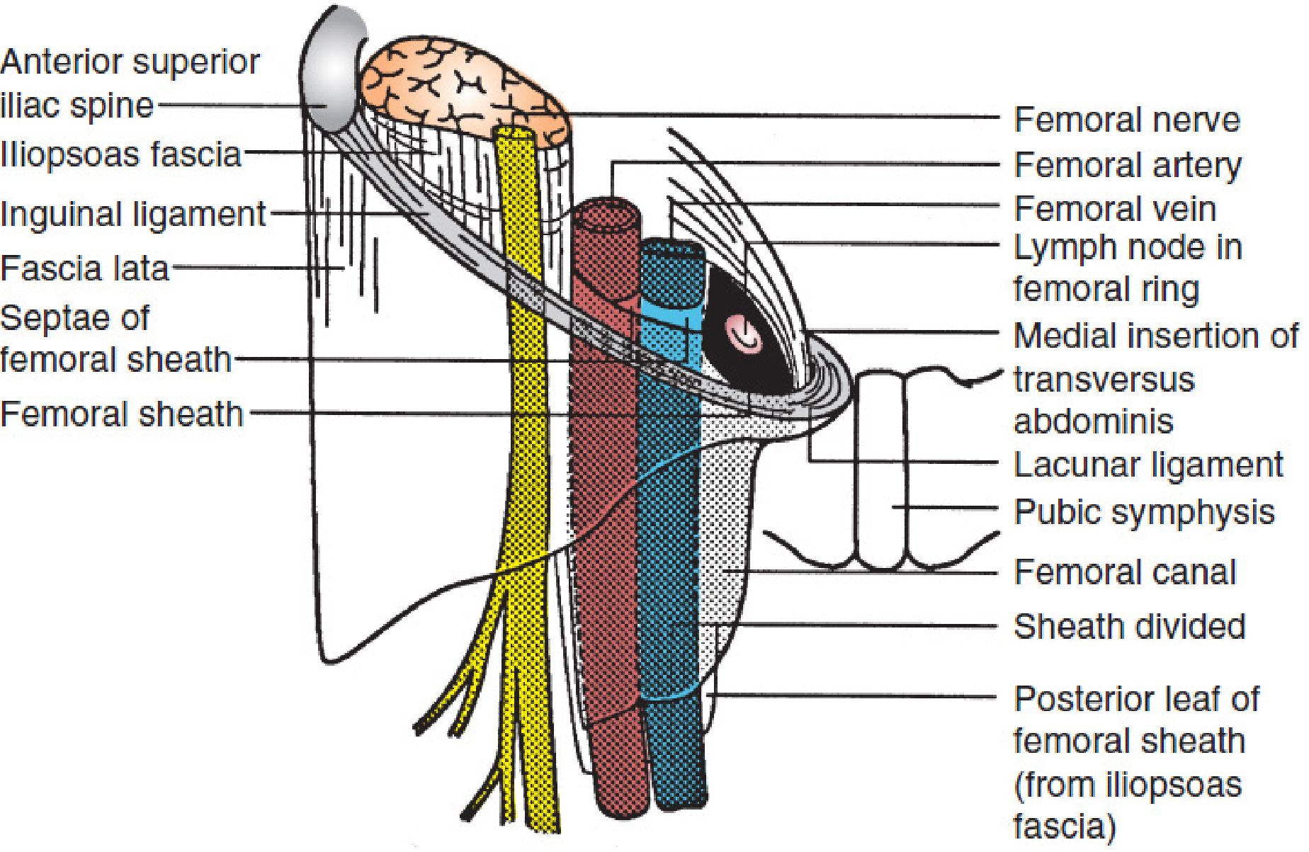
Schematic view of the femoral sheath, ring, and canal. The transversalis fascia forms the anterior portion of the sheath and the iliopsoas fascia forms the posterior portion. Septae separate the vessels from each other, and the vein from the femoral canal. - Mulholland & Greenfield's Surgery
The Femoral Canal
The femoral canal is the medial-most, cone-shaped compartment within the sheath:
- Contents: Lymphatic vessels, areolar tissue, and a lymph node (Cloquet's node / node of Rosenmuller) at its upper (proximal) end
- Normal state: Contains only wisps of connective tissue and small lymphatic nodes
- Length: Short (~1.25 cm)
- Shape: Wider superiorly, narrowing inferiorly
Femoral Ring
The femoral ring is the superior (abdominal/extraperitoneal) opening of the femoral canal. Its boundaries are:
| Boundary | Structure |
|---|---|
| Anterior | Inguinal ligament |
| Posterior | Iliopubic tract + pectineal (Cooper's) ligament |
| Lateral | Femoral vein (septum of the sheath) |
| Medial | Curved edge of transversus abdominis aponeurosis (NOT the lacunar ligament, which inserts more medially) |
The femoral ring is normally closed by the femoral septum - a plug of extraperitoneal fat and areolar tissue. It is covered superiorly by peritoneum.
Clinical Significance
Femoral Hernia
The femoral canal represents a potential weak point in the lower abdominal wall. When abdominal contents (usually a knuckle of small bowel or omentum) protrude through the femoral ring into the canal and potentially beyond, a femoral hernia results.
Key facts:
- More common in women (wider pelvis, broader femoral ring)
- Passes below and lateral to the pubic tubercle (differentiating it from inguinal hernia, which is above and medial)
- The rigid walls of the femoral ring make femoral hernias prone to strangulation
- The sharp medial boundary (lacunar ligament) makes reduction difficult
Vascular Access
The femoral artery and vein - contained within the sheath - are commonly used for:
- Cardiac catheterization (coronary angiography, angioplasty)
- Peripheral vascular procedures
- Central venous access (right heart catheterization, access to renal/gonadal veins, pulmonary artery)
- Contralateral/ipsilateral limb angiography
The femoral artery can be palpated just below the inguinal ligament at the midpoint of the inguinal ligament (midway between ASIS and pubic symphysis) as it passes over the femoral head.
Summary Diagram
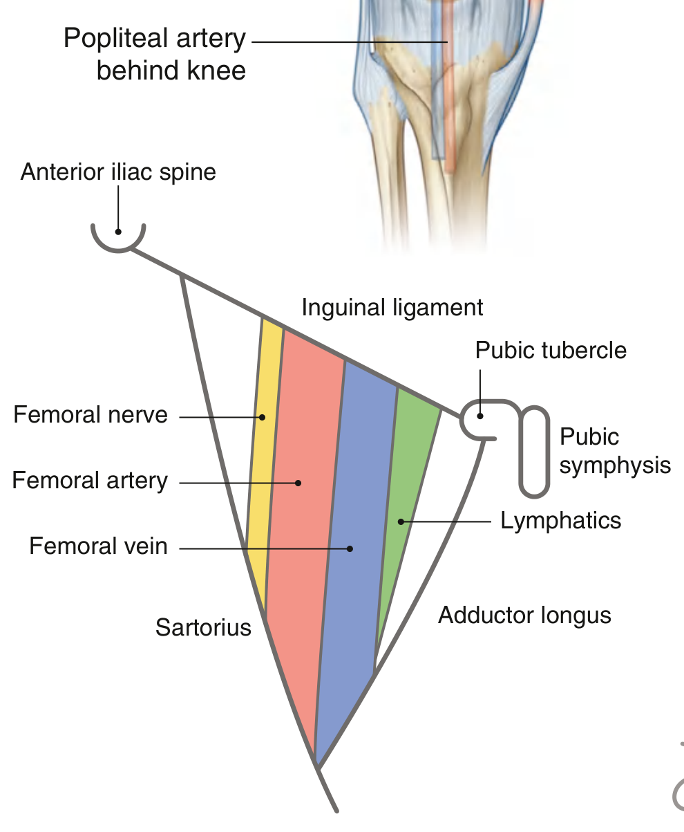
Contents of the femoral triangle showing the three compartments within the femoral sheath and the relationship of the femoral nerve lying outside it. - Gray's Anatomy for Students
Mnemonic for sheath contents (lateral to medial): NAVY
-
N - Nerve (femoral nerve - lateral but OUTSIDE the sheath)
-
A - Artery (femoral artery)
-
V - Vein (femoral vein)
-
Y - Y-fronts (femoral canal with lymphatics - the "empty" space)
-
Gray's Anatomy for Students, p. 676
-
Mulholland & Greenfield's Surgery (7e), pp. 3650-3652
This is a shared conversation. Sign in to Orris to start your own chat.