
Upper limb anatomy high yield summary
brachial plexus diagram upper limb anatomy
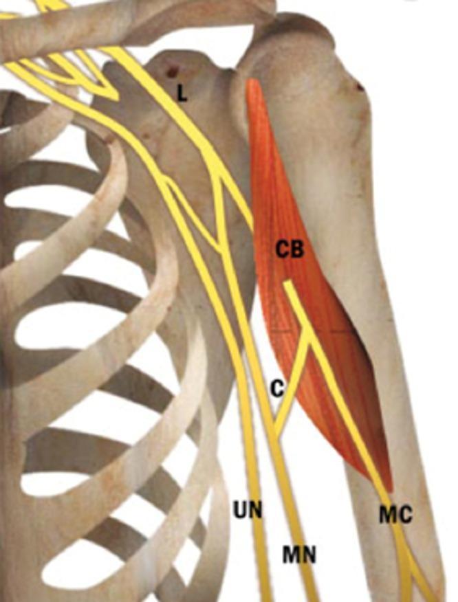
This anatomical diagram illustrates a common neuroanatomical variation of the brachial plexus in the upper limb, specifically the shoulder and proximal humerus region. The visual features the skeletal framework of the rib cage, clavicle, and humerus, with the coracobrachialis muscle (CB) highlighted in red. The neural structures are depicted in yellow, demonstrating the relationship between the musculocutaneous nerve (MC), median nerve (MN), and ulnar nerve (UN). A significant anatomical variant is shown: a communicating branch (C) originating from the musculocutaneous nerve distal to its exit from the coracobrachialis muscle. This branch travels medially and distally to join the median nerve. This illustration is an educational resource for medical students and clinicians to understand variations in peripheral nerve anatomy, which is critical for surgical planning in the axillary region and for interpreting unusual clinical presentations of peripheral nerve injuries.
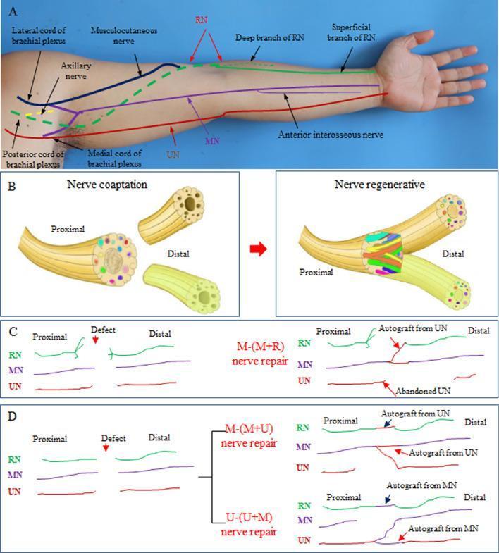
Educational medical illustration detailing upper limb nerve anatomy and surgical nerve merging repair techniques. Panel A is an anatomical diagram overlaid on a human arm showing the brachial plexus cords (lateral, posterior, medial) and the primary nerves: Musculocutaneous, Axillary, Radial (RN), Median (MN), and Ulnar (UN), including the deep and superficial RN branches and the anterior interosseous nerve. Panel B illustrates the concept of 'magnified nerve regeneration,' showing nerve coaptation where one proximal nerve trunk is sutured to two distal nerve segments to facilitate dual innervation. Panels C and D provide schematics of specific surgical protocols: M-(M+R) repair, where the proximal MN is connected to distal MN and RN segments while abandoning the UN; and M-(M+U) or U-(U+M) repairs, which utilize Y-shaped coaptations and autografts (often derived from sacrificed or donor segments) to bridge defects. The illustration serves as a clinical guide for repairing complex nerve avulsions or defects in the upper extremity by using fewer proximal donors to reinnervate multiple distal targets.
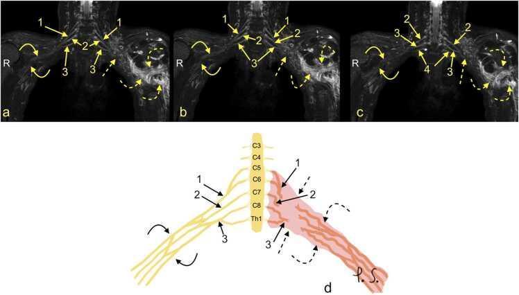
This composite educational resource comprises three Magnetic Resonance Neurography (MRN) coronal views (a, b, c) and a correlating schematic anatomical diagram (d) illustrating a severe traumatic brachial plexus injury. The right side of the images displays normal brachial plexus anatomy, with continuous nerve structures including the upper trunk (1), middle trunk (2), C8 root (3), and Th1 root (4). In contrast, the left side demonstrates a total rupture of the brachial plexus (dashed arrows). This injury is characterized by the complete discontinuity of the neural trunks and cords, accompanied by a large axillary cavity hematoma (curved dashed arrows). The schematic (d) highlights the side-to-side difference, showing the organized right-sided roots (C5-Th1) and trunks versus the disorganized, hemorrhagic area on the left. This material is designed for advanced medical education in radiology and neurosurgery, focusing on diagnostic imaging features of high-grade nerve trauma, specifically Seddon grade 3-5 injuries (neurotmesis) and the identification of postganglionic plexopathy.
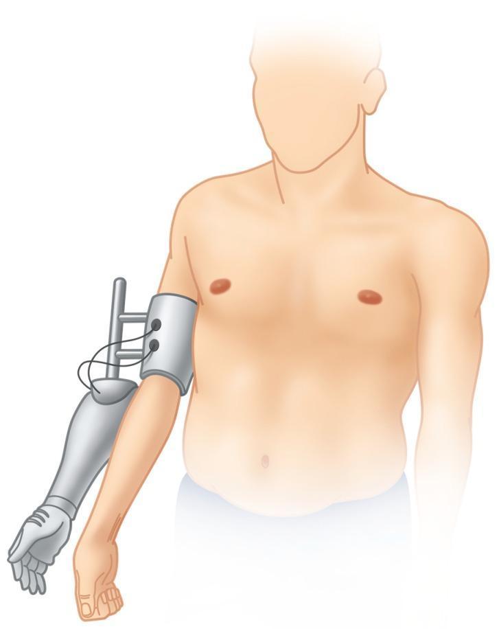
An anatomical illustration depicting a bionic reconstruction concept for a patient with a global brachial plexus injury. The diagram shows a fair-skinned male torso with a functionless biological arm and an attached hybrid prosthetic device. The prosthetic consists of a metallic upper-arm socket or cuff equipped with surface electromyography (sEMG) sensors and wiring, which interface with the underlying musculature of the denervated limb. Extending from this cuff is a mechanical frame that supports a prosthetic forearm and an articulated hand with segmented fingers. The device is positioned alongside the original, non-functional limb to allow for functional testing and signal training before a potential elective amputation. This setup is used in clinical rehabilitation to evaluate if myoelectric control of a mechatronic device can provide superior hand function compared to the original compromised extremity. The illustration emphasizes the biotechnological interface between biological signal sites and prosthetic control systems.
upper limb nerves median ulnar radial anatomy diagram
Educational medical illustration detailing upper limb nerve anatomy and surgical nerve merging repair techniques. Panel A is an anatomical diagram overlaid on a human arm showing the brachial plexus cords (lateral, posterior, medial) and the primary nerves: Musculocutaneous, Axillary, Radial (RN), Median (MN), and Ulnar (UN), including the deep and superficial RN branches and the anterior interosseous nerve. Panel B illustrates the concept of 'magnified nerve regeneration,' showing nerve coaptation where one proximal nerve trunk is sutured to two distal nerve segments to facilitate dual innervation. Panels C and D provide schematics of specific surgical protocols: M-(M+R) repair, where the proximal MN is connected to distal MN and RN segments while abandoning the UN; and M-(M+U) or U-(U+M) repairs, which utilize Y-shaped coaptations and autografts (often derived from sacrificed or donor segments) to bridge defects. The illustration serves as a clinical guide for repairing complex nerve avulsions or defects in the upper extremity by using fewer proximal donors to reinnervate multiple distal targets.
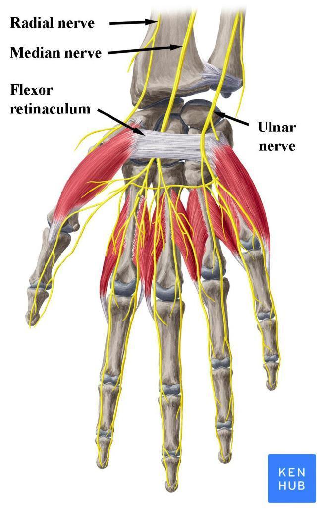
An anatomical illustration of the human hand and wrist (anterior palmar view), highlighting the peripheral nerves and the carpal tunnel. The image depicts the skeletal structure of the carpus, metacarpals, and phalanges, overlaid with musculature and major nerves in yellow. Key structures are labeled: the median nerve is shown passing deep to the flexor retinaculum (transverse carpal ligament), demonstrating its entry into the carpal tunnel. The ulnar nerve is positioned medially (ulnar side), passing superficial to the flexor retinaculum, while the superficial branch of the radial nerve is shown laterally near the radius. Distal to the wrist, the nerves are shown branching into digital nerves to provide sensory and motor innervation to the fingers. This diagram is clinically relevant for explaining the pathophysiology of Carpal Tunnel Syndrome, where the median nerve undergoes compression beneath the flexor retinaculum. Target audience: medical students and clinicians studying upper limb anatomy and entrapment neuropathies.
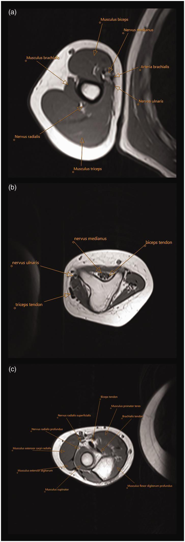
This diagnostic visual contains three labeled axial MRI T1-weighted sections (a, b, c) of the upper extremity, focusing on the musculoskeletal and neurovascular anatomy associated with ulnar dimelia (mirror hand). Section (a) shows the mid-upper arm, identifying the Musculus biceps, Musculus brachialis, and Musculus triceps, alongside the Nervus medianus, Nervus ulnaris, Arteria brachialis, and Nervus radialis in their relative anatomical compartments. Section (b) demonstrates a triangular-shaped dysplastic distal humerus, highlighting the atypical morphology of the bony structures and the positioning of the median and ulnar nerves relative to the biceps and triceps tendons. Section (c) represents a cross-section just distal to the elbow joint, showing two proximal ulnae instead of the standard radius and ulna. It labels the superficial and profound branches of the radial nerve, the biceps and brachialis tendons, and the surrounding musculature including the Musculus pronator teres, Musculus extensor carpi radialis, Musculus extensor digitorum, Musculus supinator, and Musculus flexor digitorum profundus. This series illustrates the complex neurovascular and muscular rearrangements required for surgical planning in congenital limb anomalies.
rotator cuff muscles anatomy shoulder
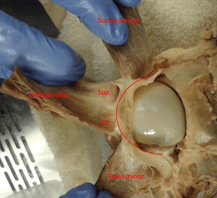
Anatomical Dissection: This clinical photograph provides a posterior view of a dissected left human shoulder, focusing on the rotator cuff complex. The supraspinatus, infraspinatus, and teres minor muscles are reflected laterally to reveal the underlying humeral head and its capsule. A curved red line overlay highlights the rotator cable, a thick fibrous band that spans the avascular zone of the rotator cuff. The infraspinatus muscle is further subdivided with labels 'Sup.' and 'Inf.', representing the superior and inferior portions of the muscle, respectively. The image demonstrates the varying degrees of adherence between the infraspinatus and the rotator cable, illustrating the functional anatomy of the shoulder's capsuloligamentous structures. This material is useful for studying musculoskeletal anatomy, specifically the insertions of the rotator cuff and the biomechanical role of the rotator cable in distributing forces across the shoulder joint.
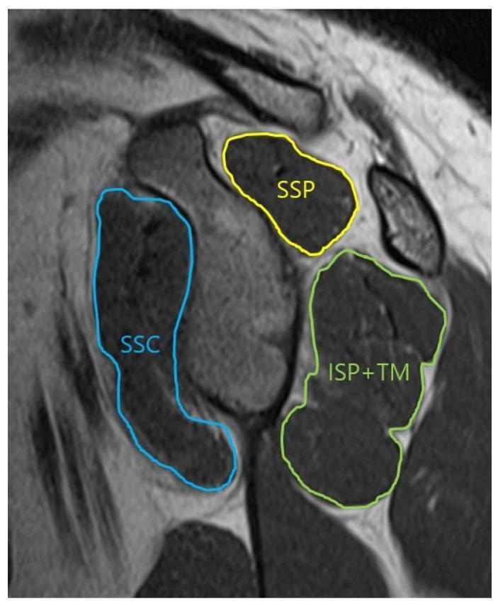
This oblique-sagittal T2 Turbo Spin Echo (TSE) MRI scan demonstrates the musculoskeletal anatomy of the shoulder in a glenoid face view, specifically for assessing the cross-sectional area of the rotator cuff muscles. The image features color-coded annotations outlining the muscular boundaries: the supraspinatus (SSP) is outlined in yellow in the superior position; the subscapularis (SSC) is traced in blue anteriorly; and the infraspinatus combined with the teres minor (ISP+TM) is outlined in green in the posterior-inferior quadrant. The musculature appears as intermediate to dark gray signal intensity, allowing for the evaluation of muscle volume and potential fatty infiltration or atrophy. This imaging view is clinically significant for orthopedic evaluation and preoperative planning in cases of rotator cuff tears or shoulder instability, providing a clear visualization of the muscular support surrounding the glenohumeral joint.
carpal tunnel hand muscles intrinsic anatomy
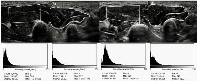
This diagnostic image displays four musculoskeletal ultrasound (US) frames and their corresponding grayscale intensity histograms, comparing the intrinsic hand muscles of a healthy control to a patient with moderate Carpal Tunnel Syndrome (CTS). Panels (a) and (c) depict the thenar muscle using a rectangular region of interest (ROI), while (b) and (d) show the hypothenar muscle with freehand traced ROIs. The visual echogenicity in the CTS patient's thenar muscle (c) appears heterogeneously increased compared to the healthy control (a). Quantitative analysis is provided through histograms below each US frame, showing pixel distribution across an intensity scale (0–255). Key metrics include Count, Mean, Standard Deviation (StdDev), and Mode. Notably, the thenar muscle in CTS (c) shows a significant rightward shift in its histogram, with a mean intensity of 40.839 compared to 20.262 in the control (a), indicating increased echo intensity (hyperechogenicity) which may correlate with denervation or fibrosis. The hypothenar histograms remain relatively similar between groups, serving as a comparative baseline. This clinical imaging demonstrates the use of quantitative grayscale analysis in evaluating muscle changes secondary to peripheral nerve compression.
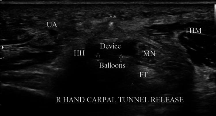
This transverse diagnostic ultrasound image captures a right hand during a carpal tunnel release procedure at the level of the hook of hamate. The image displays the carpal tunnel's internal anatomy and the precise positioning of a retrograde microknife within the transverse safe zone (TSZ). Key anatomical landmarks include the ulnar artery (UA) on the left, the hook of hamate (HH) presenting as a hyperechoic bony structure with posterior acoustic shadowing, and the median nerve (MN) on the right, which shows a characteristic speckled, hypoechoic oval appearance. Inferior to the median nerve are the flexor tendons (FT), while the thenar muscles (ThM) are visible on the far right. The surgical device is centrally located in the TSZ between the MN and HH; its deployed balloons, filled with saline, appear as hypoechoic rounded structures that physically displace neurovascular bundles to ensure a safe cutting path. A hyperechoic dot (marked with a double asterisk) represents the cross-section of the cutting blade positioned superiorly, ready to transect the transverse carpal ligament. This visual demonstrates the utility of real-time ultrasound guidance in identifying safe surgical margins during minimally invasive orthopedic procedures.
dermatomes upper limb C5 C6 C7 C8 T1
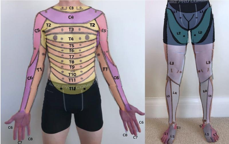
This clinical dermatome map, presented as an anatomical diagram overlaid on a human subject, illustrates the cutaneous sensory distribution of spinal nerves. The anterior trunk displays a sequential horizontal banding pattern representing thoracic dermatomes T2 through T12, with T4 roughly at the nipple line and T10 at the umbilicus. The upper extremities show cervical and upper thoracic distribution: C5 (lateral upper arm), C6 (lateral forearm and thumb), C7 (middle finger), C8 (medial hand and little finger), and T1 (medial forearm and arm). The lower extremity map details the lumbar and sacral levels: L1 (inguinal region), L2 (anterior mid-thigh), L3 (distal thigh and knee), L4 (medial lower leg), L5 (lateral lower leg and dorsal foot), and S1 (lateral foot and small toe). This resource is designed for primary care musculoskeletal medicine to assist in the neurological examination of sensory deficits, radiculopathy, or spinal cord injury level localization.
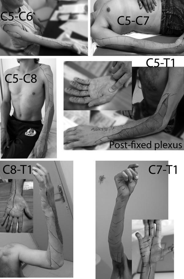
A compilation of clinical photographs depicting sensory mapping of the upper limb in different types of brachial plexus palsy. The images use skin marking to delineate zones of lost protective sensation (determined by Semmes-Weinstein monofilament testing). The series illustrates progression of sensory loss across several injury levels: C5-C6 (lateral forearm), C5-C7 (lateral arm and forearm), C5-C8 (extended lateral involvement and dorsal hand anesthesia), and C5-T1 with post-fixed plexus (near-total limb involvement with small medial zones of preservation). Lower plexus injuries are also shown, including C8-T1 (medial forearm and ulnar hand involvement) and C7-T1 (extending to the inner arm and middle finger). Key educational concepts include the correlation between brachial plexus root involvement and specific cutaneous dermatomal deficits, as well as the variability introduced by anatomical variations like post-fixation. The imagery serves as a diagnostic guide for identifying the level of nerve root avulsion or injury based on distal sensory presentation.
Upper Limb Anatomy - High Yield Summary
1. BRACHIAL PLEXUS

Structure (mnemonic: Randy Travis Drinks Cold Beer)
| Level | Structure | Formation |
|---|---|---|
| Trunks | Superior | C5 + C6 |
| Middle | C7 only | |
| Inferior | C8 + T1 | |
| Cords | Lateral | Anterior div. of Superior + Middle (C5-C7) |
| Medial | Anterior div. of Inferior (C8-T1) | |
| Posterior | All 3 posterior divisions (C5-T1) |

Pre-clavicular branches (from roots/trunks)
| Nerve | Root | Supplies |
|---|---|---|
| Dorsal scapular | C5 | Rhomboid major & minor |
| Long thoracic | C5, C6, C7 | Serratus anterior |
| Nerve to subclavius | C5, C6 | Subclavius |
| Suprascapular | C5, C6 | Supraspinatus, infraspinatus |
Terminal branches (from cords)
| Nerve | Cord Origin | Root Values |
|---|---|---|
| Musculocutaneous | Lateral | C5, C6, C7 |
| Median (lateral root) | Lateral | C6, C7 |
| Median (medial root) | Medial | C8, T1 |
| Ulnar | Medial | C8, T1 (C7) |
| Radial | Posterior | C5-T1 |
| Axillary | Posterior | C5, C6 |
2. THE FIVE MAJOR NERVES - Mnemonics & Lesion Patterns
Musculocutaneous (C5-C7) - from Lateral cord
- Motor: Coracobrachialis, biceps brachii, brachialis (anterior arm flexors)
- Sensory: Lateral cutaneous nerve of forearm
- Lesion: Weak elbow flexion/supination; sensory loss lateral forearm
Median Nerve (C6-T1) - from Lateral + Medial cords
- Motor in forearm: Most flexors (except FCU and medial FDP); pronators; AIN → FPL, lateral FDP, pronator quadratus
- Motor in hand (LOAF): Lumbricals 1&2, Opponens pollicis, Abductor pollicis brevis, Flexor pollicis brevis (superficial head)
- Sensory: Palmar surface - thumb, index, middle finger, lateral half of ring; dorsal tips of same digits
- High lesion (above elbow): "Hand of benediction" - loss of FDS/FDP to index/middle; weak pronation
- Low lesion (at wrist - carpal tunnel): Thenar wasting (ape hand); no motor loss in forearm
- Sensory: Index fingertip - "pointer finger" test
Ulnar Nerve (C8-T1) - from Medial cord
- Course: Passes posterior to medial epicondyle (cubital tunnel), then Guyon's canal at wrist
- Motor in forearm: FCU, medial half of FDP (ring & little fingers)
- Motor in hand: All intrinsics EXCEPT LOAF; hypothenar muscles; lumbricals 3&4; all interossei; adductor pollicis
- Sensory: Little finger, medial half of ring finger (palmar & dorsal)
- High lesion: "Ulnar paradox" - less clawing than low lesion because FDP also lost
- Low lesion: Claw hand (ring & little fingers); froment's sign (adductor pollicis weak - uses FPL to compensate)
- Key test: Card test (palmar interossei); abduction/adduction of fingers (dorsal interossei)
Radial Nerve (C5-T1) - from Posterior cord
- Course: Spiral (radial) groove of humerus → anterior to lateral epicondyle → splits into superficial (sensory) and deep (PIN) branches
- Motor: Triceps (before groove), brachioradialis, ECRL (at/above elbow), then all extensors via PIN
- Sensory: First dorsal web space (key sensory test)
- High lesion (axilla - "Saturday night palsy"): Wrist drop + finger drop; triceps also affected
- Mid-humerus lesion (spiral groove): Wrist drop + finger drop; triceps spared
- PIN lesion (at radial tunnel/lateral epicondyle area): Finger drop only; no wrist drop (ECRL spared); no sensory loss
- Superficial radial nerve: Sensory only - first dorsal web space
Axillary Nerve (C5, C6) - from Posterior cord
- Course: Exits axilla through quadrangular space with posterior circumflex humeral artery
- Motor: Deltoid, teres minor
- Sensory: "Regimental badge" area - lateral upper arm
- Lesion: Inability to abduct arm (after first 15° which is supraspinatus); loss of sensation over deltoid
3. PERIPHERAL NERVE INNERVATION TABLE (High Yield)
| Function | Nerve | Root |
|---|---|---|
| Shoulder abduction 0-15° | Supraspinatus | C5, C6 |
| Shoulder abduction 15-90° | Deltoid (axillary) | C5, C6 |
| Elbow flexion + supination | Musculocutaneous | C5, C6 |
| Wrist extension | Radial (ECRL/ECRB) | C6, C7 |
| Wrist flexion (radial) | Median (FCR) | C6, C7 |
| Wrist flexion (ulnar) | Ulnar (FCU) | C7, C8 |
| Thumb opposition | Median | C8, T1 |
| Finger abduction/adduction | Ulnar (interossei) | T1 |
| Finger extension | Radial (PIN) | C7, C8 |
4. ROTATOR CUFF - "SITS"
| Muscle | Nerve | Root | Action |
|---|---|---|---|
| Supraspinatus | Suprascapular | C5, C6 | Initiation of abduction (0-15°) |
| Infraspinatus | Suprascapular | C5, C6 | External rotation |
| Teres minor | Axillary | C5, C6 | External rotation |
| Subscapularis | Upper/Lower subscapular | C5, C6 | Internal rotation |
- Supraspinatus is the most commonly torn rotator cuff tendon
- All four hold the humeral head in the glenoid fossa
5. COMPARTMENTS & MUSCLES
Arm
| Compartment | Muscles | Nerve |
|---|---|---|
| Anterior | Biceps brachii, coracobrachialis, brachialis | Musculocutaneous |
| Posterior | Triceps brachii | Radial |
Forearm Anterior (Flexor) Compartment
Forearm Posterior (Extensor) Compartment
- Superficial: ECRL, ECRB, ED, EDM, ECU, brachioradialis
- Deep: APL, EPL, EPB, EI (supinator also here)
6. DERMATOMES (Upper Limb)
| Root | Sensory Area |
|---|---|
| C5 | Lateral upper arm (deltoid patch) |
| C6 | Lateral forearm, thumb, index finger |
| C7 | Middle finger (dorsal) |
| C8 | Ring and little finger, medial hand |
| T1 | Medial forearm |
| T2 | Medial upper arm, axilla |
7. REFLEXES
| Reflex | Root Level | Nerve |
|---|---|---|
| Biceps | C5, C6 | Musculocutaneous |
| Brachioradialis | C5, C6 | Radial |
| Triceps | C7 | Radial |
8. KEY INJURY PATTERNS & MNEMONICS
| Lesion | Cause | Sign | Nerve |
|---|---|---|---|
| Erb's palsy (C5, C6) | Birth trauma (shoulder dystocia), neck stretch | "Waiter's tip" - arm adducted, internally rotated, pronated | Upper trunk |
| Klumpke's palsy (C8, T1) | Arm pulled upward, cervical rib | Claw hand + Horner's syndrome | Lower trunk |
| Wrist drop | Humeral shaft fracture (spiral groove) | Loss of wrist/finger extension; spared triceps | Radial |
| Claw hand | Medial epicondyle fracture | Ring & little finger clawing, hypothenar wasting | Ulnar |
| Ape hand | Carpal tunnel | Thenar wasting, loss of opposition | Median |
| Saturday night palsy | Crutch/arm-over-chair pressure | Full radial palsy including triceps | Radial (axilla) |
| Winged scapula | Stab wound to chest wall / mastectomy | Scapula protrudes during pushing | Long thoracic (serratus ant.) |
9. AXILLARY SPACES (High Yield for Quadrangular Space)
| Space | Boundaries | Contents |
|---|---|---|
| Quadrangular space | Teres minor (sup), teres major (inf), long head triceps (medial), surgical neck of humerus (lateral) | Axillary nerve + posterior circumflex humeral artery |
| Triangular space | Teres minor (sup), teres major (inf), long head triceps (lateral) | Circumflex scapular artery |
| Triangular interval | Teres major (sup), long head triceps (medial), shaft humerus (lateral) | Radial nerve + profunda brachii artery |
10. ARTERIES - High Yield
| Artery | Key Points |
|---|---|
| Axillary artery | 3 parts divided by pectoralis minor; becomes brachial after teres major |
| Brachial artery | Medial to biceps tendon; bifurcates at neck of radius → radial + ulnar |
| Radial artery | Lateral forearm; "anatomical snuffbox" → deep palmar arch (mainly) |
| Ulnar artery | Medial forearm; Guyon's canal → superficial palmar arch (mainly) |
| Profunda brachii | From brachial; travels with radial nerve in spiral groove |
11. QUICK MUSCLE ROOT VALUES (The "Big 5")
| Action | Root |
|---|---|
| Shoulder abduction | C5 |
| Elbow flexion | C5, C6 |
| Wrist extension | C6, C7 |
| Elbow extension / Finger extension | C7 |
| Finger abduction / intrinsics | T1 |