Management of edh
Reading File
Finding Sources
Finding Sources
Searching PubMed
"epidural hematoma" AND management
Reading File
Reading File
Reading File
Reading File
Reading File
Loading Image
Reading File
I now have comprehensive information from multiple authoritative textbooks. Here is the full, detailed answer:
Management of Epidural Hematoma (EDH)
Overview
An epidural hematoma (EDH) is a neurosurgical emergency where blood collects in the potential space between the inner table of the skull and the outer (periosteal) layer of the dura mater. It occurs in approximately 2% of traumatic brain injuries and is more common in young males; it is uncommon after age 50.
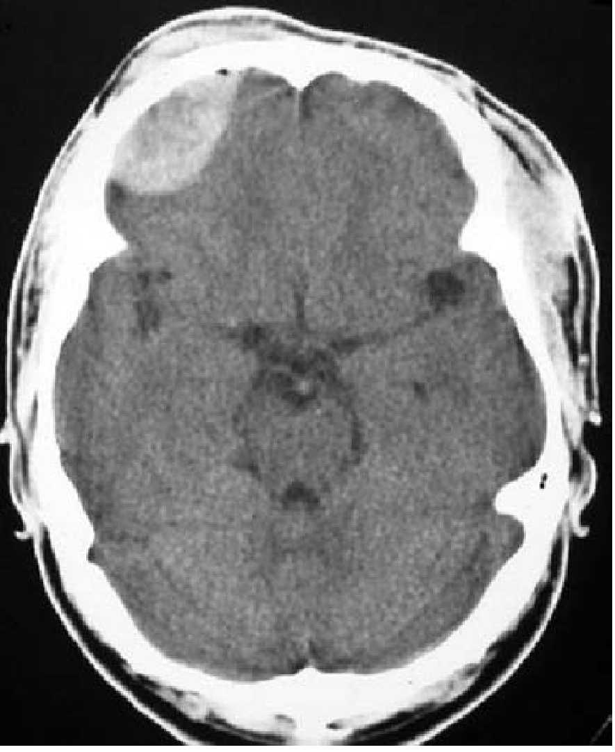
CT appearance: biconvex (lens-shaped/football-shaped) hyperdense mass, typically in the temporal region.
Mechanism & Etiology
- Most commonly: laceration of the middle meningeal artery by a temporal bone fracture (crossing the middle meningeal groove)
- Less commonly: tears of dural venous sinuses (parieto-occipital or posterior fossa trauma) - these bleed more slowly and may be self-limited
- High-pressure arterial bleeding means herniation can occur within hours of injury
Clinical Presentation
| Feature | Detail |
|---|---|
| Classic presentation | LOC → lucid interval → rapid neurologic deterioration |
| Frequency of classic presentation | Only 15-20% of cases |
| Most cases | Mild symptoms picked up on CT; only ~30% require surgical intervention |
| Signs of raised ICP | Vomiting, ipsilateral pupil dilation (uncal herniation), Cushing's triad (HTN + bradycardia + irregular breathing) |
| Skull fracture signs | Battle's sign (post-auricular ecchymosis), raccoon eyes (periorbital ecchymosis), hemotympanum |
Diagnosis
- Head CT (non-contrast) is the investigation of choice - shows biconvex hyperdense mass
- Check for skull fracture crossing the middle meningeal groove (bone windows on CT)
- Coronal reconstruction needed if vertex hematoma is suspected (may be missed on axial cuts)
- MRI is not required for EDH diagnosis but may be used to evaluate underlying brain contusions/edema
- Even if the initial CT is negative, careful follow-up is required as EDH development can be delayed
Management
A. Surgical Management (DEFINITIVE)
Indications for immediate surgical evacuation (craniotomy):
- EDH causing brain displacement with impaired consciousness
- Hematoma volume ≥30 mL
- Clot thickness (diameter) ≥15 mm
- Midline shift ≥5 mm
- GCS score ≤8
- Focal neurologic deficits
Key surgical principles:
- Surgery is an emergency - duration from injury to treatment is a critical determinant of prognosis
- Most patients operated on promptly recover, including ~one-third of those with a fixed dilated pupil preoperatively
- Factors affecting outcome: age, depth of coma, degree of midline shift, hematoma size
- EDH typically does not injure underlying brain parenchyma - full neurologic recovery is expected with timely evacuation
B. Conservative (Non-Surgical) Management
A non-surgical approach may be considered only if all of the following criteria are met:
| Criterion | Threshold |
|---|---|
| Hematoma volume | < 30 mL |
| Clot diameter | < 15 mm |
| Midline shift | < 5 mm |
| GCS score | > 8 |
| Neurologic examination | Non-focal |
Conservative management consists of:
- Correction of any underlying coagulopathy
- Close inpatient observation with serial neurologic examinations
- Repeat CT imaging to monitor hematoma size
- Patient and family education - return immediately if neurologic deterioration occurs
Note: Even with an apparently negative initial CT, any trauma patient with a skull fracture crossing the middle meningeal groove needs close surveillance for delayed EDH.
Management of Spinal EDH (if context includes this)
- Caused by mechanical disruption of epidural vessels by vertebral fracture fragments or dislocation
- Diagnosed on MRI: collection isointense on T1, hyperintense on T2, typically extending 2-3 vertebral segments
- Immediate surgical decompression for patients with neurologic deficits
- Conservative management (coagulopathy correction + observation) for patients without neurologic deficits
- 6-month outcomes are similar between surgical and conservatively managed groups
Prognosis
- Excellent if evacuated before herniation or neurologic deficits develop
- Unlike SDH, EDH usually does not injure underlying brain - full recovery is expected with early treatment
- Outcome worsens with delayed treatment, deep coma, bilateral fixed pupils, large hematoma, and advanced age
Sources:
- Goldman-Cecil Medicine International Edition, Chapter 368 (Treatment and Prognosis)
- Plum and Posner's Diagnosis and Treatment of Stupor and Coma, Chapter 4
- Rosen's Emergency Medicine: Concepts and Clinical Practice, Chapter 257
- Tintinalli's Emergency Medicine: A Comprehensive Study, Chapter 257
Recent evidence note (PubMed 2024-2026): A systematic review and meta-analysis (Daoud et al., 2024, PMID 38943702) specifically addressed operative vs. non-operative management of posterior fossa EDH - this is a nuanced subgroup where conservative criteria may differ from supratentorial EDH.
This is a shared conversation. Sign in to Orris to start your own chat.