
A 32-year-old carpenter presents with difficulty in performing screwing in movements of the examination, the clinician notices weakness of the supinator muscle. There is associated we deep extensors of forearm but actions of triceps is unaffected.
posterior interosseous nerve anatomy radial nerve forearm branches
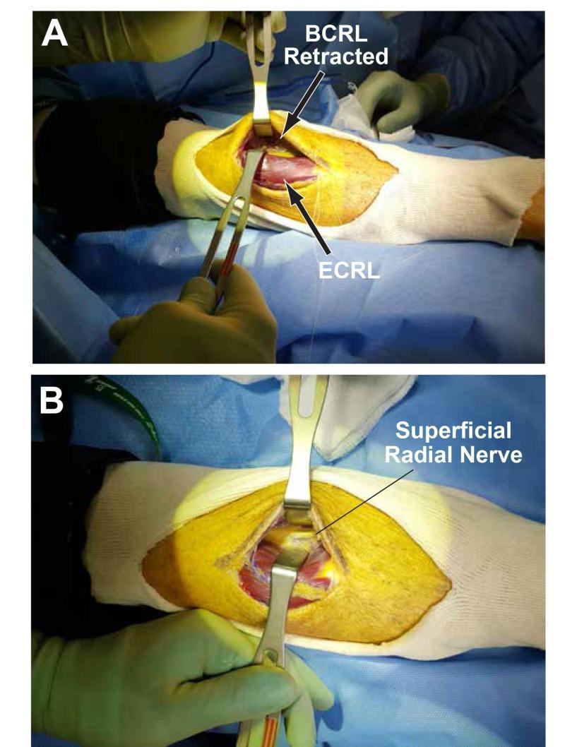
Clinical intraoperative photographs demonstrating a surgical approach to the forearm. Figure A shows an open incision with the brachioradialis (BCRL) and extensor carpi radialis longus (ECRL) muscles identified. Self-retaining or handheld retractors are used to develop the plane between these two muscle bellies using blunt dissection. Figure B illustrates deeper exposure achieved through further lateral retraction of the brachioradialis. This maneuver reveals the superficial radial nerve, which appears as a pale, cord-like longitudinal structure situated in the deep plane. The surgical field highlights the relevant anatomy for decompression of the radial nerve or its branches, such as in cases of posterior interosseous nerve (PIN) entrapment. The images serve as an educational guide for surgical planes in the proximal forearm, emphasizing the relationship between mobile wad musculature and the radial nerve's sensory branch.
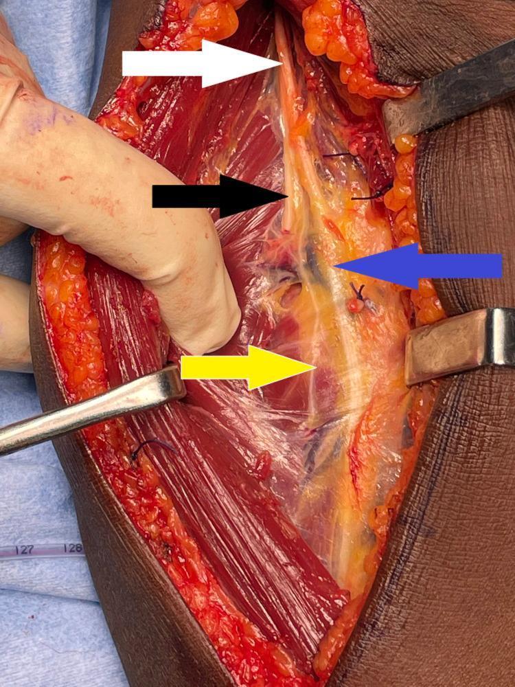
This clinical photograph displays a surgical dissection of the right proximal forearm and elbow region, utilizing an extended Henry approach to visualize the radial nerve and its distal branches. The surgical field is retracted, exposing the deep muscular and neurovascular anatomy. A white arrow identifies the main trunk of the radial nerve as it descends between the brachialis and brachioradialis muscles. The branching pattern is clearly demarcated: a black arrow points to the posterior interosseous nerve (PIN), which dives toward the supinator muscle; a blue arrow indicates the superficial radial nerve, which continues distally under the brachioradialis; and a yellow arrow points to the motor branch supplying the extensor carpi radialis brevis (ECRB). The nerves appear as glistening, off-white cord-like structures set against the deep red, striated muscle tissue and yellow lobulated subcutaneous fat. This image serves as a high-fidelity anatomical reference for peripheral nerve surgery, demonstrating the critical spatial relationships between the radial nerve bifurcation and the surrounding musculature.
Diagnosis: Posterior Interosseous Nerve (PIN) Palsy
Anatomical Basis - Why These Specific Muscles?
- Superficial branch (purely sensory - dorsum of hand/lateral 3.5 fingers)
- Deep branch / Posterior Interosseous Nerve (PIN) (purely motor to forearm extensors)
- Extensor carpi radialis brevis (ECRB)
- Extensor digitorum communis (EDC)
- Extensor digiti minimi (EDM)
- Extensor carpi ulnaris (ECU)
- Extensor indicis proprius (EIP)
- Abductor pollicis longus (APL)
- Extensor pollicis brevis (EPB)
- Extensor pollicis longus (EPL)
Why Triceps is SPARED
Why Supinator is Affected
"PIN innervates the ECRB, supinator, EIP, ECU, EDC, EDM, APL, EPB, and EPL" - Miller's Review of Orthopaedics, 9th Ed.
Why This Patient (Carpenter)?
"Repetitive pronation and supination of the forearm can predispose to compression of the deep/posterior interosseous nerve branch of the radial nerve at the fibrous arcade of Frohse." - Imaging Anatomy: Bones, Joints, Vessels and Nerves
Key Clinical Features - PIN Palsy
| Feature | Findings |
|---|---|
| Wrist drop | Present but with radial deviation (ECRL spared, as it is innervated by radial nerve proper before bifurcation) |
| Finger extension | Lost at MCP joints |
| Thumb extension/abduction | Lost |
| Wrist extension | Partially preserved with radial deviation |
| Triceps power | Normal |
| Brachioradialis | Normal (innervated before bifurcation) |
| Sensation | Normal - PIN is purely motor |
Sites of Compression (Mnemonic: LEAFS)
- L - Recurrent Leash of Henry (radial recurrent artery fan)
- E - Proximal edge of Extensor carpi radialis brevis
- A - Arcade of Frohse (most common - proximal edge of supinator)
- F - Fascial band at the radial head
- S - Distal edge of Supinator
- Miller's Review of Orthopaedics, 9th Ed., p. 694
Differentiating PIN Palsy from Higher Radial Nerve Injuries
| Level of Injury | Triceps | Brachioradialis | ECRL | Supinator | Deep extensors | Sensation |
|---|---|---|---|---|---|---|
| Axilla | Weak | Weak | Weak | Weak | Weak | Lost |
| Spiral groove (Saturday night palsy) | Spared | Weak | Weak | Weak | Weak | Lost |
| PIN (this case) | Spared | Spared | Spared | Weak | Weak | Normal |
Management
-
Conservative (first-line): Activity modification (rest from repetitive pro/supination), splinting, NSAIDs - for 3 months
-
Surgical decompression: If no recovery by 3 months - release at the arcade of Frohse; good to excellent results in ~85% of patients
-
Tendon transfer: For irreversible paralysis - FCR to EDC + palmaris longus to EPL
-
Campbell's Operative Orthopaedics, 15th Ed. (2026); Miller's Review of Orthopaedics, 9th Ed.
Surgical Anatomy Image