

9d9d964b-2938-4e41-9358-1b0648c842c5.jpeg
probable diagnose and treatment
analyse_media
Reading File
The image shows a classic annular (ring-shaped) erythematous lesion on the upper arm - highly characteristic of Erythema Migrans from Lyme disease or an early tinea corporis. Let me consult the medical library and search for relevant images.
Searching sources...
Finding Sources
Searching Images
erythema migrans annular ring-shaped rash upper arm Lyme disease
Searching PubMed
erythema migrans Lyme disease treatment
Now let me pull the relevant textbook passages on diagnosis and treatment:
Reading File
Reading File
Let me fetch the textbook reference images and get the treatment section:
Loading Image
Reading File
Reading File
I now have everything needed for a comprehensive answer. Let me compile it.
Probable Diagnosis: Erythema Migrans (Lyme Disease - Early Localized Stage)
The image shows a solitary, large annular (ring-shaped) erythematous plaque on the upper arm/deltoid region, with a well-demarcated raised outer ring of pink-red erythema and partial central clearing - the hallmark appearance of Erythema Migrans (EM), the primary skin manifestation of Lyme disease (caused by Borrelia burgdorferi).
Compare the patient's lesion to this textbook reference case of erythema migrans:
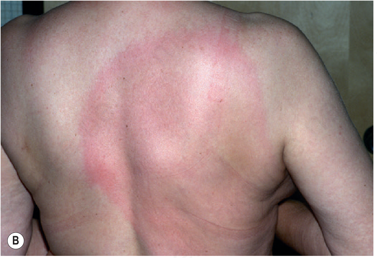
Why This Fits
| Feature | This Lesion | Erythema Migrans |
|---|---|---|
| Shape | Annular/oval ring | Annular, expanding ring |
| Color | Pinkish-red border, central clearing | Erythematous border, partial central clearing |
| Borders | Well-demarcated, smooth | Well-demarcated |
| Scaling | Absent | Absent (key distinction from tinea) |
| Vesicles | None prominent | Usually none |
| Location | Upper arm/deltoid | Any area; common on limbs, groin, axilla |
| Satellite papules | Present nearby | Secondary lesions possible in disseminated disease |
- Per [Andrews' Diseases of the Skin](Andrews' Diseases of the Skin, p. 334): EM begins as a red macule or papule expanding outward over days to weeks; "the advancing border is usually slightly raised, warm, red to bluish-red, and free of any scale." The median diameter reaches ~15 cm (range 3-68 cm).
- The lesion is typically painless and nonpruritic (which differentiates it from contact dermatitis). About 50% have a burning sensation.
- Per the [Red Book 2021](Red Book 2021, p. 774): EM is diagnosed clinically based on appearance in the correct seasonal and geographic context. No serology is needed if EM is present - it's the most characteristic manifestation.
Differential Diagnoses to Consider
- Tinea corporis - would show scaling along the advancing border (absent here); KOH prep positive
- Erythema Annulare Centrifugum (EAC) - slower expanding, often has a trailing inner scale
- Southern Tick-Associated Rash Illness (STARI) - if in southern US states, caused by Amblyomma americanum tick; clinically identical to EM, etiology unknown
- Cellulitis - does not form rings; more painful, no central clearing
- Granuloma annulare - skin-colored papules in a ring, not erythematous; no tick bite history
- Allergic reaction to tick bite - smaller (<5 cm), more pruritic, does not expand
Clinical Assessment
Key questions to confirm:
- History of tick bite (~50% of patients recall one) - 3-32 days prior (median 11 days)
- Recent outdoor/wooded area exposure (April-October season)
- Constitutional symptoms: malaise, headache, mild neck stiffness, myalgia, arthralgia, low-grade fever?
- Geographic location - endemic areas include northeastern/mid-Atlantic US, upper Midwest, Europe, Asia
Serology is not required if EM is clinically evident; in fact, serology is often negative in early localized disease.
Treatment
Per the 2025 Guidelines for Lyme borreliosis treatment and Red Book 2021:
First-Line (Early Localized/Uncomplicated EM)
| Drug | Dose | Duration |
|---|---|---|
| Doxycycline (adults, >8 yrs) | 100 mg PO twice daily | 14 days |
| Amoxicillin (children <8 yrs, pregnant) | 500 mg PO three times daily (or 50 mg/kg/day divided TID in children) | 14 days |
| Cefuroxime axetil (alternative) | 500 mg PO twice daily | 14 days |
- Doxycycline is preferred in adults - it also covers co-infections like Anaplasma
- A 10-day course of doxycycline is accepted in some guidelines if EM is the only manifestation
- Do not use macrolides (azithromycin, erythromycin) - less effective, higher relapse rate
If Early Disseminated Disease is Present (multiple EM lesions, Bell's palsy, meningitis, carditis):
- Extend oral antibiotics to 21 days, or use IV ceftriaxone 2g/day x 14-21 days for neurological or severe cardiac involvement
Adjunctive
- NSAIDs for constitutional symptoms (arthralgia, myalgia, headache)
- Monitor for treatment response: EM should begin fading within days of antibiotic initiation
- If fever persists >24-48 hours after starting treatment, consider co-infection (babesiosis, anaplasmosis)
Important Notes
- Do not delay treatment waiting for serology - treat clinically if EM is present
- Follow-up at 2-4 weeks to confirm resolution
- If lesion expands or new lesions appear, or neurological/cardiac symptoms develop, reassess for disseminated disease
- A formal in-person evaluation by a clinician is necessary for definitive diagnosis and prescription
Disclaimer: This analysis is based on image appearance and published clinical criteria. It is not a substitute for in-person clinical evaluation. A qualified healthcare provider must examine the patient and confirm the diagnosis.
This is a shared conversation. Sign in to Orris to start your own chat.