
Protein chemistry
Reading File
"Protein chemistry" is a broad topic. Let me clarify what you're looking for before diving in.
Asking for Preferences
What aspect of protein chemistry are you interested in? · What level of depth do you need?
Searching sources...
Finding Sources
Finding Sources
Finding Sources
Finding Sources
Good - found the key sources. Let me read the most relevant sections from Lippincott (the gold standard for USMLE biochemistry) and Basic Medical Biochemistry.
Reading File
Reading File
Finding Sources
Reading File
Reading File
Now I have rich textbook content. Let me also fetch the key protein structure diagram for visual clarity.
Loading Image
I have everything needed for a thorough, exam-focused answer. Here it is:
Protein Folding & Denaturation - USMLE/Medical School Review
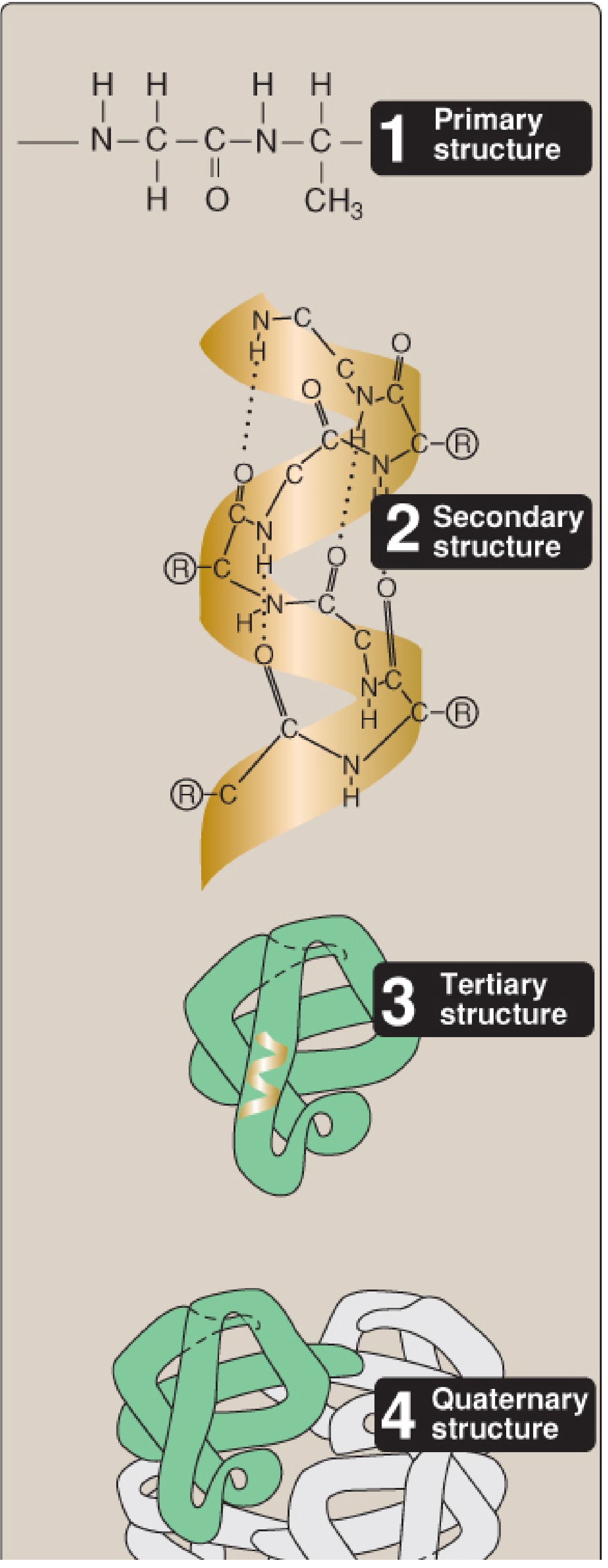
Figure 2.1 - Four hierarchies of protein structure (Lippincott Illustrated Reviews: Biochemistry, 8th ed.)
1. Four Levels of Protein Structure
| Level | Definition | Bonds Involved |
|---|---|---|
| Primary | Linear sequence of amino acids (N-term → C-term) | Peptide bonds (covalent) |
| Secondary | Local repeating folding patterns (α-helix, β-sheet, β-turn) | Hydrogen bonds (backbone) |
| Tertiary | 3D folding of the entire polypeptide chain | Hydrophobic interactions, H-bonds, ionic bonds, disulfide bonds |
| Quaternary | Arrangement of multiple polypeptide subunits | Primarily noncovalent (H-bonds, ionic, hydrophobic) |
Key exam point: Quaternary structure requires MORE than one polypeptide. Proteins with only one chain (monomers) do NOT have quaternary structure.
2. Secondary Structures
α-Helix
- Right-handed spiral; stabilized by intrachain hydrogen bonds between the carbonyl oxygen of one peptide bond and the NH of a residue 4 positions ahead
- Each turn = 3.6 amino acids
- Side chains project outward from the core
- Examples: keratin (structural), myoglobin (~75% α-helical)
Amino acids that DISRUPT α-helices:
- Proline - rigid secondary amine, creates a kink (most tested)
- Glycine - too flexible (H as R group)
- Clustered charged residues or bulky branched residues
β-Sheet
- Extended, pleated strands aligned side-by-side
- Stabilized by interchain hydrogen bonds (between separate strands) and intrachain H-bonds between regions of a single polypeptide
- Can be parallel or antiparallel
- Example: silk fibroin
β-Turn (β-Bend)
- Allows the chain to reverse direction (found at protein surface)
- Proline and glycine are commonly found here (remember: proline disrupts helices but can appear in turns)
3. Forces Stabilizing Tertiary Structure
- Hydrophobic interactions - the PRIMARY driving force for protein folding; nonpolar side chains cluster in the interior away from water
- Hydrogen bonds - between polar side chains
- Ionic bonds (salt bridges) - between oppositely charged R groups (e.g., Glu and Lys)
- Disulfide bonds - covalent bonds between cysteine residues; the only covalent bond stabilizing tertiary structure (other than peptide bonds)
High-yield: The two Cys residues forming a disulfide bond may be far apart in the primary sequence but are brought together by 3D folding.
4. Protein Folding
- The information for correct folding is encoded entirely in the primary structure (amino acid sequence)
- The primary driving force is the hydrophobic effect (burial of nonpolar residues)
- Many proteins require assistance from molecular chaperones
Chaperones (Heat-Shock Proteins, HSPs)
- Prevent premature or incorrect folding during translation
- Bind exposed hydrophobic regions of nascent/denatured polypeptides
- Folding requires ATP hydrolysis
| Chaperone | Location/Function |
|---|---|
| Hsp70 | Cytosol; binds elongating polypeptide during synthesis, keeps it unfolded until synthesis is complete |
| Hsp60 (GroEL in bacteria) | Forms a barrel-shaped cage; partially folded protein enters, folds inside the cage, is released |
High-yield: Chaperones are induced by heat stress (hence "heat-shock proteins") - an example of a cellular stress response.
5. Protein Denaturation
Definition: Unfolding and disorganization of secondary and tertiary structures without hydrolysis of peptide bonds. Primary structure (amino acid sequence) is preserved.
Denaturing Agents
| Agent | Mechanism |
|---|---|
| Heat | Disrupts hydrogen bonds and hydrophobic interactions |
| Urea (high conc.) | Disrupts H-bonds and hydrophobic interactions |
| Organic solvents | Disrupt hydrophobic interactions |
| Strong acids/bases | Disrupt ionic bonds and H-bonds |
| Detergents (SDS) | Disrupt hydrophobic interactions |
| Heavy metal ions (Pb²⁺, Hg²⁺) | React with sulfhydryl groups; disrupt disulfide bonds |
Reversibility
- Reversible denaturation: The protein can refold into native conformation when the denaturing agent is removed (e.g., ribonuclease in classical experiments)
- Irreversible denaturation: Most denatured proteins do NOT refold correctly - they often aggregate and precipitate out of solution
Key exam point: Peptide bonds are NOT broken during denaturation. Only the 3D conformation is disrupted.
6. High-Yield Clinical Correlations
| Disease/Concept | Protein Chemistry Connection |
|---|---|
| Sickle cell disease | Single amino acid change in primary structure (Val replaces Glu at position 6 of β-globin) → abnormal tertiary structure → sickling |
| Prion diseases (CJD, scrapie) | Normal PrP^C undergoes conformational change to abnormal β-sheet-rich PrP^Sc → misfolded protein accumulates |
| Amyloidosis | Misfolded proteins aggregate into β-sheet-rich fibrils → organ damage |
| α₁-Antitrypsin deficiency | Mutation → misfolding → protein retained in ER of hepatocytes → liver damage + loss of function in lungs |
| Alzheimer's disease | Misfolded amyloid-β (β-sheet rich) and tau protein aggregates |
7. USMLE Practice Questions (with answers)
Q1. The primary driving force for protein folding is:
- A. Hydrophobic effect ✓ - nonpolar residues cluster away from the aqueous environment
Q2. A mutation of methionine → proline in the middle of an α-helix would most likely:
- Disrupt the helix - proline's ring structure prevents the backbone geometry needed for an α-helix
Q3. Denaturation by boiling leads to:
- Unfolding of secondary and tertiary structure WITHOUT hydrolysis of peptide bonds - primary structure is intact
Q4. β-sheets differ from α-helices in that they:
- Are stabilized by interchain hydrogen bonds (between separate strands, not only intrachain)
Source: Biochemistry, 8th ed. Lippincott Illustrated Reviews, Chapter 2 (pp. 51-87)
This is a shared conversation. Sign in to Orris to start your own chat.