
I want you to read the pdf very carefully and make notes from this pdf nothing should be from outside explain to me this pdf also exclude blood supply, innervations and lymphatics also skip the clinical correlations also for muscles the name and their functions only
python3 << 'EOF' import pdfplumber pdf_path = "/tmp/workspace/attachments/d33c03ea-6b2a-495b-a9eb-49ad47250a17/Lecture 13.pdf" with pdfplumber.open(pdf_path) as pdf: print(f"Total pages: {len(pdf.pages)}") for i, page in enumerate(pdf.pages): text = page.extract_text() print(f"\n===== PAGE {i+1} =====") print(text) EOF
📚 Lecture 13 Notes - Orbit (Head & Neck)
1. OVERVIEW OF THE ORBIT
- Below the anterior cranial fossa
- Anterior to the middle cranial fossa
- Eyeball
- Optic nerve
- Extra-ocular muscles
- Lacrimal apparatus
- Adipose tissue and fascia
- Nerves and vessels
2. BONY ORBIT
Bones (7 total)
- Wide base opens anteriorly onto the face
- Apex extends in a posteromedial direction
- Has 4 walls: medial, lateral, superior (roof), inferior (floor)
Orbital Rim (Base of pyramid)
| Side | Bone |
|---|---|
| Superiorly | Frontal bone |
| Medially | Frontal process of maxilla |
| Inferiorly | Zygomatic process of maxilla + zygomatic bone |
| Laterally | Zygomatic bone + frontal process of zygomatic bone + zygomatic process of frontal bone |
Apex
3. WALLS OF THE BONY ORBIT
Roof (Superior Wall)
- Made of: orbital part of the frontal bone + small contribution from sphenoid bone
- This thin plate separates orbit contents from the brain (anterior cranial fossa)
- Unique features:
- Anteromedially: trochlear fovea (for pulley of superior oblique muscle) + possible frontal sinus intrusion
- Anterolaterally: lacrimal fossa (depression for lacrimal gland)
- Posteriorly: lesser wing of sphenoid completes the roof
Medial Wall
- Bones: Maxilla, Lacrimal, Ethmoid, Sphenoid (in order, anteriorly to posteriorly)
- Largest contributor: orbital plate of ethmoid bone (contains ethmoidal cells)
- At the junction of roof and medial wall (along the frontoethmoidal suture): anterior and posterior ethmoidal foramina - transmit anterior and posterior ethmoidal nerves and vessels
- Anterior to ethmoid: lacrimal bone (small)
- Completing the anterior medial wall: frontal process of the maxilla
- Lacrimal bone + frontal process of maxilla form the lacrimal groove, which contains the lacrimal sac:
- Posterior lacrimal crest = part of lacrimal bone
- Anterior lacrimal crest = part of maxilla
- Posterior to ethmoid: small part of sphenoid forms part of the medial wall of optic canal
Floor (Inferior Wall)
- Also the roof of the maxillary sinus
- Primarily: orbital surface of the maxilla
- Small contributions from: zygomatic and palatine bones
- Along lateral boundary: inferior orbital fissure (begins posteriorly)
- Beyond the anterior end of the fissure: zygomatic bone completes the floor
- Near the junction of maxilla, ethmoid, and sphenoid: small contribution from orbital process of palatine bone
Lateral Wall
- Anteriorly: zygomatic bone
- Posteriorly: greater wing of sphenoid bone
- Superior orbital fissure lies between the greater wing and lesser wing of sphenoid (which forms part of the roof)
4. FISSURES AND FORAMINA
Optic Canal
- Round opening at the apex of the bony orbit
- Opens into the middle cranial fossa
- Bounded medially by the body of sphenoid, laterally by the lesser wing of sphenoid
- Transmits: Optic nerve + Ophthalmic artery
Superior Orbital Fissure
- Triangular-shaped gap between the roof and lateral wall of the bony orbit
- Communication between orbit and the middle cranial fossa
- Transmits:
- Superior and inferior branches of oculomotor nerve [III]
- Trochlear nerve [IV]
- Abducent nerve [VI]
- Lacrimal, frontal, nasociliary branches of ophthalmic nerve [V1]
- Superior ophthalmic vein
Inferior Orbital Fissure
- Separates lateral wall from the floor of the orbit
- Borders: greater wing of sphenoid + maxilla + palatine + zygomatic bones
- Allows communication between:
- Orbit and pterygopalatine fossa (posteriorly)
- Orbit and infratemporal fossa (middle)
- Orbit and temporal fossa (posterolaterally)
- Transmits: maxillary nerve [V2] + zygomatic branch, infra-orbital vessels, and a vein communicating with the pterygoid plexus
Infra-orbital Groove and Canal
- Beginning posteriorly across ~two-thirds of the inferior orbital fissure
- Groove continues anteriorly across the floor of the orbit
- Connects with the infra-orbital canal → opens onto the face at the infra-orbital foramen
- Transmits: infra-orbital nerve (part of V2) and vessels
Anterior and Posterior Ethmoidal Foramina
- At the junction between superior and medial walls
- Exits from orbit into the ethmoid bone for anterior and posterior ethmoidal nerves and vessels
Nasolacrimal Canal
- In the lower part of the medial wall anteriorly
- Formed by lacrimal bone + frontal process of maxilla (forms the lacrimal sac depression)
- Continuous with the nasolacrimal canal, which leads to the inferior nasal meatus
- Contains: nasolacrimal duct (part of the lacrimal apparatus)
5. FASCIAL SPECIALIZATIONS
Periorbita
- The periosteum lining the bones of the orbit
- Continuous at the orbital margins with the periosteum on the outer surface of the skull
- Sends extensions into upper and lower eyelids = orbital septa
- At openings communicating with the cranial cavity: continuous with the periosteal layer of dura mater
- In the posterior orbit: thickens around the optic canal and the central part of the superior orbital fissure
- This thickened area = common tendinous ring (origin of the four rectus muscles)
6. EYELIDS
Layers (Anterior to Posterior)
- Skin
- Subcutaneous tissue (thin, loose - accounts for fluid/blood accumulation after injury)
- Voluntary muscle (orbicularis oculi)
- Orbital septum
- Tarsus
- Conjunctiva
Muscles
| Muscle | Function |
|---|---|
| Orbicularis oculi (palpebral part) | Closes the eyelids |
| Levator palpebrae superioris | Raises/opens the upper eyelid |
| Superior tarsal muscle (smooth muscle) | Assists in elevating the upper eyelid (contributes to upper eyelid position) |
- Orbital part: surrounds the orbit (circular fibers)
- Palpebral part: within the eyelids; thin, anchored medially by the medial palpebral ligament (attaches to anterior lacrimal crest) and laterally blends with the lateral palpebral ligament
- Lacrimal part: fibers on the medial border, pass deeply to attach to the posterior lacrimal crest; may be involved in drainage of tears
Orbital Septum
- Extension of periosteum into both upper and lower eyelids from the margin of the orbit
- Extends downward into upper eyelid and upward into lower eyelid
- Continuous with periosteum outside and inside the orbit
- In upper eyelid: attaches to the tendon of the levator palpebrae superioris muscle
- In lower eyelid: attaches to the tarsus
Tarsal Plates
- Superior tarsus: large, in the upper eyelid
- Inferior tarsus: smaller, in the lower eyelid
- Both attached:
- Medially: to the anterior lacrimal crest of the maxilla via the medial palpebral ligament
- Laterally: to the orbital tubercle on the zygomatic bone via the lateral palpebral ligament
Levator Palpebrae Superioris
- Origin: posterior part of the roof of the orbit, just superior to the optic foramen
- Insertion: anterior surface of the superior tarsus (a few fibers may attach to skin)
- Associated with it: superior tarsal muscle (smooth muscle on its inferior surface, passing to upper edge of superior tarsus)
Conjunctiva
- Thin membrane covering the posterior surface of each eyelid
- Reflects onto the outer surface (sclera) of the eyeball
- Attaches at the junction of sclera and cornea
- Creates the conjunctival sac when eyelids are closed
- Extensions of this sac: superior and inferior conjunctival fornices
Glands
- Tarsal glands: embedded in tarsal plates; modified sebaceous glands; secrete an oily substance that increases viscosity of tears and decreases evaporation; empty onto the free margin of each eyelid
- Blockage + inflammation = chalazion (on inner surface of eyelid)
- Sebaceous and sweat glands: associated with eyelash follicles
- Blockage + inflammation = stye (on edge of the eyelid)
7. LACRIMAL APPARATUS
- Lacrimal gland and its ducts
- Lacrimal canaliculi
- Lacrimal sac
- Nasolacrimal duct
Lacrimal Gland
- Located: anterior in the superolateral region of the orbit
- Divided into two parts by the levator palpebrae superioris:
- Orbital part (larger): in the lacrimal fossa of the frontal bone
- Palpebral part (smaller): inferior to the levator palpebrae superioris in the superolateral eyelid
- Numerous ducts empty secretions into the lateral part of the superior fornix of the conjunctiva
- Fluid is continuously secreted and moved across the eyeball surface from lateral to medial as the eyelids blink
Lacrimal Lake and Drainage
- Fluid accumulates medially in the lacrimal lake
- Drained from the lake by lacrimal canaliculi (one per eyelid)
- Opening into each canaliculus = lacrimal punctum
- Canaliculi join the lacrimal sac (which lies between the anterior and posterior lacrimal crests, posterior to the medial palpebral ligament and anterior to the lacrimal part of orbicularis oculi)
- When orbicularis oculi contracts during blinking, the lacrimal part may dilate the lacrimal sac and draw tears through the canaliculi from the conjunctival sac
- Lacrimal sac drains into the nasolacrimal duct, which leads to the inferior nasal meatus
8. EYEBALL
- The outward bulge anteriorly = cornea (transparent; about one-sixth of total eyeball area)
- From front to back: anterior chamber → iris/pupil → posterior chamber → lens → postremal (vitreous) chamber → retina
Anterior and Posterior Chambers
- Anterior chamber: directly posterior to the cornea, anterior to the iris
- Pupil: central opening in the iris
- Posterior chamber: posterior to the iris, anterior to the lens (smaller)
- Both chambers are continuous via the pupillary opening
- Both filled with aqueous humor:
- Secreted into the posterior chamber
- Flows into the anterior chamber through the pupil
- Absorbed into the scleral venous sinus (canal of Schlemm) - circular venous channel at the cornea-iris junction
- Functions: supplies nutrients to the avascular cornea and lens; maintains intra-ocular pressure
- If production > absorption: increased intra-ocular pressure = glaucoma
Lens
- Separates the anterior one-fifth from the posterior four-fifths of the eyeball
- Transparent, biconvex, elastic disc
- Attached circumferentially (via zonular fibers) to muscles of the outer wall
- This lateral attachment allows the lens to change its refractive ability to maintain visual acuity (accommodation)
- Opacity of the lens = cataract
Postremal (Vitreous) Chamber
- Occupies posterior four-fifths of the eyeball (lens to retina)
- Filled with vitreous body (vitreous humor): transparent, gelatinous substance
- Unlike aqueous humor, it cannot be replaced
9. WALLS OF THE EYEBALL (Three Layers)
Outer Fibrous Layer
| Part | Location | Description |
|---|---|---|
| Sclera | Posterior and lateral (~5/6 of eyeball) | Opaque, dense connective tissue; "white of the eye"; pierced by vessels/nerves including optic nerve; provides attachment for extraocular muscles |
| Cornea | Anterior (~1/6 of eyeball) | Transparent; allows light to enter |
Middle Vascular Layer
- Represents approximately two-thirds of the vascular layer
- Thin, highly vascular, pigmented layer
- Smaller vessels adjacent to the retina; larger vessels more peripherally
- Firmly attached to the retina internally; loosely attached to sclera externally
- Extends from the anterior border of the choroid
- Triangular-shaped; forms a complete ring around the eyeball
- Two components:
- Ciliary muscle: smooth muscle fibers arranged longitudinally, circularly, and radially
- Ciliary processes: longitudinal ridges projecting from the inner surface of the ciliary body; give rise to zonular fibers that attach to the lens (= suspensory ligament of the lens)
- Ciliary processes also contribute to formation of aqueous humor
- Circular structure, projecting outward from the ciliary body
- Colored part of the eye with a central opening (the pupil)
- Contains two smooth muscle structures:
| Muscle | Arrangement | Function |
|---|---|---|
| Sphincter pupillae | Circular fibers in the iris | Constricts (decreases) the pupillary opening |
| Dilator pupillae | Radial fibers in the iris | Dilates (increases) the pupillary opening |
Intrinsic Muscles of the Eye (Summary Table)
| Muscle | Location | Function |
|---|---|---|
| Ciliary muscle | Muscle fibers in the ciliary body | Constricts ciliary body; relaxes tension on the lens; lens becomes more rounded (accommodation for near vision) |
| Sphincter pupillae | Circularly arranged fibers in the iris | Constricts pupil |
| Dilator pupillae | Radially arranged fibers in the iris | Dilates pupil |
Inner Retinal Layer
- Optic part of the retina (posterior and lateral) - sensitive to light
- Nonvisual part (anterior) - covers internal surface of ciliary body and iris
- Junction between these two parts = ora serrata (irregular line)
| Layer | Description |
|---|---|
| Pigmented layer (outer) | Firmly attached to choroid; continues anteriorly over internal surface of ciliary body and iris |
| Neural layer (inner) | Only attached to the pigmented layer at two points: around the optic nerve and at the ora serrata; this is the layer that separates in a detached retina |
- Optic disc: where the optic nerve leaves the retina; lighter than surrounding retina; branches of the central retinal artery spread from this point; no light-sensitive receptor cells here = blind spot
- Macula lutea: lateral to the optic disc; small area with yellowish coloration; has:
- Fovea centralis: central depression; thinnest area of retina; highest visual sensitivity
- Fewer rods (function in dim light, insensitive to color)
- More cones (respond to bright light, sensitive to color)
KEY TERMS SUMMARY
| Term | Definition |
|---|---|
| Palpebral fissure | Space between the eyelids when open |
| Conjunctival sac | Space formed when eyelids are closed |
| Lacrimal punctum | Opening into each lacrimal canaliculus |
| Lacrimal lake | Medial accumulation of lacrimal fluid |
| Ora serrata | Junction between optic and nonvisual retina |
| Optic disc | Where optic nerve exits the retina (blind spot) |
| Fovea centralis | Central depression of macula; highest acuity |
| Macula lutea | Area lateral to optic disc; yellowish; highest visual sensitivity |
| Aqueous humor | Fluid in anterior/posterior chambers; nourishes cornea and lens |
| Vitreous humor | Gelatinous fluid in postremal chamber; cannot be replaced |
| Common tendinous ring | Origin of the four rectus muscles; formed by thickened periorbita |
Can you include daigrams with the explanation for easier understanding and did you miss anything?
mkdir -p /tmp/workspace/lecture13-orbit/images && python3 << 'EOF' import pypdf import pdfplumber from pypdf import PdfReader import io, os pdf_path = "/tmp/workspace/attachments/d33c03ea-6b2a-495b-a9eb-49ad47250a17/Lecture 13.pdf" out_dir = "/tmp/workspace/lecture13-orbit/images" reader = PdfReader(pdf_path) count = 0 for page_num, page in enumerate(reader.pages): if "/XObject" in page["/Resources"]: xobjects = page["/Resources"]["/XObject"].get_object() for obj_name, obj_ref in xobjects.items(): obj = obj_ref.get_object() if obj.get("/Subtype") == "/Image": try: width = obj.get("/Width", 0) height = obj.get("/Height", 0) # Only save reasonably sized images (diagrams, not tiny icons) if width > 100 and height > 100: data = obj.get_data() color_space = obj.get("/ColorSpace", "") filter_type = obj.get("/Filter", "") fname = f"{out_dir}/page{page_num+1}_{obj_name.strip('/')}_{width}x{height}.jpg" with open(fname, "wb") as f: f.write(data) count += 1 print(f"Saved: {fname} | {width}x{height} | filter: {filter_type}") except Exception as e: print(f" Error on page {page_num+1} {obj_name}: {e}") print(f"\nTotal images extracted: {count}") EOF
python3 << 'EOF' # Convert the extracted raw image data to proper JPEGs using Pillow from PIL import Image import os, io img_dir = "/tmp/workspace/lecture13-orbit/images" files = sorted(os.listdir(img_dir)) for f in files: path = os.path.join(img_dir, f) try: img = Image.open(path) print(f"{f}: {img.size} mode={img.mode} format={img.format}") # Re-save as proper jpeg to ensure readability img_rgb = img.convert("RGB") img_rgb.save(path, "JPEG", quality=90) except Exception as e: print(f"{f}: ERROR - {e}") EOF
cd /tmp/workspace/lecture13-orbit/images && python3 -m http.server 8765 & sleep 1 echo "Server started" curl -s -o /dev/null -w "%{http_code}" http://localhost:8765/page1_Im2_460x387.jpg
bones of the orbit seven bones pyramid shaped bony orbit
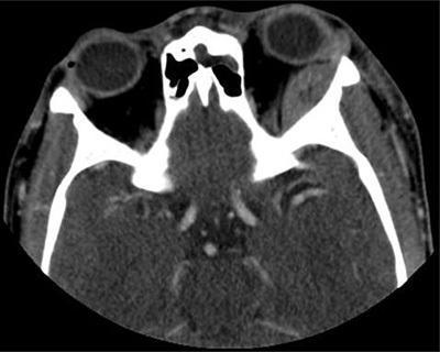
This diagnostic image is an axial contrast-enhanced CT scan of the orbits and skull base. The primary finding is a well-defined, biconcave or lens-shaped extraconal mass located along the superolateral wall of the left orbit. The lesion displays high attenuation, which is characteristic of acute hemorrhage (subperiosteal hematoma) or dense proteinaceous material. This mass exerts a mass effect on the adjacent intraorbital structures, resulting in slight proptosis. The contralateral (right) orbit and its contents appear normal. The bony margins of the frontal and sphenoid bones in this section appear intact without evidence of destructive lesions or gross fractures. Centrally, the ethmoid sinus and the anatomical structures of the skull base are visible. In the clinical context of sickle cell disease, this appearance is highly suggestive of a subperiosteal orbital hematoma, likely occurring secondary to bone infarction. The image serves as an educational example of orbital pathology and the radiological presentation of acute subperiosteal collections in the setting of hematological disorders.
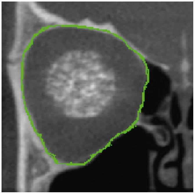
This diagnostic image is a coronal cross-section of the human bony orbit obtained via Cone Beam Computed Tomography (CBCT). The image demonstrates the internal anatomy of the orbital cavity, showing the heterogeneous density of the surrounding facial bones in varying shades of gray. A prominent green digital trace outlines the bony orbital border, providing a clear demarcation of the orbital rim and walls. This automated or semi-automated segmentation encompasses the orbital volume and highlights specific anatomical contours, including the superior and inferior orbital margins. The trace follows the complex geometry of the bone, including slight indentations that correspond to the locations of neurovascular passages, such as the superior or inferior orbital fissures and foramina. Centrally, a blurred hyperdense region represents the soft tissue contents of the orbit. This visualization is used in clinical workflows for orbital volume measurement, assessment of post-traumatic deformities, and planning for orbital implants or reconstructive surgery.
eyelid layers cross section orbicularis oculi tarsus conjunctiva levator palpebrae
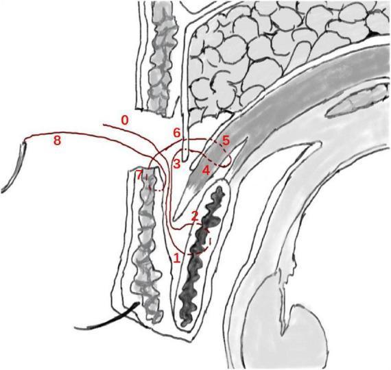
This medical anatomical diagram illustrates a sagittal cross-section of the upper eyelid, detailing the 'flexible-rigid fixation' technique used in small-incision blepharoplasty (double eyelid surgery). The illustration shows the surgical pathway of a mattress suture, color-coded in red and numbered 0 through 8 to indicate the sequence of needle passage. The suture begins at the incision site (0), engages the anterior tarsus fascia or tarsal plate (1-2), and then traverses superiorly through the levator aponeurosis and the retro-orbital septum complex (3-6). The needle is then reversed to anchor the suture to the lower lip of the orbicularis oculi muscle (7) before exiting (8). Key anatomical structures depicted include the skin, pretarsal and preseptal orbicularis oculi muscles, tarsal plate, levator aponeurosis, orbital septum, and preaponeurotic fat pads. This educational diagram demonstrates how the technique establishes an indirect link between the levator mechanism and the skin to create a natural-looking eyelid crease through both stable (rigid) and dynamic (flexible) tissue approximation.
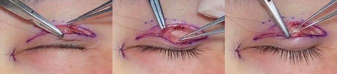
This composite of three clinical photographs captures key intraoperative steps of an upper eyelid blepharoplasty. The images show a surgical field with purple antiseptic marking lines delineating the supratarsal crease. A needle holder and fine forceps are visible, manipulating tissues within a partial-thickness incision. The primary educational focus is the formation of a tarsus linkage mechanism, where a 7-0 nylon suture is passed through the orbicularis oculi muscle and the tarsal plate to facilitate the creation of a stable double eyelid fold. In the central frame, the removal of pre-tarsal fascia and herniated orbital fat is demonstrated, exposing the deeper anatomical layers. The transparent monofilament sutures are used both for internal tissue fixation (levator aponeurosis-tarsus-orbicularis-skin linkage) and subsequent skin closure. This visual illustrates a specialized technique in plastic and reconstructive surgery aimed at achieving aesthetic eyelid contouring and reinforcing structural adhesion during scar healing.
lacrimal apparatus gland canaliculi sac nasolacrimal duct
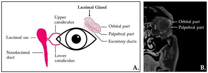
This dual-panel educational figure presents the anatomy and radiographic appearance of the human lacrimal apparatus. Panel A is an anatomical diagram illustrating the secretory and excretory components, including the almond-shaped lacrimal gland situated superolaterally to the eye, its orbital and palpebral parts, and excretory ducts. The drainage system is shown via the upper and lower canaliculi leading into the lacrimal sac and nasolacrimal duct. Panel B is a coronal, contrast-enhanced T1-weighted MR image with fat saturation of the left orbit. It demonstrates the normal radiological appearance of the lacrimal gland within the extraconal space of the superolateral orbit. The image clearly differentiates the superior orbital lobe and the inferior palpebral lobe. This material is designed for medical students and clinicians to understand the structural organization of the tear production and drainage system, which is critical for evaluating conditions like Sjögren's syndrome, dacryoadenitis, or nasolacrimal duct obstruction.
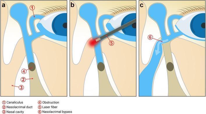
This three-panel medical schematic (Pathophysiology Diagram and Surgical Algorithm) illustrates the mechanism and surgical correction of primary acquired nasolacrimal duct obstruction (PANDO) via laser-assisted dacryocystorhinostomy (DCR). Panel A depicts the pre-operative state where an infrasaccal obstruction (4) in the nasolacrimal duct (2) blocks tear drainage, despite patent canaliculi (1), leading to epiphora. Panel B shows the intra-operative procedure, where a transcanalicular laser fiber (5) is advanced into the lacrimal sac and directed medially toward the nasal wall to create a bypass. Panel C illustrates the post-operative outcome: a newly formed nasolacrimal bypass or ostium (6) allows tears to drain directly from the lacrimal sac into the nasal cavity (3), effectively bypassing the original site of obstruction. Key anatomical structures labeled include the canaliculi, lacrimal sac, nasolacrimal duct, and nasal cavity. This educational visual is intended for ophthalmology and oculoplastic surgery training to demonstrate the transcanalicular laser approach for treating distal lacrimal system blockages.
eyeball cross section anatomy cornea lens retina iris ciliary body choroid sclera
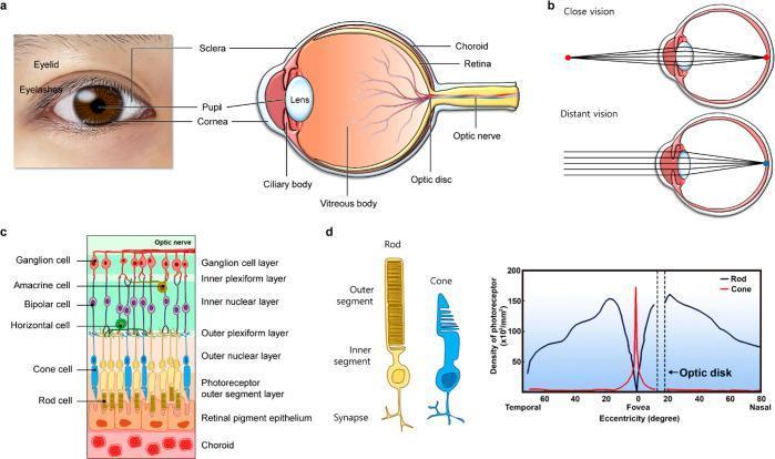
Educational medical illustration of ocular anatomy and the visual process. (a) A clinical photograph of a human eye paired with a sagittal schematic of the eyeball, labeling the cornea, sclera, iris/pupil, lens, ciliary body, vitreous body, choroid, retina, optic disc, and optic nerve. (b) A comparison of visual accommodation, showing the lens thickening for close vision and thinning for distant vision to focus light precisely on the retina. (c) A cross-sectional anatomical diagram of the retina, detailing the cellular layers from the choroid and retinal pigment epithelium through the photoreceptor layer (rods and cones), horizontal, bipolar, amacrine, and ganglion cell layers. (d) A specialized illustration of rod and cone cell morphology (left) and a comparative line graph showing photoreceptor density across the retinal eccentricity. The graph highlights the sharp peak of cone density at the fovea and the high density of rods in the peripheral (nasal and temporal) retina, with both photoreceptor types absent at the optic disc (blind spot).
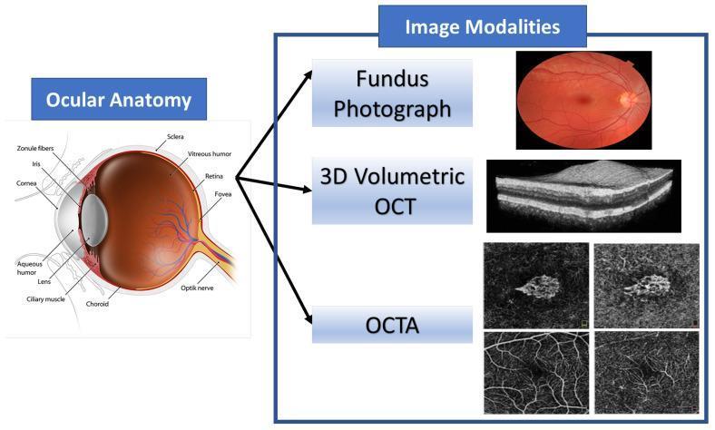
This educational graphic combines an anatomical diagram of the human eye with corresponding ophthalmic imaging modalities. The 'Ocular Anatomy' section features a detailed sagittal cross-section identifying key structures: the cornea, aqueous humor, iris, pupil, biconvex lens, zonule fibers, and ciliary muscle in the anterior segment; and the vitreous humor, retina, fovea, choroid, sclera, and optic nerve in the posterior segment. The 'Image Modalities' section illustrates diagnostic tools used to visualize these structures. A color Fundus Photograph provides a surface view of the retina, optic disc, and vasculature. A 3D Volumetric Optical Coherence Tomography (OCT) scan displays a high-resolution, grayscale cross-sectional view of retinal layers, highlighting tissue stratification. Finally, Optical Coherence Tomography Angiography (OCTA) panels show non-invasive, depth-resolved images of the retinal and choroidal microvasculature. This composite serves as a reference for correlating anatomical landmarks with clinical diagnostic imaging used in the management of retinal diseases such as Age-related Macular Degeneration (AMD).
superior orbital fissure optic canal contents nerves orbit
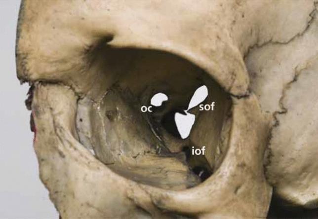
This clinical photograph of a human dry skull specimen provides an anterior view of the right orbit, highlighting the critical foramina and fissures of the orbital apex and walls. Three primary openings are labeled: the optic canal (OC), the superior orbital fissure (SOF), and the inferior orbital fissure (IOF). The optic canal is depicted as a rounded opening in the lesser wing of the sphenoid bone, positioned superiorly and medially. Inferolateral to the optic canal is the superior orbital fissure, which exhibits a characteristic comma-shaped or 'racket' morphology, wider at its base and tapering as it extends superolaterally between the greater and lesser wings of the sphenoid. Positioned below the SOF, along the floor and lateral wall junction, is the inferior orbital fissure, shown as an elongated, slit-like gap. The image serves as an educational anatomical reference for understanding the spatial relationships of the orbital apex, specifically the arrangement of neurovascular passages including the optic nerve, ophthalmic artery, and cranial nerves III, IV, V1, and VI.
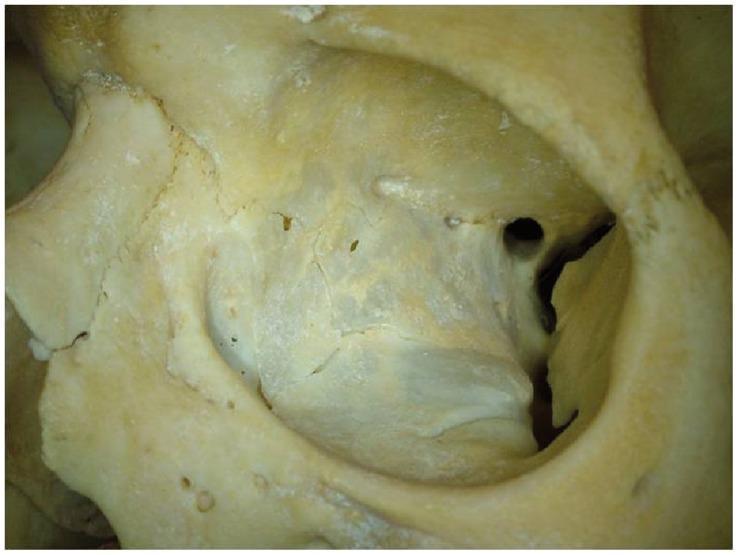
This clinical photograph displays a close-up, anterolateral view of a human bony orbital cavity, likely from a dry skull specimen. The image provides a detailed anatomical view of the internal surfaces and apertures of the orbit. Centrally located at the posterior aspect is the optic canal, appearing as a rounded foramen. Adjacent to it, the superior orbital fissure is visible as an elongated, narrow cleft between the greater and lesser wings of the sphenoid bone. The bony walls—comprising the frontal, zygomatic, and sphenoid bones—exhibit a characteristic pale yellowish-white color with varied textures, including smooth articular areas and roughened sites for muscular and ligamentous attachments. Visible sutures and several smaller accessory foramina (such as ethmoidal foramina) are present along the medial wall and floor. This visual material is used in medical education to demonstrate normal cranial osteology, orbital volume analysis, and the structural relationship between cranial nerves and the orbital apex.
retina optic disc macula fovea ophthalmoscopy fundus
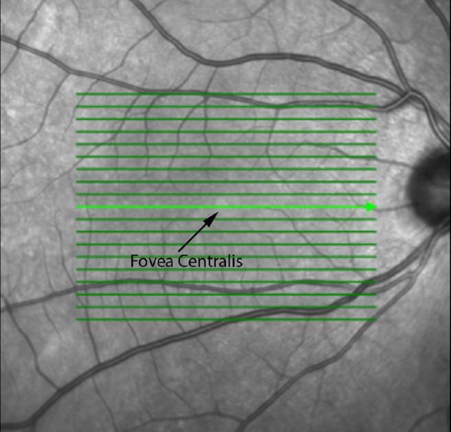
This en-face Scanning Laser Ophthalmoscopy (SLO) image provides a grayscale topographic view of the fundus, commonly used as a reference for Optical Coherence Tomography (OCT) scanning. The image depicts the posterior pole of the retina, including the macula and optic disc. The fovea centralis is visualized as a centrally located, relatively hyporeflective (darker) circular area compared to the surrounding parafoveal tissue, marked here with a black arrow and label. To the right of the macula, the optic disc appears as a vertically oriented, darker oval structure where the major retinal vasculature converges. The retinal blood vessels are visible as dark, branching tubular structures emerging from the optic nerve head and traversing the retinal surface. A series of green horizontal lines are overlaid across the macular region, representing the acquisition grid and specific scan locations (B-scans) for sectional OCT imaging. This imaging modality is essential for anatomical localization and quantitative measurement of retinal structures like the fovea in relation to the optic nerve head.
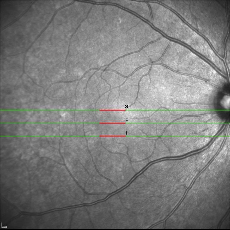
This diagnostic image is a high-resolution, grayscale scanning laser ophthalmoscopy (SLO) fundus image used as a reference for Optical Coherence Tomography (OCT) raster scanning. The image displays the posterior pole of the human retina, with the optic disc visible on the far right and the retinal vasculature radiating toward the macula. Three horizontal green lines indicate the orientation of specific B-scans across the macular region. Overlaid on these green lines are red segments (labeled S, F, and I) centered horizontally. The central line 'F' (Fovea) passes through the foveal center, while 'S' (Superior) and 'I' (Inferior) represent scans located 1,270 microns superior and inferior to the fovea, respectively. The 1,000-micron red segments signify the specific lateral zones where retinal layer thickness measurements (segmentation analysis) were performed. This visual is used in ophthalmology to demonstrate the methodology for mapping retinal morphology and quantifying regional thickness variations in clinical research.
ciliary body iris sphincter pupillae dilator pupillae zonular fibers lens suspension
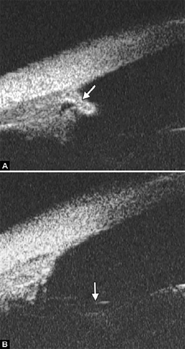
This composite image presents two diagnostic ultrasound biomicroscopy (UBM) scans of the ocular anterior segment, illustrating different manifestations of aniridia. Image (A) shows a maldeveloped, vestigial iris (indicated by the white arrow). The iris appears as an amorphous, truncated structure with irregular borders and heterogeneous reflectivity, alongside malformation of the ciliary body. Image (B) demonstrates a more severe clinical presentation where the iris is entirely absent (aniridia). The white arrow in this section highlights the exposed lens zonules, which appear as thin, highly reflective linear structures that are normally obscured by the iris. These images demonstrate the utility of high-frequency ultrasound in evaluating iris hypoplasia and identifying anatomical landmarks like the scleral spur, ciliary body, and zonular fibers when corneal opacity or severe structural defects prevent adequate slit-lamp visualization. The findings are characteristic of congenital aniridia and its associated anterior segment dysgenesis.
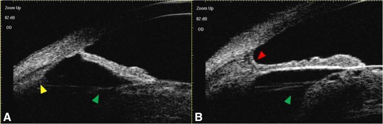
Educational ultrasound biomicroscopy (UBM) images (Figures A and B) of the right eye anterior segment, demonstrating congenital megalophthalmos and cataract. The images show a deep anterior chamber with an open iridocorneal angle. Key findings include: 1. Lens: An opacified, cataractous lens is visible as an echogenic structure. 2. Suspensory Ligaments: Prominent, elongated zonular fibers (green arrowheads) connect the lens equator to the ciliary body, a hallmark of anterior segment enlargement. 3. Ciliary Processes: Small, hyperechoic ciliary processes (yellow arrowhead in Fig A) are visualized posterior to the iris. 4. Pectinate Ligament: A distinct pectinate ligament (red arrowhead in Fig B) is noted at the anterior chamber angle, appearing as a bridge of tissue spanning the angle recess. This diagnostic imaging illustrates the structural abnormalities associated with congenital ocular enlargement, specifically focusing on the anatomical relationship between the lens, zonules, and ciliary body, and is intended for ophthalmology residents and specialists studying lens subluxation risk or glaucoma in megalophthalmos.
tarsal plate orbicularis oculi medial lateral palpebral ligament
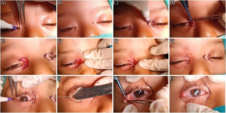
This composite of clinical photographs illustrates a 12-step surgical sequence (A-L) for the simultaneous correction of epicanthus and telecanthus, typically associated with Blepharophimosis-ptosis-epicanthus inversus syndrome (BPES). Panels A-C show the preoperative marking of the C-U medial canthoplasty, utilizing points A-D to delineate the desired horizontal palpebral fissure length (HPFL). Panel D demonstrates the excision of skin and underlying orbicularis oculi muscle. Panels E-G depict the exposure, plication, and shortening of the medial canthal tendon to reduce inter-isthmus distance. Panel H shows the completed medial wound closure. The final row (I-L) focuses on lateral canthoplasty, showing the marking of the lateral skin, surgical widening of the lateral canthal wound to lengthen the HPFL, and suturing of the undermined conjunctiva to form a sharp lateral canthal angle. This educational material highlights the specialized oculoplastic techniques required to achieve anatomical correction of both the medial and lateral ocular structures.
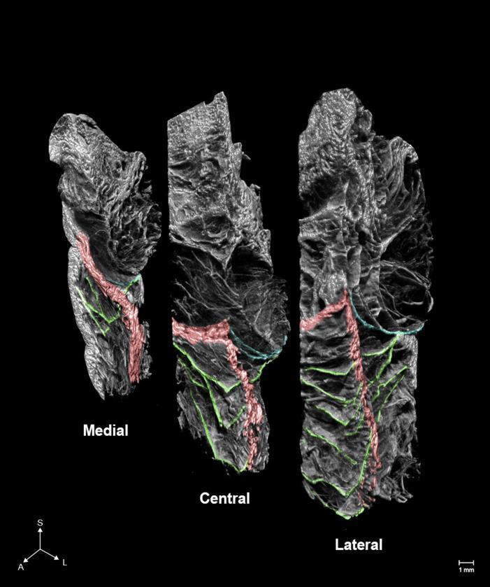
This diagnostic image consists of three high-resolution micro-computed tomography (microCT) slices depicting the anatomical architecture of the periorbital region in the medial, central, and lateral planes. The visual highlights the orbicularis retaining ligament (ORL) and its spatial relationship with adjacent structures using false-color overlays. The ORL (green) is shown as a complex network of multilayered plexiform fibers that arborize horizontally. The image demonstrates a progressive increase in the number, complexity, and branching of ORL fibers from the medial to the lateral aspect. The orbital septum is highlighted in blue, while sections of the orbicularis oculi muscle are indicated in red. The spatial arrangement reveals the ORL is positioned anterior to the orbital septum and interweaves with the muscle fibers of the orbicularis oculi. Anatomical orientation is provided by an axis indicator showing Superior (S), Anterior (A), and Lateral (L) directions, along with a 1 mm scale bar. This content is used to illustrate the delicate ligamentous structures involved in facial aging and surgical rejuvenation.
📚 Lecture 13 - Complete Notes: ORBIT (Head & Neck)
1. OVERVIEW OF THE ORBIT
- Below the anterior cranial fossa
- Anterior to the middle cranial fossa
- Eyeball, optic nerve
- Extra-ocular muscles
- Lacrimal apparatus
- Adipose tissue and fascia
- Nerves and vessels
2. BONY ORBIT
Seven bones: Maxilla · Zygomatic · Frontal · Ethmoid · Lacrimal · Sphenoid · Palatine
- Wide base opens anteriorly onto the face
- Apex points posteromedially
Orbital Rim (Base)
| Side | Bone |
|---|---|
| Superior | Frontal bone |
| Medial | Frontal process of maxilla |
| Inferior | Zygomatic process of maxilla + zygomatic bone |
| Lateral | Zygomatic bone + frontal process of zygomatic + zygomatic process of frontal |
3. WALLS OF THE BONY ORBIT
Roof (Superior Wall)
- Orbital part of the frontal bone + small part of the sphenoid
- Thin plate separates the orbit from the anterior cranial fossa (brain above)
- Special features:
- Anteromedially: trochlear fovea (pulley for superior oblique) ± frontal sinus
- Anterolaterally: lacrimal fossa (depression for lacrimal gland)
- Posteriorly: lesser wing of sphenoid completes the roof
Medial Wall (from anterior to posterior)
- Largest contributor: orbital plate of ethmoid (contains ethmoidal air cells)
- At roof-medial wall junction (frontoethmoidal suture): anterior & posterior ethmoidal foramina
- Lacrimal groove: formed by lacrimal bone (posterior lacrimal crest) + frontal process of maxilla (anterior lacrimal crest) → houses the lacrimal sac
- Posterior part completed by a small bit of sphenoid (forms part of the medial wall of the optic canal)
Floor (Inferior Wall)
- = roof of the maxillary sinus
- Primarily: orbital surface of maxilla
- Small contributions: zygomatic + palatine bones
- Along lateral boundary: inferior orbital fissure (begins posteriorly)
- Near the maxilla-ethmoid-sphenoid junction: small contribution from orbital process of the palatine bone
Lateral Wall
- Anteriorly: zygomatic bone
- Posteriorly: greater wing of sphenoid
- Superior orbital fissure lies between the greater wing and lesser wing of sphenoid
4. FISSURES AND FORAMINA
Optic Canal
- Round opening at the apex of the orbit
- Opens into the middle cranial fossa
- Bounded: medially by body of sphenoid, laterally by lesser wing of sphenoid
- Contents: Optic nerve [II] + Ophthalmic artery
Superior Orbital Fissure
- Triangular gap between the roof and lateral wall
- Communicates orbit with the middle cranial fossa
- Contents:
- Oculomotor nerve [III] - superior and inferior branches
- Trochlear nerve [IV]
- Abducent nerve [VI]
- Lacrimal, frontal, nasociliary branches of ophthalmic nerve [V1]
- Superior ophthalmic vein
Inferior Orbital Fissure
- Separates lateral wall from the floor
- Borders: greater wing of sphenoid + maxilla + palatine + zygomatic
- Communicates:
- Orbit → pterygopalatine fossa (posteriorly)
- Orbit → infratemporal fossa (middle)
- Orbit → temporal fossa (posterolaterally)
- Contents: maxillary nerve [V2] + zygomatic branch, infra-orbital vessels, vein to pterygoid plexus
Infra-orbital Groove → Canal → Foramen
- Groove begins at posterior two-thirds of inferior orbital fissure
- Continues anteriorly as the infra-orbital canal → opens onto face at the infra-orbital foramen
- Contents: infra-orbital nerve (V2) and vessels
Anterior & Posterior Ethmoidal Foramina
- At the junction of the superior and medial walls
- Exit from orbit into ethmoid bone
- Transmit anterior and posterior ethmoidal nerves and vessels
Nasolacrimal Canal (Medial wall, anteriorly)
- Depression for lacrimal sac (lacrimal bone + frontal process of maxilla)
- Continuous with the nasolacrimal canal → leads to inferior nasal meatus
- Contains: nasolacrimal duct
5. FASCIAL SPECIALIZATIONS
Periorbita
- Periosteum lining the bones of the orbit
- Continuous with periosteum on the outer surface of the skull
- Sends extensions into upper and lower eyelids = orbital septa
- At openings communicating with the cranial cavity: continuous with the periosteal layer of dura mater
- Posteriorly, thickens around the optic canal and central part of the superior orbital fissure = Common Tendinous Ring (origin of all four rectus muscles)
6. EYELIDS
Layers (Anterior → Posterior)
Skin → Subcutaneous tissue → Orbicularis oculi (voluntary)
→ Orbital septum → Tarsus → Conjunctiva
Note: The subcutaneous tissue is very thin and loosely arranged. This is why blood/fluid accumulates easily after injury to this area.
Muscles of the Eyelid
| Muscle | Function |
|---|---|
| Orbicularis oculi | Closes the eyelids |
| Levator palpebrae superioris | Opens/raises the upper eyelid |
| Superior tarsal muscle (smooth) | Assists in maintaining elevation of the upper eyelid |
- Orbital part - circular fibers surrounding the orbit
- Palpebral part - within the eyelids; anchored by medial and lateral palpebral ligaments
- Lacrimal part - medial border fibers attached to posterior lacrimal crest; may be involved in tear drainage by dilating the lacrimal sac during blinking
Orbital Septum
- Extension of periosteum from orbital margin into both eyelids
- Extends downward into upper eyelid and upward into lower eyelid
- Continuous with periosteum inside and outside the orbit
- Upper eyelid: attaches to the tendon of levator palpebrae superioris
- Lower eyelid: attaches to the tarsus
Tarsal Plates
- Superior tarsus (large) - upper eyelid
- Inferior tarsus (small) - lower eyelid
- Dense connective tissue - major structural support for eyelids
- Attached medially via medial palpebral ligament → to anterior lacrimal crest of maxilla
- Attached laterally via lateral palpebral ligament → to orbital tubercle of zygomatic bone
Levator Palpebrae Superioris
- Origin: posterior roof of the orbit, just superior to the optic foramen
- Insertion: anterior surface of the superior tarsus (± skin of upper eyelid)
- Associated: Superior tarsal muscle (smooth muscle) runs from the inferior surface of the levator to the upper edge of the superior tarsus
Conjunctiva
- Thin membrane covering the posterior surface of each eyelid
- Reflects onto the outer surface of the eyeball (sclera)
- Attaches at the sclero-corneal junction
- When eyelids closed = conjunctival sac with superior and inferior conjunctival fornices
Glands of the Eyelid
| Gland | Location | Secretion | Blockage/Inflammation |
|---|---|---|---|
| Tarsal glands (modified sebaceous) | Embedded in tarsal plates | Oily substance - increases tear viscosity, decreases evaporation | Chalazion (inner surface of eyelid) |
| Sebaceous + sweat glands | Associated with eyelash follicles | Normal sebum/sweat | Stye (edge of eyelid) |
7. LACRIMAL APPARATUS
Components
- Lacrimal gland + ducts
- Lacrimal canaliculi
- Lacrimal sac
- Nasolacrimal duct
Lacrimal Gland
- Position: anterior superolateral region of the orbit
- Divided into two parts by the levator palpebrae superioris:
| Part | Location |
|---|---|
| Orbital part (larger) | In the lacrimal fossa of the frontal bone |
| Palpebral part (smaller) | Inferior to the levator palpebrae superioris in the superolateral part of the eyelid |
- Ducts empty into the lateral part of the superior conjunctival fornix
Flow of Tears
Lacrimal gland (secreted)
→ Lateral superior conjunctival fornix
→ Across the eye surface (lateral to medial during blinking)
→ Lacrimal lake (medial accumulation)
→ Lacrimal canaliculi (one per eyelid, entering via lacrimal punctum)
→ Lacrimal sac (between anterior & posterior lacrimal crests,
   posterior to medial palpebral ligament, anterior to lacrimal part of orbicularis oculi)
→ Nasolacrimal duct
→ Inferior nasal meatus
Key mechanism: When orbicularis oculi contracts during blinking, the lacrimal part may dilate the lacrimal sac, drawing tears in through the canaliculi.
8. ⚠️ WHAT WAS MISSED IN THE FIRST NOTES: CILIARY GANGLION
Ciliary Ganglion (in the orbit)
1. Motor Root (Parasympathetic)
- From the inferior branch of the oculomotor nerve [III]
- Carries preganglionic parasympathetic fibers
- These synapse in the ganglion on postganglionic neurons
- Postganglionic fibers travel in the short ciliary nerves to the eyeball
- Innervates: ciliary muscle and sphincter pupillae
2. Sensory Root
- From the nasociliary nerve (branch of V1)
- Enters the posterosuperior aspect of the ganglion
- Sensory fibers pass through (do not synapse) and continue in the short ciliary nerves to the eyeball
- Responsible for sensory innervation of all parts of the eyeball
3. Sympathetic Root (most variable)
- Contains postganglionic sympathetic fibers from the superior cervical ganglion
- Travels up the internal carotid artery, leaves the plexus in the cavernous sinus
- Enters orbit through the common tendinous ring
- Enters the posterior aspect of the ciliary ganglion, crosses through, continues in short ciliary nerves to the eyeball
- Alternatively: sympathetic fibers may NOT enter the ganglion and instead travel directly in the long ciliary nerves to the eyeball
- Innervates: dilator pupillae muscle
9. EYEBALL
Anterior and Posterior Chambers
- Anterior chamber: behind the cornea, in front of the iris
- Pupil: central opening in the iris
- Posterior chamber: behind the iris, in front of the lens (smaller)
- Both filled with aqueous humor:
- Secreted into posterior chamber by ciliary processes
- Flows through pupil → into anterior chamber
- Absorbed into scleral venous sinus (canal of Schlemm) - circular channel at the cornea-iris junction
- Functions: nourishes the avascular cornea and lens; maintains intra-ocular pressure
- Imbalance (production > absorption) = glaucoma → raised intra-ocular pressure → visual problems
Lens
- Separates anterior 1/5 from posterior 4/5 of the eyeball
- Transparent, biconvex, elastic disc
- Attached circumferentially via zonular fibers (= suspensory ligament of the lens) to the ciliary processes
- Changes refractive ability for visual acuity (accommodation)
- Opacity of lens = cataract
Postremal (Vitreous) Chamber
- Posterior 4/5 of eyeball (lens to retina)
- Filled with vitreous body (vitreous humor): transparent, gelatinous
- Cannot be replaced (unlike aqueous humor)
10. WALLS OF THE EYEBALL (3 Layers)
Layer 1 - Outer Fibrous Layer
| Structure | Location | Description |
|---|---|---|
| Sclera | Posterior + lateral (~5/6) | Opaque dense connective tissue; "white of the eye"; pierced by optic nerve posteriorly; attachment for extraocular muscles |
| Cornea | Anterior (~1/6) | Transparent; allows light to enter |
- Fascial sheath of the eyeball: covers sclera externally from the optic nerve entrance to the corneoscleral junction; loosely attached internally to the choroid
Layer 2 - Middle Vascular Layer (3 continuous parts, posterior to anterior)
- ~2/3 of the vascular layer
- Thin, highly vascular, pigmented
- Smaller vessels adjacent to retina; larger vessels more peripherally
- Firmly attached to retina internally; loosely attached to sclera externally
- Extends from the anterior border of the choroid
- Triangular-shaped; forms a complete ring around the eyeball
- Two components:
- Ciliary muscle: smooth muscle (longitudinal, circular, radial fibers)
- Ciliary processes: longitudinal ridges projecting inward; give rise to zonular fibers (= suspensory ligament of the lens)
- Ciliary processes also contribute to formation of aqueous humor
How accommodation works: Ciliary muscle contracts → ciliary ring gets smaller → reduces tension on suspensory ligament → lens becomes more rounded → focused for near vision
- Circular structure projecting outward from the ciliary body
- Colored part of the eye with central opening = pupil
- Contains two smooth muscle structures controlling pupil size:
| Muscle | Fiber Arrangement | Function |
|---|---|---|
| Sphincter pupillae | Circular | Constricts the pupil (decreases size) |
| Dilator pupillae | Radial | Dilates the pupil (increases size) |
Intrinsic Muscles of the Eye (Full Table from PDF)
| Muscle | Location | Function |
|---|---|---|
| Ciliary muscle | Ciliary body | Constricts ciliary body ring; relaxes tension on lens; lens becomes more rounded → accommodation for near vision |
| Sphincter pupillae | Circular fibers in the iris | Constricts the pupil |
| Dilator pupillae | Radial fibers in the iris | Dilates the pupil |
Layer 3 - Inner Retinal Layer
- Optic retina (posterior + lateral) - sensitive to light
- Nonvisual retina (anterior) - covers ciliary body and iris internally
- Junction = ora serrata (irregular line)
| Layer | Attachment | Notes |
|---|---|---|
| Pigmented layer (outer) | Firmly attached to the choroid | Continues anteriorly over the ciliary body and iris |
| Neural layer (inner) | Only attached at two points: around the optic nerve and at the ora serrata | This is the layer that detaches in retinal detachment |
| Landmark | Description |
|---|---|
| Optic disc | Where optic nerve exits; lighter than surrounding retina; central retinal artery branches spread from here; no light-sensitive receptors = blind spot |
| Macula lutea | Lateral to optic disc; small yellowish area; highest visual sensitivity |
| Fovea centralis | Central depression of macula; thinnest area of retina; fewest rods, most cones; highest visual acuity |
| Rods | Light-sensitive; function in dim light; insensitive to color |
| Cones | Light-sensitive; function in bright light; sensitive to color |
COMPLETE QUICK-REFERENCE SUMMARY
Bones of Bony Orbit (7)
What passes through each opening?
| Opening | Contents |
|---|---|
| Optic canal | Optic nerve [II] + Ophthalmic artery |
| Superior orbital fissure | CN III (sup + inf branches), CN IV, CN VI, V1 (lacrimal + frontal + nasociliary), superior ophthalmic vein |
| Inferior orbital fissure | V2 (maxillary nerve + zygomatic branch), infra-orbital vessels, vein to pterygoid plexus |
| Infra-orbital foramen | Infra-orbital nerve (V2) + vessels |
| Anterior ethmoidal foramen | Anterior ethmoidal nerve + vessels |
| Posterior ethmoidal foramen | Posterior ethmoidal nerve + vessels |
| Nasolacrimal canal | Nasolacrimal duct |
Muscles - Name and Function Only
| Muscle | Function |
|---|---|
| Orbicularis oculi | Closes the eyelids |
| Levator palpebrae superioris | Opens (elevates) the upper eyelid |
| Superior tarsal muscle | Assists elevation of the upper eyelid |
| Ciliary muscle | Accommodates lens for near vision (constricts ciliary body, relaxes tension on lens → lens rounds up) |
| Sphincter pupillae | Constricts the pupil |
| Dilator pupillae | Dilates the pupil |
Key Clinical Terms from the PDF
| Term | Meaning |
|---|---|
| Ptosis | Drooping of the upper eyelid |
| Glaucoma | Raised intraocular pressure due to excess aqueous humor |
| Cataract | Opacity of the lens |
| Chalazion | Blocked/inflamed tarsal gland (on inner surface of lid) |
| Stye | Blocked/inflamed sebaceous or sweat gland of eyelash (on lid edge) |
| Blind spot | Optic disc - no photoreceptors present |
| Detached retina | Separation of the neural layer from the pigmented layer |
| Palpebral fissure | Space between open eyelids |
| Conjunctival sac | Space between eyelids and eyeball (when closed) |
| Lacrimal punctum | Entry opening of each lacrimal canaliculus |
| Ora serrata | Junction between optic and nonvisual retina |