
Severe herpes simplex management
"herpes simplex" AND management AND severe
herpes simplex encephalitis MRI temporal lobe
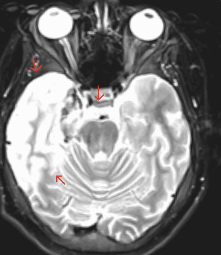
An axial T2-weighted MRI scan of the brain with contrast, demonstrating significant findings characteristic of Herpes Simplex Encephalitis (HSE). The image reveals marked signal alteration in the right hemisphere, specifically involving the mesial temporal lobe and limbic system. Key features include contrast enhancement and evidence of hemorrhagic transformation, indicated by mixed signal intensities within the affected parenchyma. Red arrows highlight the involvement of the right limbic structures, the fornix, and the right optic radiation. Anatomical consequences include mild effacement of the right lateral ventricle and a minimal leftward midline shift, suggesting localized mass effect and cerebral edema. This diagnostic image illustrates the classic predilection of HSV-1 for the temporal lobes and its potential for hemorrhagic evolution, which can lead to complications such as increased intracranial pressure and visual pathway deficits.
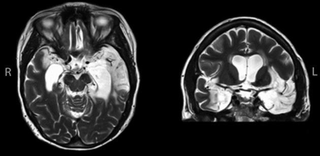
High-resolution T2-weighted Magnetic Resonance Imaging (MRI) of the brain in axial (left) and coronal (right) sections, demonstrating profound bilateral neuroanatomical abnormalities secondary to herpes simplex encephalitis. The images reveal extensive tissue loss and encephalomalacia within the medial temporal lobes and hippocampi. The damage is asymmetric, with significantly more pronounced involvement of the left hemisphere, where structural obliteration extends into the anterior temporal pole, lateral temporal regions, and fusiform gyrus. The coronal section highlights compensatory ventriculomegaly (hydrocephalus ex vacuo), particularly involving the temporal horns of the lateral ventricles, due to severe parenchymal atrophy. Large hyperintense regions on T2 imaging indicate fluid-filled cystic spaces where healthy neural tissue once resided. This diagnostic image serves as a classic illustration of the long-term sequelae of viral encephalitis, specifically the preferential destruction of the limbic system and temporal lobe structures, which clinically correlates with profound memory and cognitive deficits.
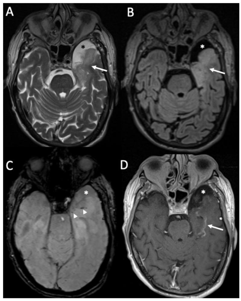
This composite diagnostic image displays four axial MRI views (A-D) of the brain at the level of the midbrain and temporal lobes, illustrating the subacute phase of Herpes Simplex Virus Type 1 (HSV-1) encephalitis. Panel A (T2-weighted) and Panel B (FLAIR) demonstrate significant hyperintensity and swelling within the left temporal lobe (white arrows), characteristic of inflammatory edema. Panel C (Susceptibility-Weighted Imaging, SWI) reveals small, dark punctate areas (white arrowheads) representing hemorrhagic foci, a common complication of necrotizing encephalitis. Panel D (Contrast-enhanced T1-weighted) shows curvilinear leptomeningeal or parenchymal enhancement (white arrow) in the affected temporal region, indicating blood-brain barrier breakdown. Additionally, a well-circumscribed temporo-polar arachnoid cyst is consistently visible across all sequences (asterisk), appearing hyperintense on T2 (A), suppressed on FLAIR (B), and following CSF signal intensity on SWI (C) and T1 (D). These findings emphasize the classic limbic system involvement and hemorrhagic potential of HSV encephalitis.
herpes simplex virus skin lesions vesicles
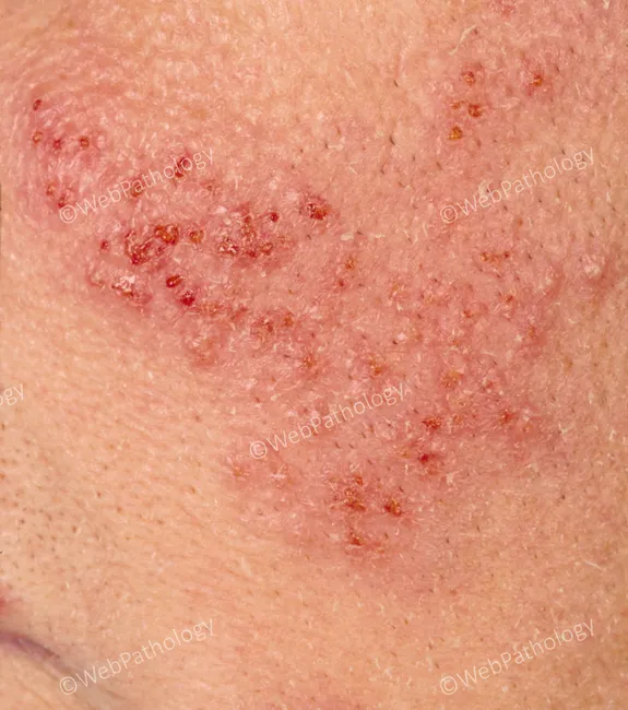
This dermatologic image shows a perioral, facial eruption consistent with recurrent herpes simplex virus infection (HSV-1). The modality is a close‑up clinical photograph under standard white light to capture surface morphology. The perioral skin demonstrates clustered, vesicular lesions evolving on an erythematous base, with shallow erosions and crusting at multiple discrete sites. The vesicles are small to medium in size, often grouped in crops, with clear or hemorrhagic fluid content and surrounding mild edema. The distribution is localized to the perioral region and adjacent cheek, with a frontal facial view that emphasizes symmetry and lesion layering. The lesions reflect the reactivation pattern of HSV, showing papulovesicular to vesicular stages with crusted crusts as they heal. Clinically, these findings correspond to HSV-1 reactivation rather than varicella or other vesiculobullous dermatoses, though differential diagnoses include impetigo, eczema herpeticum, contact dermatitis, or herpes zoster in atypical locations. The image highlights characteristic features such as grouped vesicles on an erythematous base, crusted fissures, and superficial erosions, which inform antiviral therapy decisions and patient counseling regarding contagion. This representation is valuable for education, differential diagnosis training, and clinical reference in dermatology, primary care, and infectious disease contexts.
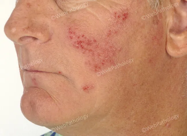
Clinical photography of recurrent herpes simplex virus infection with perioral facial involvement. The image depicts grouped vesicles on an erythematous base positioned around the mouth and adjacent cheek, with focal erosions and crusting consistent with an active HSV-1 reactivation in the perioral distribution. Lesions are small to medium in size, discrete to coalescing, fluid-filled in the vesicular stage, then rupturing to form shallow erosions that crust as they heal. The surrounding skin shows mild edema and diffuse erythema; no purulent discharge or satellite lesions are evident. The left perioral region is the predominant site; this is a common clinical pattern, often precipitated by sun exposure, stress, or viral reactivation. The appearance is classic for herpes labialis variants, though HSV-2 less commonly affects this region. In this context, diagnosis is largely clinical, supported by morphology of grouped vesicles on an erythematous base and characteristic crusting. Differential considerations include impetigo, contact dermatitis, eczema herpeticum, and varicella-zoster in atypical presentations; confirmation can be obtained by PCR or viral culture if needed. Clinically, this presentation guides antiviral therapy (acyclovir, valacyclovir) and symptomatic care; patient counseling on contagion reduction and recurrences is essential. This image is suitable for educational demonstrations of herpes simplex skin lesions and for comparison studies of lesion evolution over time.
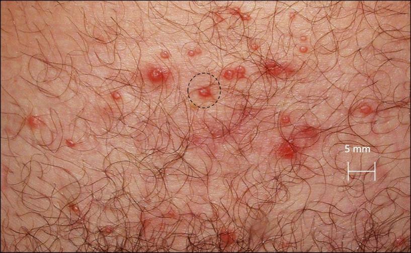
This clinical photograph displays a close-up view of a cutaneous outbreak in a hair-bearing genital region, characteristic of Herpes Simplex Virus type 2 (HSV-2). The primary findings consist of multiple small, erythematous vesicles and shallow round ulcers clustered together. The individual lesions are predominantly less than 5 mm in diameter, as indicated by the accompanying scale bar. The lesions exhibit a spectrum of progression, from intact fluid-filled vesicles (blisters) to open, weeping sores with surrounding inflammatory erythema. A black dotted circle highlights a representative cluster of these spatial features, demonstrating the tendency of the virus to manifest as concurrent, spatially grouped foci of infection. The surrounding skin shows localized inflammation but lacks extensive scarring, suggesting an acute or recurrent shedding episode. This image serves as an educational tool to illustrate the morphology, distribution, and spatial dynamics of genital herpes lesions during a symptomatic reactivation phase.
Severe Herpes Simplex Management
Overview of Severe Presentations
- HSV encephalitis (HSV-1; mortality >70% untreated)
- Neonatal HSV (disseminated, CNS, or SEM disease)
- Immunocompromised host - disseminated or visceral HSV (pneumonitis, hepatitis, esophagitis)
- Severe primary genital/orolabial infection with systemic symptoms
- Acyclovir-resistant HSV (especially in HSCT/HIV patients)
- Ocular HSV (keratitis, acute retinal necrosis)
Antiviral Backbone
Mechanism
Syndrome-Specific Treatment Regimens
1. HSV Encephalitis - EMERGENCY
| Parameter | Regimen |
|---|---|
| Drug | IV Acyclovir |
| Dose | 10 mg/kg every 8 hours |
| Duration | 14-21 days |
| Route | Intravenous only |
"High-dose parenteral acyclovir is the therapy of choice for treatment of herpes simplex encephalitis." - Goldman-Cecil Medicine
"Treatment with acyclovir...yields the best results when administered before patients become comatose." - Plum and Posner's Diagnosis and Treatment of Stupor and Coma
- CSF PCR for HSV DNA - gold standard (more sensitive than culture)
- CSF: typically elevated WBC (lymphocyte predominance), elevated RBCs (hemorrhagic necrosis), elevated protein, normal glucose
- MRI (preferred over CT): T2/FLAIR hyperintensity in medial temporal lobes, insula, cingulate cortex
- EEG: periodic 1-Hz sharp waves from temporal lobes - highly characteristic
- If initial CSF PCR is negative but suspicion remains high (especially early or immunocompromised), continue treatment and repeat PCR in 72 hours
2. Neonatal HSV
| Indication | Dose |
|---|---|
| Neonatal HSV (all forms) | IV Acyclovir 20 mg/kg every 8 hours |
| Duration (SEM disease) | 14 days |
| Duration (CNS or disseminated) | 21 days |
3. Severe Mucocutaneous or Disseminated HSV in Immunocompromised Patients
- Goldman-Cecil Medicine: "Parenteral acyclovir is indicated for the initial treatment of mucosal or cutaneous herpes simplex infection in immunocompromised patients...and disseminated or organ-invasive infections in immunocompetent patients."
4. HSV Esophagitis (Severe Odynophagia)
5. Severe Primary Genital Herpes
| Agent | Dose | Duration |
|---|---|---|
| Oral Acyclovir | 400 mg TID (or 200 mg 5x/day) | 7-10 days |
| Valacyclovir | 1 g BID | 7-10 days |
| Famciclovir | 250 mg TID | 7-10 days |
| If hospitalization needed | IV Acyclovir 5 mg/kg q8h | Until able to take PO |
6. Herpes Keratitis
- Acyclovir 400 mg 5x/day OR valacyclovir 500 mg TID (oral)
- Topical options: trifluridine, acyclovir, or ganciclovir eye drops (equally effective)
- Acute retinal necrosis: IV acyclovir 10-15 mg/kg q8h for 10-14 days, then oral valacyclovir
Acyclovir Resistance - Severe/Refractory Disease
| Agent | Dose | Notes |
|---|---|---|
| IV Foscarnet | 40 mg/kg q8h or 60 mg/kg q12h | First choice for acyclovir-resistant HSV; inhibits viral DNA polymerase directly without TK phosphorylation |
| IV Cidofovir | 5 mg/kg once weekly x2, then every 2 weeks | Reserve for acyclovir- AND foscarnet-resistant HSV; phosphorylated by host enzymes |
Recent evidence (PMID 39090526) - a 2024 systematic review on refractory HSV in HSCT recipients highlights foscarnet as the primary salvage agent, with brincidofovir and combination regimens under investigation.
Acyclovir Toxicity (Relevant for High-Dose/IV Use)
- Renal dysfunction - crystalline nephropathy; mitigate with adequate hydration and slow infusion
- CNS toxicity - tremor, myoclonus, confusion, agitation, hallucinations (more common with renal impairment)
- Neutropenia/bone marrow toxicity - rare
- Nausea, vomiting, headache common. - Goldman-Cecil Medicine
Summary Table
| Syndrome | Preferred Agent | Dose | Route | Duration |
|---|---|---|---|---|
| HSV encephalitis | Acyclovir | 10 mg/kg q8h | IV | 14-21 days |
| Neonatal HSV (CNS/disseminated) | Acyclovir | 20 mg/kg q8h | IV | 21 days |
| Neonatal HSV (SEM) | Acyclovir | 20 mg/kg q8h | IV | 14 days |
| Disseminated/visceral HSV (immunocompromised) | Acyclovir | 5-10 mg/kg q8h | IV | Until clinical improvement, then PO |
| Severe primary genital/orolabial | Acyclovir → valacyclovir | 5 mg/kg q8h IV → 1 g BID PO | IV → PO | 7-10 days total |
| Acyclovir-resistant HSV | Foscarnet | 40 mg/kg q8h | IV | Until healed |
| Foscarnet-resistant HSV | Cidofovir | 5 mg/kg/week | IV | As needed |
Key Clinical Pearls
- Start IV acyclovir empirically in any suspected HSV encephalitis - waiting for CSF PCR confirmation costs lives; mortality without treatment exceeds 70%.
- A negative initial CSF PCR does not exclude HSV encephalitis early in illness - continue treatment and repeat at 72 hours.
- In immunocompromised patients, suspect acyclovir resistance when there is clinical deterioration or failure to improve after 5-7 days of adequate IV acyclovir.
- Renal dosing adjustments for acyclovir are mandatory (dose interval extended based on CrCl) - always check renal function before dosing.
- Severe temporal lobe edema can cause transtentorial herniation even with treatment - neurosurgical decompression may be required in extreme cases.
- Valacyclovir is the preferred oral agent for most severe-but-oral-eligible situations due to superior bioavailability (3-5x greater than oral acyclovir).