
Necrosis pathology
I'll read the medical library skill first, then research necrosis pathology comprehensively.
Reading File
Now let me search the medical library for necrosis pathology - running parallel searches for headings and paragraphs:
Finding Sources
Finding Sources
Excellent! The Robbins pathology books are the primary source. Let me read both in parallel - the Robbins Basic Pathology section on necrosis and the Robbins Pathologic Basis of Disease section:
Reading File
Reading File
Excellent content. Now let me read more of the necrosis types section from both Robbins books:
Reading File
Reading File
I can see necrosis type images. Let me now read the necrosis types section from Robbins Basic Pathology and fetch the key images:
Reading File
Reading File
I have comprehensive content. Now let me fetch the key necrosis images from Robbins to include in the answer:
Loading Image
Loading Image
Loading Image
Loading Image
I now have rich content and images. Let me also get the coagulative necrosis image and the biochemical mechanisms section:
Finding Sources
Reading File
I now have comprehensive, authoritative content from both Robbins textbooks with pathological images. Let me compile the full answer.
Necrosis - Pathology
Definition
Necrosis is a pathologic form of cell death resulting from severe, irreparable injury. It is distinct from apoptosis (programmed cell death) in that it is always pathological, triggers inflammation, and involves uncontrolled breakdown of cellular membranes.
Main causes include: ischemia, microbial toxins, burns and chemical/physical injury, and situations where active proteases leak out of cells (e.g., pancreatitis).
- Robbins, Cotran & Kumar Pathologic Basis of Disease, p. 54-56
- Robbins & Kumar Basic Pathology, p. 21-23
Pathogenesis
Necrosis follows a sequence:
- Severe injury damages membranes and mitochondria irreparably
- Lysosomal enzymes enter the cytoplasm and digest the cell from within
- Cellular contents leak through the damaged plasma membrane into extracellular space
- DAMPs (Damage-Associated Molecular Patterns) are released - including ATP (from damaged mitochondria) and uric acid (DNA breakdown product)
- DAMPs are recognized by receptors on macrophages, triggering phagocytosis and cytokine-driven inflammation
- Inflammatory cells produce more proteolytic enzymes, leading to clearance of necrotic debris
This necrosis-associated leakage of intracellular proteins forms the basis of blood biomarkers - cardiac troponins (detectable as early as 2 hours after myocardial necrosis), transaminases in hepatocyte necrosis, and alkaline phosphatase in bile duct damage.
Microscopic (Morphologic) Changes
Cytoplasmic Changes
- Increased eosinophilia (pink on H&E) - due to denatured cytoplasmic proteins binding eosin dye, plus loss of basophilic RNA
- Glassy, homogeneous appearance - from loss of glycogen particles
- Vacuolated, "moth-eaten" cytoplasm - after enzymatic digestion of organelles
- Myelin figures - phospholipid whorls from disrupted membranes; may calcify (dystrophic calcification)
- EM: discontinuities in plasma and organelle membranes; marked mitochondrial dilation with amorphous intramitochondrial densities; disrupted lysosomes
Nuclear Changes (3 Patterns)
| Pattern | Description |
|---|---|
| Pyknosis | Nuclear shrinkage + increased basophilia (DNA condenses into dark, shrunken mass) |
| Karyorrhexis | Fragmentation of the pyknotic nucleus |
| Karyolysis | Fading of basophilia due to DNase-mediated digestion of DNA; nucleus dissolves within 1-2 days |
Morphologic Patterns of Tissue Necrosis
There are 6 recognized patterns, each with distinct causes and appearances:
1. Coagulative Necrosis
- Most common type
- Tissue architecture is preserved for days because the injurious stimulus denatures structural proteins AND enzymes, blocking proteolysis
- Affected tissue becomes firm in texture
- Histology: intensely eosinophilic, anucleate "ghost cells" with preserved cell outlines
- Caused by: ischemia (infarction) in all solid organs except the brain
- A localized area is called an infarct
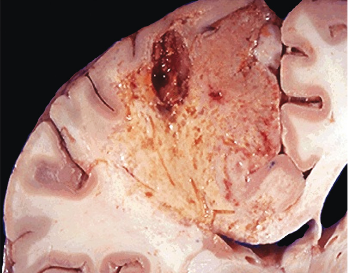
2. Liquefactive Necrosis
- Dead cells are completely digested, transforming tissue into a viscous liquid
- Caused by: bacterial or fungal infections (leukocytes accumulate and release enzymes); also seen in CNS ischemia (brain infarcts) for unknown reasons
- When due to acute bacterial infection: material is creamy yellow = pus; localized pus collection = abscess
3. Gangrenous Necrosis
- Not a distinct pattern - essentially coagulative necrosis of a limb (usually lower leg) that has lost blood supply, involving multiple tissue planes
- Dry gangrene: ischemic coagulative necrosis
- Wet gangrene: superimposed bacterial infection converts it to liquefactive necrosis
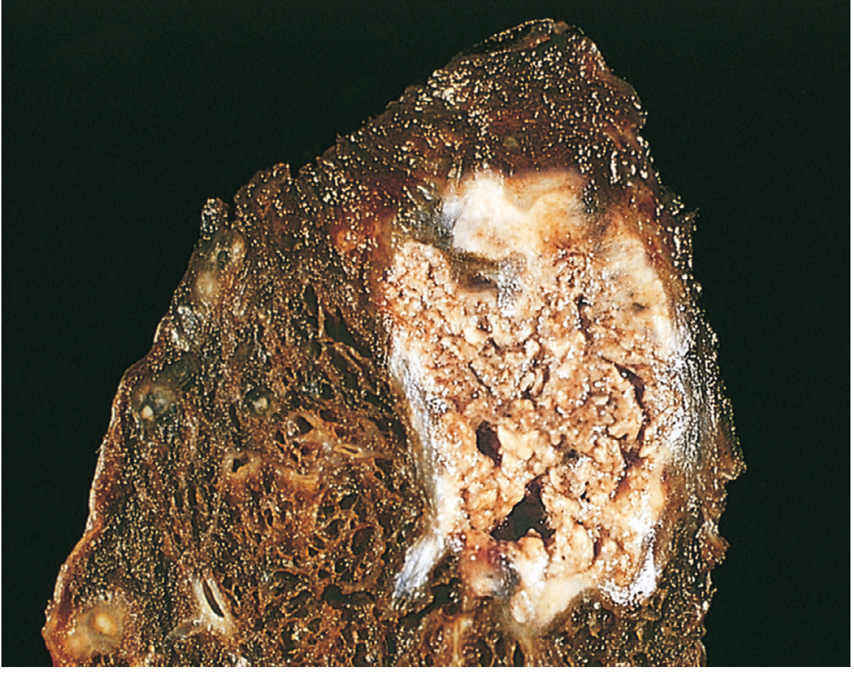
4. Caseous Necrosis
- Classic feature of tuberculous infection (and other mycobacterial/fungal granulomatous diseases)
- Gross: friable, yellow-white, "cheese-like" appearance ("caseous" = cheeselike)
- Histology: structureless collection of fragmented/lysed cells with amorphous granular pink debris; tissue architecture is completely obliterated (unlike coagulative necrosis)
- Surrounded by a collection of macrophages (epithelioid cells) = granuloma
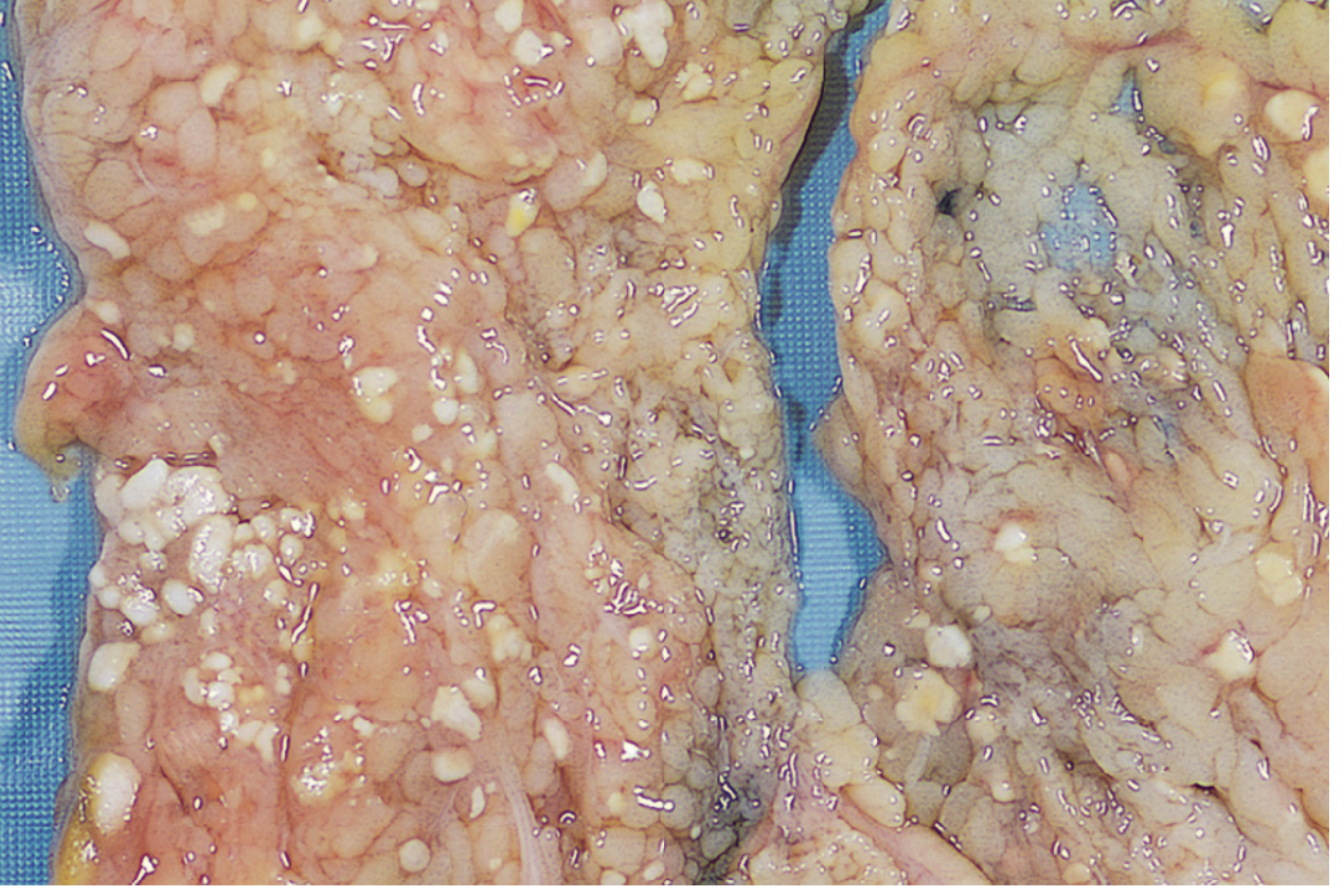
5. Fat Necrosis
- Focal areas of fat destruction
- Acute pancreatitis is the prototypical cause - pancreatic lipases leak from damaged acinar cells and digest peritoneal fat cells, releasing triglycerides
- Fatty acids combine with calcium -> chalky white deposits (saponification = calcium soap formation)
- Histology: shadowy outlines of necrotic fat cells + basophilic calcium deposits + inflammatory reaction
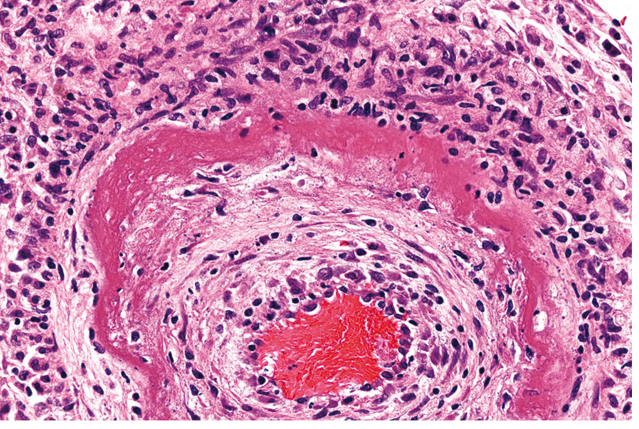
6. Fibrinoid Necrosis
- Detected only microscopically (no distinctive gross appearance)
- Seen in immune complex vasculitis (antigen-antibody complexes deposited in vessel walls) and malignant hypertension
- Histology: bright pink (eosinophilic), amorphous "fibrin-like" material in vessel walls = "fibrinoid" appearance
- Associated with: certain forms of vasculitis, glomerulonephritis
Summary Table
| Type | Cause | Key Feature | Example |
|---|---|---|---|
| Coagulative | Ischemia (all organs except brain) | Preserved architecture; ghost cells | Myocardial infarct |
| Liquefactive | Bacterial infection; CNS ischemia | Complete dissolution; pus | Brain abscess, brain infarct |
| Gangrenous | Vascular occlusion of limb | Coagulative ± liquefactive | Diabetic foot gangrene |
| Caseous | Tuberculosis, fungi | Cheesy; granuloma; architecture lost | Pulmonary TB |
| Fat | Acute pancreatitis; trauma | Chalky white saponification | Peritoneal fat in pancreatitis |
| Fibrinoid | Immune vasculitis; malignant HTN | Bright pink vessel wall deposits | Polyarteritis nodosa |
Necrosis vs. Apoptosis (Key Distinction)
| Feature | Necrosis | Apoptosis |
|---|---|---|
| Nature | Always pathologic | Physiologic or pathologic |
| Membrane integrity | Lost early | Maintained until late |
| Cell size | Swelling | Shrinkage |
| Nuclear change | Pyknosis/karyorrhexis/karyolysis | Chromatin condensation, fragmentation |
| Inflammation | Yes (DAMPs released) | No |
| Mechanism | Uncontrolled; enzymatic digestion | Regulated; caspase-mediated |
Other Regulated Forms of Necrosis (Modern Concepts)
- Necroptosis - programmed necrosis with features of both necrosis and apoptosis; regulated by specific signaling pathways (RIP kinases)
- Pyroptosis - cell death associated with release of pro-inflammatory cytokines (IL-1β, IL-18); important in innate immunity and infection
Sources: Robbins & Kumar Basic Pathology, 10th ed., pp. 21-24 | Robbins, Cotran & Kumar Pathologic Basis of Disease, pp. 54-57
Bwm disposal and treatment microbiology
Asking for Preferences
What does 'Bwm' stand for in your question?
Biomedical waste management - this covers disposal, treatment methods, and the microbiology of infectious agents in healthcare waste. Let me search the medical library and web simultaneously.
Searching sources...
Reading File
Searching the Web
biomedical waste management disposal treatment methods microbiology infectious agents
Now let me search the medical library for biomedical waste management and microbiology content in parallel:
Finding Sources
Finding Sources
Reading Web Page
https://www.cdc.gov/infection-control/hcp/environmental-cont…
Park's Preventive Medicine has a detailed table. Let me read that section alongside a search for infectious microorganisms in biomedical waste:
Reading File
Finding Sources
Excellent - great content from Park's. Let me read more of this section including the biomedical waste categories table and the microbiology/infectious agent content:
Reading File
Finding Sources
Reading File
Reading File
Excellent content from Park's. Let me now read the earlier section on BMW definitions and categories, and also look for the microbiology-specific content:
Reading File
Let me look for the BMW definition/introduction section in Park's:
Finding Sources
Finding Sources
Reading File
Let me read a bit earlier to get the BMW definition:
Reading File
Reading File
I now have comprehensive content. Let me compile the full, well-organized answer on Biomedical Waste Management - Disposal and Treatment, with the microbiology angle.
Biomedical Waste Management (BMW) - Disposal, Treatment & Microbiology
Definition
Bio-medical waste means any waste generated during the diagnosis, treatment, or immunization of human beings or animals, or in research activities, or in the production or testing of biologicals.
(BMW Management and Handling Rules, 1998, India - as cited in Park's Textbook of Preventive & Social Medicine)
Composition of Health-Care Waste
| Fraction | Percentage |
|---|---|
| General (non-hazardous) waste | 75-90% |
| Pathological and infectious waste | ~15% |
| Sharps waste | ~1% |
| Chemical and pharmacological waste | ~3% |
| Special waste (radioactive, cytotoxic, etc.) | <1% |
Hospital waste generation: 0.5-4 kg per bed per day in government hospitals; 0.5-2 kg per bed per day in private hospitals.
Categories & Colour Coding System (India - BMW Rules)
| Colour | Category | Examples | Treatment/Disposal |
|---|---|---|---|
| Yellow | Human anatomical waste | Tissues, organs, body parts, foetuses | Incineration, plasma pyrolysis, or deep burial |
| Yellow | Animal anatomical waste | Experimental animal carcasses | Incineration/plasma pyrolysis |
| Yellow | Soiled waste | Blood-soaked dressings, plaster casts, swabs, blood bags | Incineration/plasma pyrolysis; or autoclaving + shredding |
| Yellow | Expired/discarded medicines | Antibiotics, cytotoxic drugs, ampoules, vials | Return to manufacturer; incineration at >1200°C; encapsulation |
| Yellow | Chemical waste | Disinfectants, production chemicals | Incineration/plasma pyrolysis/encapsulation |
| Yellow | Chemical liquid waste | Formalin, X-ray developer, floor washings | Effluent treatment system; pre-treat before discharge |
| Yellow | Discarded linen/mattresses | Blood/body fluid contaminated | Chemical disinfection + incineration; or shredding |
| Yellow | Microbiology/biotech lab waste | Cultures, microbial stocks, vaccines, cell cultures, toxins | On-site sterilization (autoclaving/chemical), then incineration |
| Red | Contaminated recyclable waste | IV tubes, catheters, syringes without needles, gloves | Chemical disinfection/autoclaving/microwaving, then recycling |
| White (Translucent) | Waste sharps | Needles, syringes with needles, scalpels, broken glass | Autoclaving/dry heat sterilization/microwaving + shredding, or encapsulation |
| Blue | Glassware | Broken/discarded glass from labs | Disinfection, then disposal in landfill or reuse |
Microbiology of Biomedical Waste
Key Infectious Agents
Pathogens in infectious waste enter the body via:
- Puncture, abrasion, or cut in skin
- Mucous membranes
- Inhalation
- Ingestion
Bacteria
- HIV-associated opportunistic bacterial pathogens
- Hepatitis B and C viruses - strongest evidence for transmission via healthcare waste (especially sharps)
- Mycobacterium tuberculosis - in sputum, respiratory specimens
- Antibiotic-resistant organisms (MRSA, VRE, ESBL-producers) - these resist both antibiotics and some chemical disinfectants
- Clostridium species (endospores survive many disinfection methods)
Viruses
- HIV (blood, body fluids)
- Hepatitis B virus (HBV) - can survive on surfaces up to 7 days
- Hepatitis C virus (HCV)
- SARS-CoV-2 and other respiratory viruses
Other Pathogens
- Prions (Creutzfeldt-Jakob disease) - extremely resistant; require special incineration or prolonged autoclaving (134°C for 18 minutes or longer)
Highest Risk Waste Streams (Microbiologically)
Of all categories of regulated medical waste, microbiological wastes (untreated cultures, stocks, amplified microbial populations) pose the greatest potential for infectious disease transmission. Sharps pose the greatest risk for injury.
Untreated stocks and cultures must be considered for on-site decontamination, preferably within the laboratory itself.
Treatment Methods
1. Autoclaving (Steam Sterilization / Wet Thermal Treatment)
- Uses high-pressure steam at 121°C (250°F) for up to 90 minutes (depending on load size)
- Kills bacteria, viruses, fungi, and most spores
- Best for: Microbiological waste, sharps, soiled/solid waste, lab waste
- Not suitable for: Anatomical waste, cytotoxic drugs, certain chemical waste
- After autoclaving, residue can be safely handled as non-hazardous solid waste
2. Incineration
- Burns waste at high temperatures (standard ≥850°C; cytotoxics require >1200°C)
- Types:
- Rotary kiln - handles all infectious, most chemical/pharmaceutical waste; expensive
- Pyrolytic (two-chamber) incineration - very high disinfection efficiency; preferred method for sharps; most pharmaceutical and chemical waste
- Single-chamber incineration - good efficiency, lower cost, but atmospheric pollutant emissions
- Drum/brick incinerator - low cost; destroys ~99% of microorganisms; emits black smoke and toxic flue gas
- Preferred for: Sharps (eliminates microorganisms AND puncture risk), anatomical waste, pathological waste
- Note: Improper incineration of high-moisture waste (e.g., pathology waste) causes emission problems
3. Chemical Disinfection
- Common agents: 1% sodium hypochlorite (kills most vegetative bacteria), formaldehyde, glutaraldehyde
- Best for: Liquid/chemical waste, blood specimens, some microbiological waste
- Limitations: Requires qualified operators; hazardous chemicals; inadequate for cytotoxic/pharmaceutical/some infectious waste
- Effectiveness depends on type and concentration of microorganisms, organic load, contact time, and pH
4. Microwave Irradiation
- High-frequency waves cause intramolecular vibration generating heat - kills pathogens from within
- Good disinfection efficiency; environmentally sound
- Best for: Microbiological waste, sharps, solid waste
- Limitations: High cost; maintenance problems; not suitable for large volumes
5. Plasma Pyrolysis
- Converts organic waste into commercially useful by-products using intense plasma heat
- Environment-friendly; handles all types including municipal solid waste
- High investment cost
6. Encapsulation
- Sharps and chemical waste are immobilized in cement, plastic foam, or bituminous sand in containers
- Simple, low-cost, safe
- Not recommended for non-sharp infectious waste
7. Safe Burial / Deep Burial
- Used where no other option is available
- A pit >2 m deep, lined with lime; waste covered with lime and soil after each deposit
- Only suitable for anatomical waste in remote areas with limited resources
8. Inertization
- Waste mixed with cement + lime + water (ratio ~65:15:15:5) → pellets buried at suitable storage sites
- Only for: Pharmaceutical waste
- Not applicable to infectious waste
Advantages & Disadvantages Summary
| Method | Key Advantage | Key Disadvantage |
|---|---|---|
| Rotary kiln | Handles all infectious + most chemical waste | Very high cost |
| Pyrolytic incineration | Very high disinfection; handles pharmaceutical waste | Incomplete destruction of cytotoxics |
| Chemical disinfection | Highly efficient under good conditions | Hazardous chemicals; inadequate for some waste types |
| Autoclave (wet thermal) | Environmentally sound; relatively cheap | Inadequate for anatomical, pharmaceutical, chemical waste |
| Microwave irradiation | Good disinfection; reduces volume; eco-friendly | High cost; maintenance issues |
| Encapsulation | Simple, safe, low-cost | Only for sharps/chemicals |
| Safe burying | Very low cost | Only safe with restricted access and dry site |
| Inertization | Inexpensive | Not for infectious waste |
Health Hazards from BMW
Groups at Risk
- Healthcare workers (doctors, nurses, lab staff, housekeeping)
- Patients
- Visitors
- Hospital support workers (laundry, transport)
- Workers at waste disposal facilities and landfills (including scavengers)
Types of Hazard
- Infectious - pathogens in waste (HIV, HBV, HCV, TB, antibiotic-resistant bacteria)
- Sharps injury - needlestick transmission of bloodborne pathogens
- Chemical/pharmaceutical - toxic, corrosive, flammable, genotoxic agents
- Genotoxic - cytotoxic drug residues (inhalation of dust/aerosols; skin absorption)
- Radioactive - from diagnostic and therapeutic radionuclides
Key Principles of BMW Management
- Segregation at source - the single most important step; reduces volume of hazardous waste
- Proper containment - colour-coded bags and containers; sharps in puncture-proof containers (never overfill >3/4 full)
- Safe transport - within facility and off-site with labelled, leak-proof containers
- Treatment - reduce microbial load to safe levels
- Disposal - in approved, regulated facilities
- Record keeping and training - staff must be trained; regular audits
Sources: Park's Textbook of Preventive and Social Medicine, 26th ed. | CDC Regulated Medical Waste Guidelines | WHO Health-Care Waste Management | BMW (Management and Handling) Rules, 1998 / BMW Rules 2016 (India)
This is a shared conversation. Sign in to Orris to start your own chat.