
histology of all three tips muscle
The "three tips muscle" refers to the triceps brachii (Latin: triceps = three heads). Let me look up its histology from the medical library.
Searching sources...
Reading File
Now let me search for triceps histology in the library, running parallel searches:
Finding Sources
Finding Sources
Finding Sources
Let me search for muscle histology (skeletal muscle tissue) and also read the triceps sections from the anatomy atlases:
Finding Sources
Finding Sources
Reading File
Now let me read the Junqueira's Histology section on skeletal muscle (the definitive histology textbook):
Reading File
Finding Sources
Excellent! Now let me read more from Junqueira's Histology chapter on skeletal muscle in detail, and also the Wheater's/Ross Histology book:
Reading File
Reading File
Now let me read more of the Junqueira's detailed skeletal muscle histology section, including fiber types, connective tissue layers, and the key images:
Reading File
I now have rich content. Let me fetch the key histology images to include in the answer:
Loading Image
Loading Image
Reading File
Now let me read the skeletal muscle fiber types section which is critical for a complete histology answer:
Reading File
Let me get the fiber types section now:
Reading File
Reading File
I now have all the information needed to provide a comprehensive answer. Let me also quickly fetch the fiber types image:
Loading Image
I now have all the content needed. Here is the full answer:
Histology of the Triceps Brachii (Three-Headed/Tips Muscle)
The triceps brachii (Latin: tri = three, caput = head) is a skeletal muscle - so its histology is the histology of skeletal muscle tissue. All three heads (long, lateral, medial) are composed of identical tissue; the "three heads" distinction is anatomical (different origins), not histological.
1. Basic Tissue Classification
The triceps brachii is striated voluntary skeletal muscle - the same as all limb muscles. Its three heads simply have different proximal attachment points on the scapula/humerus but converge onto the same olecranon tendon. Microscopically, no head is distinguishable from another.
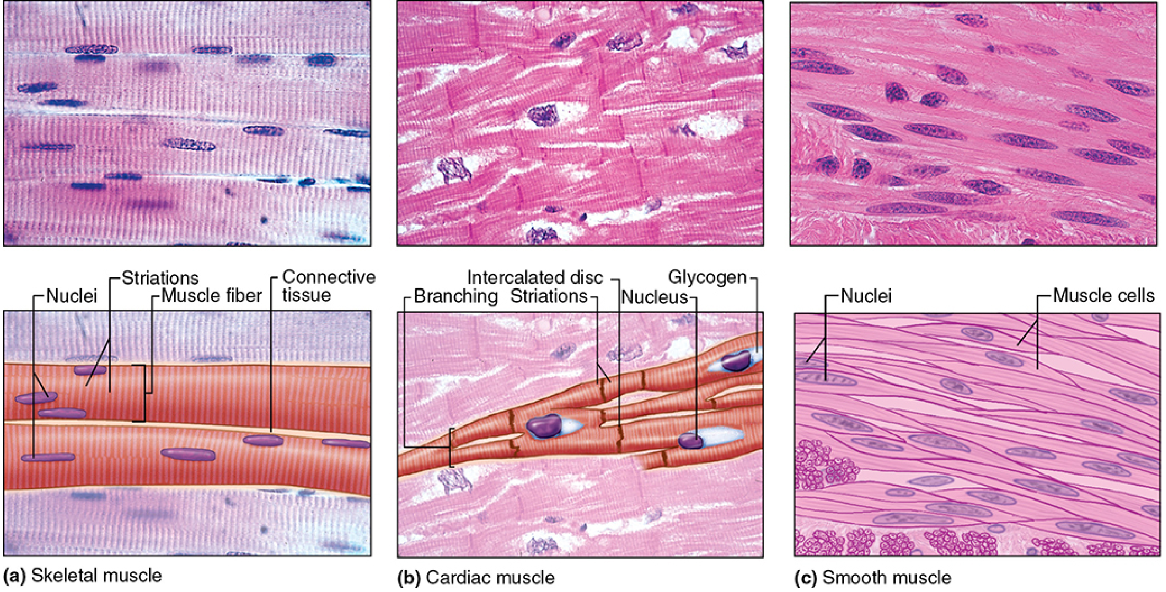
2. Connective Tissue Framework (Three Layers)
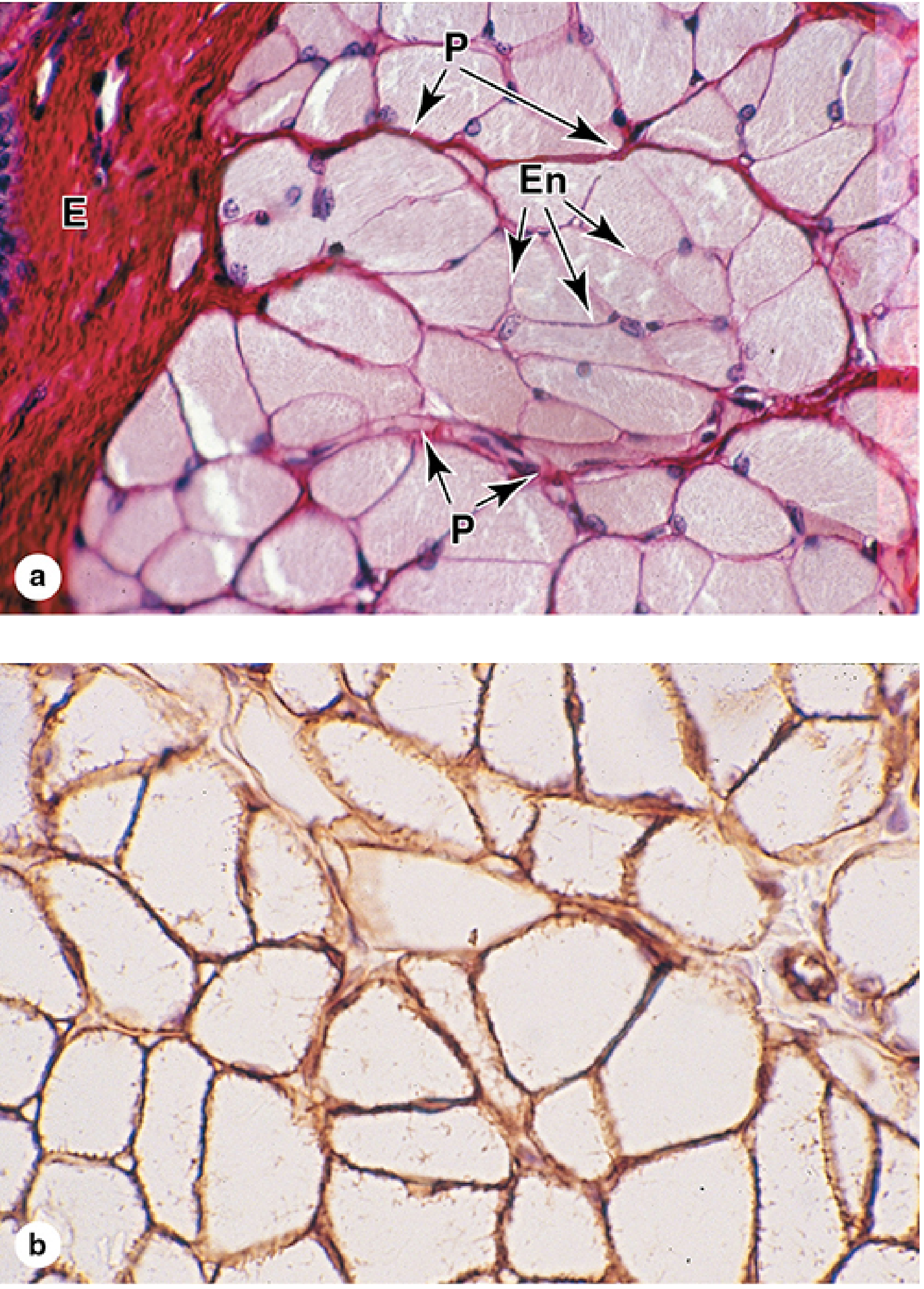
The entire muscle and its fascicles are organized by three concentric connective tissue sheaths:
| Layer | Location | Composition | Function |
|---|---|---|---|
| Epimysium | Encloses the entire muscle | Dense irregular connective tissue (collagen I + III) | Merges with fascia and tendon; protects and binds the whole muscle |
| Perimysium | Surrounds each fascicle (bundle of fibers) | Thin connective tissue; carries nerves, blood vessels, lymphatics | Groups fibers into functional units |
| Endomysium | Envelops each individual muscle fiber | Delicate reticular fibers (type III collagen) + scattered fibroblasts | Supports capillary network around each fiber; fuses with the fiber's external lamina |
The external (basal) lamina directly coats each fiber's sarcolemma and is demonstrable by laminin immunohistochemistry (brown staining in the cross-section image above, panel b).
3. The Muscle Fiber (Cell)
- Shape: Long, cylindrical
- Size: Diameter 10-100 µm; lengths up to several centimeters
- Nuclei: Multiple (multinucleated), positioned peripherally just under the sarcolemma - this is the hallmark that distinguishes skeletal muscle from cardiac (central nuclei) and smooth muscle (single central nucleus)
- Cross-striations: Prominent, due to the regular alternation of A-bands (dark, anisotropic) and I-bands (light, isotropic)
- Sarcoplasm: Rich in myofibrils, mitochondria, glycogen granules, and myoglobin
- Satellite cells: Small reserve progenitor cells lie between the sarcolemma and external lamina; responsible for muscle regeneration after injury
4. Internal Ultrastructure: The Sarcomere
Each myofibril is divided into repeating units called sarcomeres (Z disc to Z disc, ~2.5 µm at rest):
| Structure | Contents | Appearance |
|---|---|---|
| A-band | Thick myosin filaments (1.5 µm × 15 nm), overlapping with thin actin filaments | Dark (anisotropic) |
| I-band | Thin actin filaments only (non-overlapping portions) | Light (isotropic) |
| Z-disc | α-actinin; anchors actin filaments | Dense line bisecting I-band |
| H-zone | Thick filaments only, no overlap; disappears during contraction | Lighter zone within A-band |
| M-line | Myomesin proteins stabilizing thick filaments | Central line in H-zone |
Key proteins:
- Myosin (thick filaments): ~500 kDa; two heavy chains + 4 light chains; globular heads have ATPase activity and actin-binding sites
- F-actin (thin filaments): Double helix of G-actin monomers, each 8 nm wide
- Tropomyosin: lies in the groove of the actin helix; blocks myosin-binding sites at rest
- Troponin (TnT, TnC, TnI): TnC binds Ca²⁺ on neural stimulation, shifting tropomyosin to expose binding sites
- Titin: Largest protein in the body (3700 kDa); elastic scaffold connecting thick filaments to Z-disc
- Nebulin: Templates thin filament length
5. The Sarcoplasmic Reticulum and T-Tubule System
- T-tubules (transverse tubules): Invaginations of the sarcolemma at A-I junctions; propagate action potentials deep into the fiber
- Sarcoplasmic reticulum (SR): Modified smooth ER; encircles myofibrils; its terminal cisternae store and release Ca²⁺
- A T-tubule flanked by two terminal cisternae = a triad (the functional coupling unit for excitation-contraction)
6. Innervation
- Myelinated motor axons branch in the perimysium, then become unmyelinated terminals in the endomysium
- Each axonal terminal forms a neuromuscular junction (NMJ) / motor end plate on the muscle fiber surface
- The synaptic cleft lies between the axon terminus (containing ACh vesicles) and the sarcolemma, which is thrown into deep junctional folds to increase receptor surface area
- One motor neuron + all the fibers it innervates = a motor unit
7. Three Skeletal Muscle Fiber Types (in Triceps and All Skeletal Muscles)
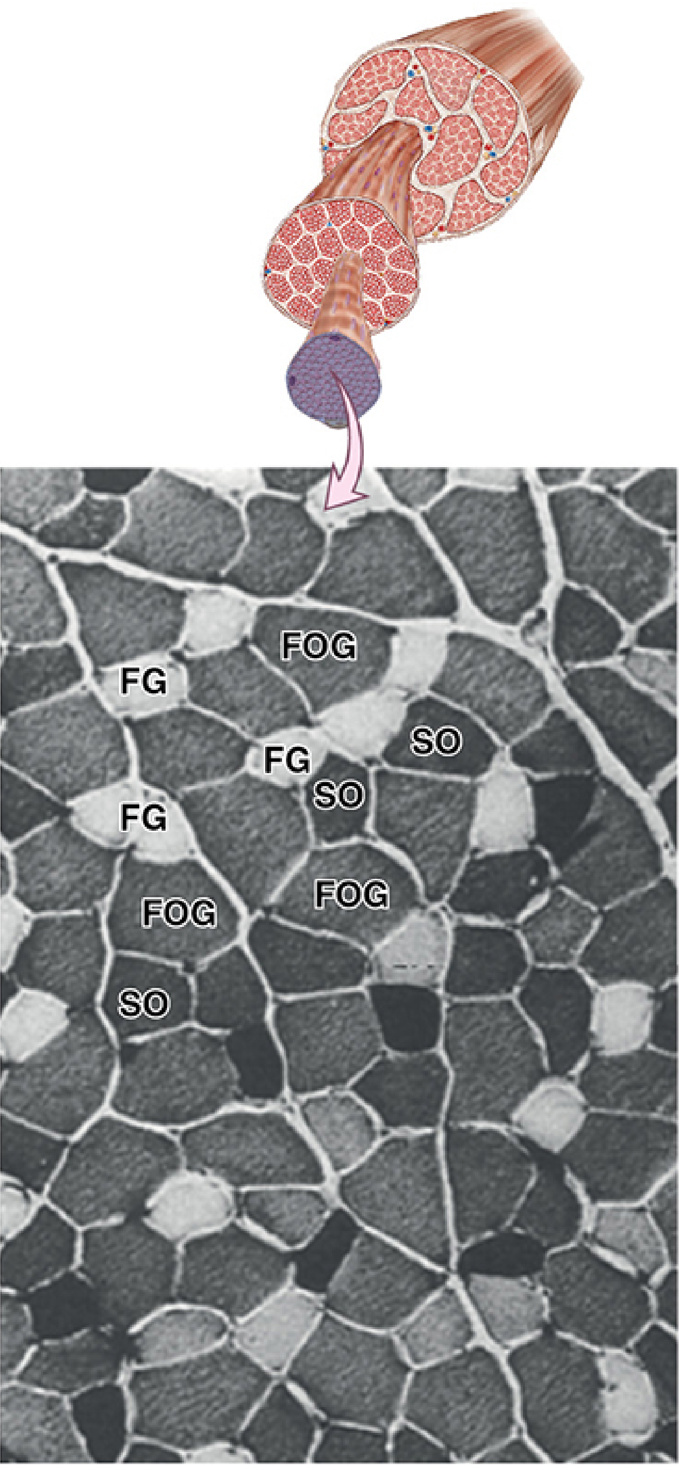
The triceps, like most limb muscles, contains a mixture of all three fiber types:
| Feature | Type I (Slow Oxidative, SO) | Type IIa (Fast Oxidative-Glycolytic, FOG) | Type IIb (Fast Glycolytic, FG) |
|---|---|---|---|
| ATPase staining (pH 4.2) | Dark | Intermediate | Light (palest) |
| Mitochondria | Numerous | Numerous | Sparse |
| Capillaries | Numerous | Numerous | Sparse |
| Myoglobin | High (red) | Intermediate | Low (white) |
| Fiber diameter | Small | Intermediate | Large |
| Glycogen | Low | Intermediate | High |
| Fatigue resistance | High | Intermediate | Low (fatigues fast) |
| Contraction speed | Slow | Fast | Fastest |
| Metabolism | Aerobic oxidative | Mixed | Anaerobic glycolytic |
| Motor unit size | Small | Intermediate | Large |
Fiber type is determined by the innervating motor neuron's firing pattern - all fibers in a motor unit are the same type.
8. Sensory Receptors Within Skeletal Muscle
- Muscle spindles: Capsule of modified perimysium; contain intrafusal fibers (nuclear bag and nuclear chain types); detect stretch/length changes; supplied by Ia and II sensory afferents
- Golgi tendon organs: Located at myotendinous junctions; detect tension (force)
Summary
The triceps brachii's three heads are architecturally distinct (different origins, pennation angles) but histologically identical - all are skeletal muscle composed of multinucleated, cross-striated fibers organized into fascicles by epimysium/perimysium/endomysium, containing a mixture of Type I, IIa, and IIb fibers, and innervated through neuromuscular junctions using acetylcholine.
Sources: Junqueira's Basic Histology 17e, Chapter 10 (Muscle Tissue); Ross & Pawlina Histology 8e; General Anatomy and Musculoskeletal System (THIEME Atlas)
histology of cardiac muscle,skeetan muscle and smooth muscle
I'll look up the detailed histology of all three muscle types from the medical library simultaneously.
Searching sources...
Finding Sources
Finding Sources
Finding Sources
Junqueira's Histology has all three. Let me read the cardiac and smooth muscle sections in parallel:
Reading File
Finding Sources
Reading File
Reading File
Reading File
Now let me get the smooth muscle section and fetch all key images in parallel:
Reading File
Loading Image
Loading Image
Now let me fetch the smooth muscle images:
Loading Image
Loading Image
I now have all the content needed. Let me also get the smooth muscle contraction diagram URL properly:
Reading File
Loading Image
I now have comprehensive content and images for all three muscle types. Here is the complete, detailed answer:
Histology of the Three Muscle Types
Source: Junqueira's Basic Histology 17e, Chapter 10 - Muscle Tissue
QUICK COMPARISON AT A GLANCE
| Feature | Skeletal Muscle | Cardiac Muscle | Smooth Muscle |
|---|---|---|---|
| Shape | Long cylindrical fiber | Short, branched cell | Fusiform (spindle-shaped) |
| Nuclei | Multiple, peripheral | Single (or 2), central | Single, central |
| Striations | Yes (prominent) | Yes (less distinct) | No |
| Control | Voluntary | Involuntary | Involuntary |
| Special junction | Neuromuscular junction | Intercalated discs | Gap junctions |
| T-tubule system | Triads (A-I junction) | Dyads (Z-disc level) | Absent (caveolae instead) |
| Regeneration | Via satellite cells | Very limited | Good (mitosis) |
| Location | Attached to bone | Heart wall | Viscera, vessels, skin |
PART 1 — SKELETAL (STRIATED) MUSCLE
Cell (Fiber) Morphology
- Shape: Very long cylindrical fibers, diameter 10-100 µm, length up to several centimeters
- Nuclei: Multiple (multinucleated) - located peripherally just under the sarcolemma - the single most diagnostic histological feature
- Striations: Prominent cross-striations (alternating A and I bands) due to organized sarcomeres
- Origin: Derived from fusion of mesenchymal myoblasts into multinucleated myotubes during embryogenesis
Connective Tissue Layers
| Layer | Surrounds | Composition |
|---|---|---|
| Epimysium | Entire muscle | Dense irregular collagen I + III |
| Perimysium | Each fascicle (bundle) | Thin CT; carries NV bundles |
| Endomysium | Each individual fiber | Reticular (collagen III) fibers + fibroblasts + capillary network |
Each fiber is also surrounded by an external (basal) lamina containing laminin and type IV collagen (stainable by immunohistochemistry).
Internal Structure - The Sarcomere
Each fiber is packed with myofibrils made up of sarcomeres (Z disc to Z disc):
| Band/Zone | Contents | Staining |
|---|---|---|
| A-band | Thick myosin + overlapping thin actin | Dark (anisotropic) |
| I-band | Thin actin only (no overlap) | Light (isotropic) |
| Z-disc | α-actinin; anchors thin filaments | Dense transverse line |
| H-zone | Myosin only (no actin overlap) | Lighter zone in A-band |
| M-line | Myomesin stabilizing thick filaments | Central line in H-zone |
Key myofibrillar proteins:
- Myosin (thick, 15 nm wide): 2 heavy chains + 4 light chains; globular heads = ATPase + actin-binding sites
- F-actin (thin, 8 nm wide): Double helix of G-actin
- Tropomyosin: Sits in actin groove; blocks myosin binding at rest
- Troponin (TnT/TnC/TnI): TnC binds Ca²⁺ to initiate contraction
- Titin: Elastic scaffold connecting thick filaments to Z-disc (3700 kDa, largest body protein)
- Nebulin: Templates thin filament length
Sarcoplasmic Reticulum and T-Tubules
- T-tubules: Sarcolemma invaginations at A-I junctions; propagate action potentials deep into the fiber
- Terminal cisternae: SR expansions flanking each T-tubule
- Triad = 1 T-tubule + 2 terminal cisternae (the Ca²⁺ release unit)
Fiber Types (histochemistry distinguishes three types)
| Type I (Slow Oxidative, SO) | Type IIa (Fast Oxidative-Glycolytic, FOG) | Type IIb (Fast Glycolytic, FG) | |
|---|---|---|---|
| ATPase at pH 4.2 | Dark (high) | Intermediate | Light (low) |
| Mitochondria | Numerous | Numerous | Sparse |
| Myoglobin | High (red) | Intermediate | Low (white) |
| Fiber diameter | Small | Intermediate | Large |
| Fatigue | Resistant | Intermediate | Fatigues fast |
| Metabolism | Aerobic | Mixed | Anaerobic glycolysis |
Satellite Cells and Regeneration
Quiescent muscle satellite cells sit between the sarcolemma and external lamina. They activate after injury to proliferate and form new fibers - the primary mechanism of skeletal muscle regeneration.
PART 2 — CARDIAC MUSCLE
Cell Morphology
- Shape: Short (15-30 µm diameter), often branched cells that interlock and interweave
- Nuclei: Single (sometimes 2), centrally located - large, euchromatic (pale-staining) oval nuclei; perinuclear lipofuscin pigment granules may surround the nucleus in older hearts
- Striations: Present but less prominent and regular than skeletal muscle
- Development: Mesenchymal cells align and form chainlike arrays but do NOT fuse - they remain as individual cells joined by junctional complexes
The Hallmark Feature: Intercalated Discs
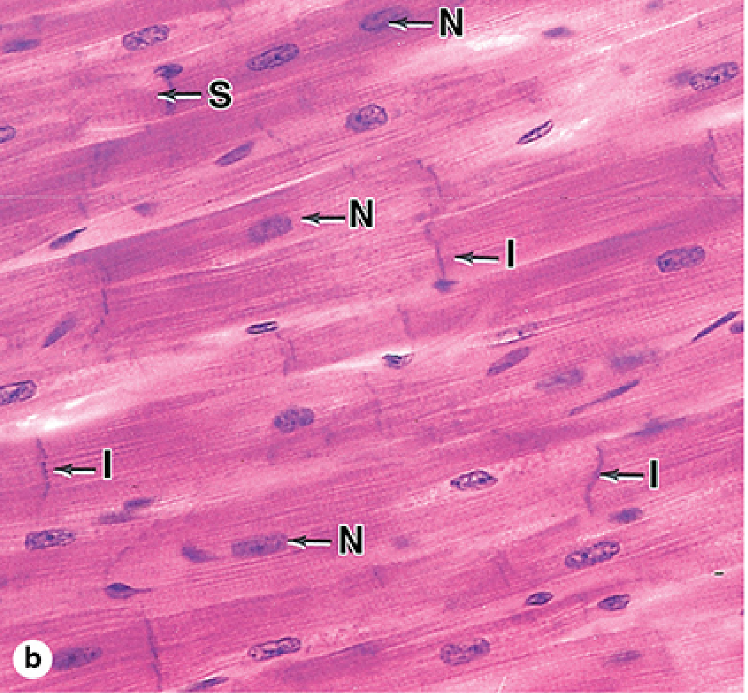
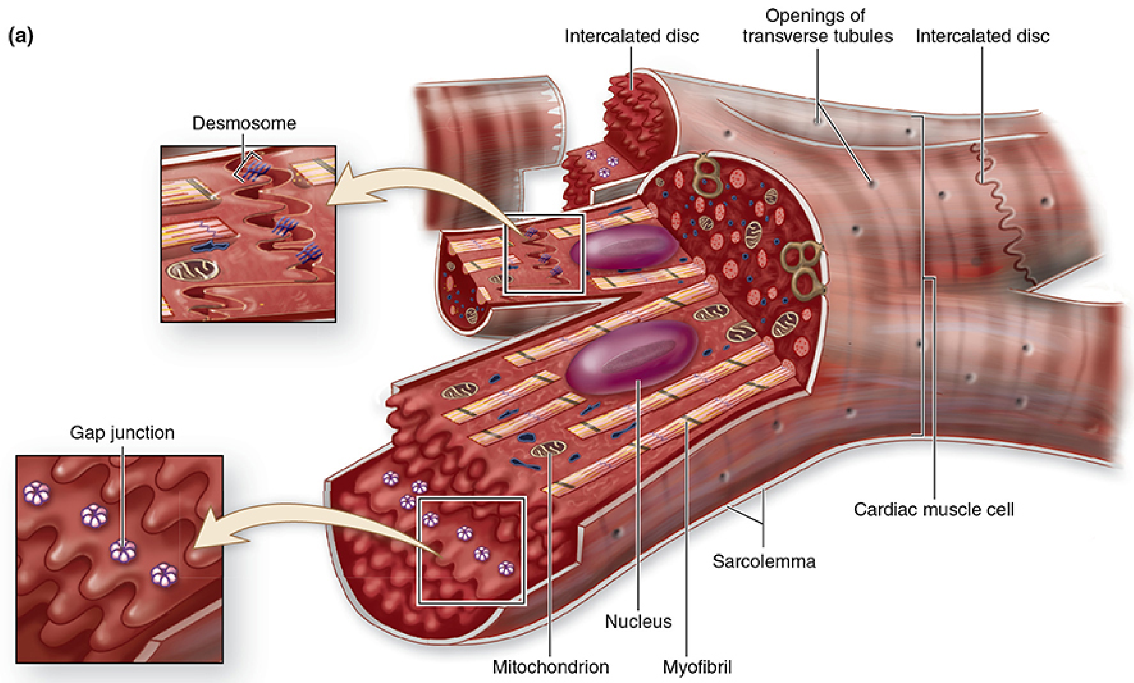
Intercalated discs are step-like transverse lines crossing cardiac fibers at irregular intervals - they are the cell-cell junctions between adjacent cardiomyocytes. They have two distinct regions:
Transverse region (perpendicular to fiber axis):
- Desmosomes: Spot welds providing strong mechanical adhesion
- Fascia adherens: Adhering junctions where actin filaments insert (functionally like Z-discs)
Lateral region (parallel to fiber axis):
- Gap junctions (nexuses): Provide ionic/electrical continuity between cells; act as "electrical synapses" enabling the heart to function as a functional syncytium - action potentials propagate rapidly through many cells simultaneously
Internal Ultrastructure
- Myofibrils: Same sarcomere organization as skeletal muscle, but slightly less regular
- Mitochondria: Extremely abundant - up to 40% of cell volume (much more than skeletal); reflects the heart's continuous aerobic demand; fatty acids are the main fuel, stored as lipid droplets
- Sarcoplasmic reticulum: Less well-organized than in skeletal muscle
- T-tubules: Present in ventricular muscle (at Z-disc level), small or absent in atrial muscle. Form dyads (1 T-tubule + 1 terminal cistern) rather than triads
- Atrial secretory granules: Membrane-bound granules in atrial cells (especially right atrium, ~600/cell) containing atrial natriuretic factor (ANF) precursor - ANF promotes natriuresis and diuresis opposing aldosterone/ADH
Connective Tissue
- Endomysium of reticular fibers surrounds each cardiomyocyte (same principle as skeletal muscle, but no perimysium divisions into distinct fascicles)
Regeneration
- Very limited in adults - cardiac muscle lacks satellite cells
- Damage (e.g., myocardial infarction) is repaired by fibroblast proliferation → fibrous scar (not functional muscle)
PART 3 — SMOOTH MUSCLE
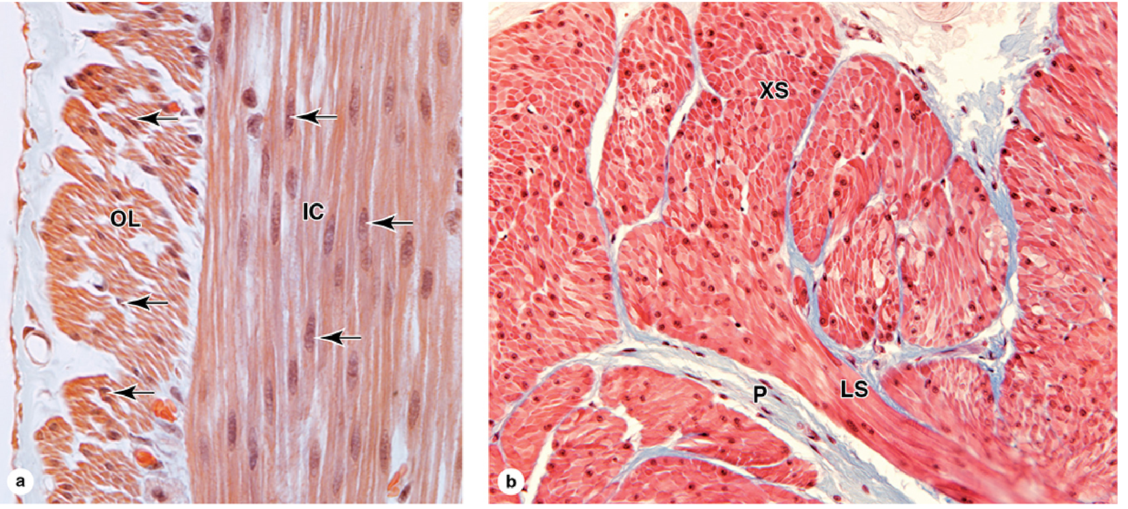
Cell Morphology
- Shape: Long, tapering fusiform cells (spindle-shaped), narrowing at both ends
- Size: 20 µm (small blood vessels) to 500 µm (pregnant uterus) in length; 3-8 µm diameter at widest point
- Nuclei: Single, elongated, centrally located at the cell's widest point; nucleus twists/spirals when cell contracts (producing a "corkscrew" appearance on H&E - a useful sign of contraction)
- No striations: Myofilaments are not organized into regular sarcomeres, so no banding pattern
- Organization: Cells are arranged in sheets or bundles; in the GI tract, two layers exist - inner circular and outer longitudinal - which work in concert for peristalsis
Internal Structure: Dense Bodies instead of Z-Discs
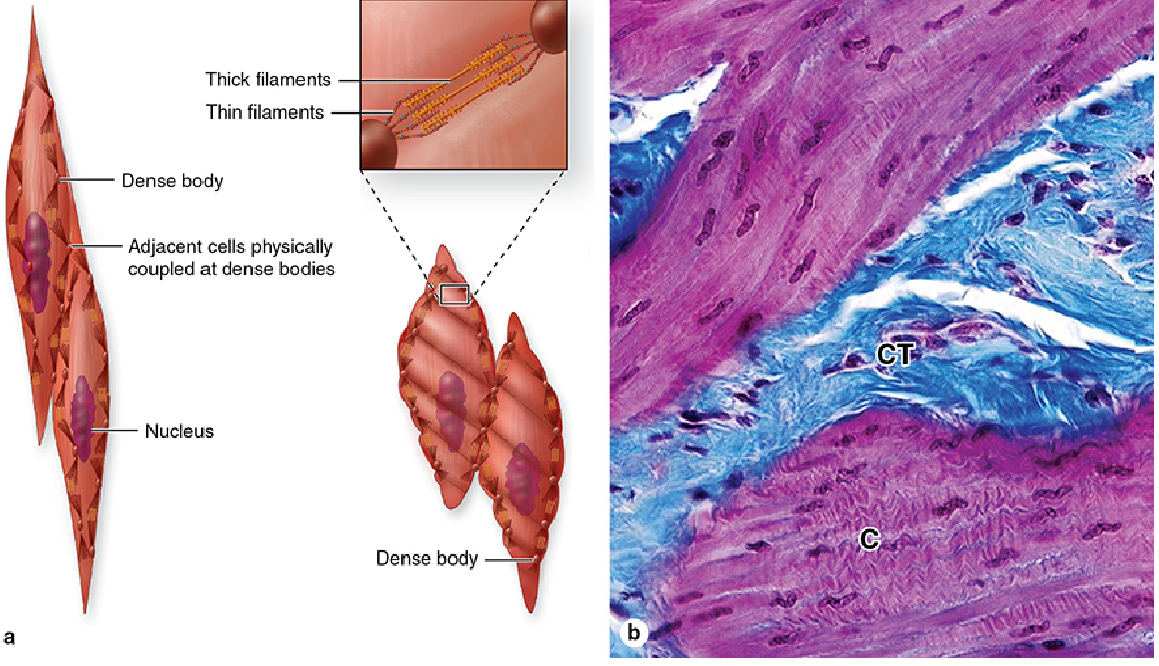
- Thick myosin and thin actin filaments run obliquely (not parallel) to the cell's long axis
- Thin filaments attach to dense bodies - electron-dense structures scattered throughout the cytoplasm and at the cell membrane
- Dense bodies contain α-actinin and function like Z-discs of striated muscle
- Also serve as attachment points for intermediate filaments (desmin) and intercellular adhesive junctions (cadherins)
- When contraction occurs, cells twist (like wringing out a cloth) due to the oblique filament orientation
- Adjacent cells are physically coupled at dense bodies via cadherins, transmitting contractile force across the tissue
Regulatory Proteins (Differ from Striated Muscle)
Smooth muscle thin filaments lack troponin. Instead, contraction is regulated by:
- Calmodulin: Binds Ca²⁺ (which enters via caveolae-associated channels and from SR cisternae)
- Myosin light-chain kinase (MLCK): Ca²⁺-calmodulin activates MLCK → phosphorylates myosin light chains → activates myosin ATPase → contraction
T-Tubules and Sarcoplasmic Reticulum
- No T-tubules - unnecessary because cells are small enough for Ca²⁺ diffusion
- Caveolae: Sarcolemmal invaginations containing Ca²⁺ channels and ion channels; serve as the functional equivalent of T-tubules
- Rudimentary SR (sarcoplasmic cisternae) present near caveolae
- Gap junctions electrically couple adjacent smooth muscle cells for coordinated contraction
Connective Tissue
- Each cell is surrounded by an external lamina
- Endomysium of collagen I and III reticular fibers (visible with silver stain) holds the cells together
Regeneration
- Good regenerative capacity - smooth muscle cells retain the ability to undergo mitosis
- After injury, viable smooth muscle cells proliferate and replace damaged tissue
- Pericytes from small blood vessels also contribute to vascular smooth muscle repair
MASTER COMPARISON TABLE
| Feature | Skeletal | Cardiac | Smooth |
|---|---|---|---|
| Cell shape | Long cylinder | Short, branched | Fusiform |
| Nucleus position | Peripheral, multiple | Central, single (1-2) | Central, single |
| Striations | Prominent | Present, less distinct | Absent |
| Special cell junction | NMJ (motor end plate) | Intercalated discs | Gap junctions |
| T-tubule arrangement | Triads (A-I junction) | Dyads (Z-disc) | Caveolae (no T-tubules) |
| SR organization | Very well organized | Less organized | Rudimentary |
| Ca²⁺ regulation | Troponin-tropomyosin | Troponin-tropomyosin | Calmodulin-MLCK |
| Z-disc equivalent | Z-disc (α-actinin) | Z-disc | Dense bodies |
| Mitochondria | Moderate-many (varies by fiber type) | Abundant (40% cell vol) | Few |
| Regeneration | Via satellite cells | Very poor | Good (mitosis) |
| Control | Voluntary | Involuntary | Involuntary |
| Contraction | Fast, forceful | Rhythmic, sustained | Slow, sustained |
Source: Junqueira's Basic Histology: Text and Atlas, 17th Edition, Chapter 10 (Muscle Tissue)
This is a shared conversation. Sign in to Orris to start your own chat.