
Role of calcium as secondary messenger for 8 marks
calcium calmodulin second messenger signaling pathway IP3 DAG
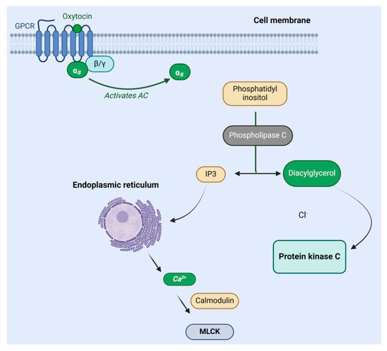
A pathophysiology diagram illustrating the oxytocin signaling pathway within a cell. The mechanism begins at the cell membrane where oxytocin binds to a G protein-coupled receptor (GPCR). This binding triggers the G-protein subunits (αs and β/γ), leading to the activation of adenylyl cyclase (AC). Downstream, phospholipase C (PLC) catalyzes the conversion of phosphatidylinositol into two secondary messengers: diacylglycerol (DAG) and inositol triphosphate (IP3). DAG is shown activating protein kinase C (PKC). Simultaneously, IP3 interacts with the endoplasmic reticulum, prompting the release of calcium ions (Ca2+). These calcium ions bind to calmodulin, which subsequently activates myosin light chain kinase (MLCK). This biochemical cascade is fundamental to understanding myometrial contraction and milk ejection. The diagram uses standard medical icons and labels to represent intracellular components and molecular interactions, suitable for undergraduate to advanced medical education.
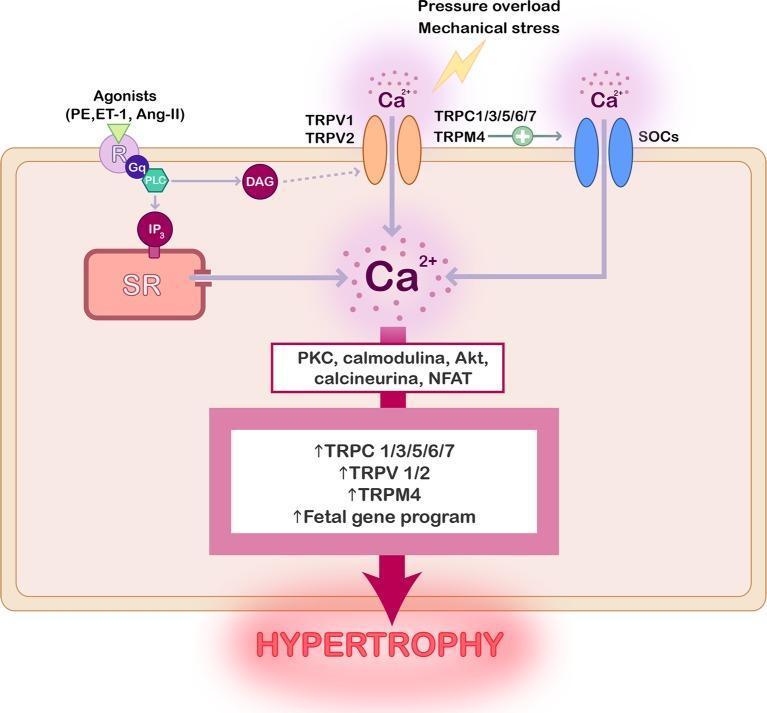
A pathophysiology diagram illustrating the signaling pathways of TRP channels in cardiac hypertrophy. The diagram depicts the plasma membrane containing several ion channels: TRPV1/2, TRPC1/3/5/6/7, TRPM4, and SOCs. Activation occurs via two main stimuli: mechanical stress/pressure overload and hypertrophic agonists (PE, ET-1, Ang-II) binding to Gq-protein coupled receptors (GPCR). The GPCR pathway activates PLC, generating DAG and IP3; IP3 triggers calcium release from the sarcoplasmic reticulum (SR). Simultaneously, mechanical stress and receptor-mediated signaling promote calcium influx through the TRP channels. The resulting elevation in intracellular calcium (Ca2+) concentration activates a downstream cascade of signaling proteins, including PKC, calmodulin, Akt, calcineurin, and NFAT. This cascade leads to the upregulation of TRP channel expression and the activation of a fetal gene program, ultimately resulting in cellular hypertrophy. The illustration uses color-coded icons and directional arrows to trace the metabolic and mechanical pathways contributing to pathological heart remodeling.
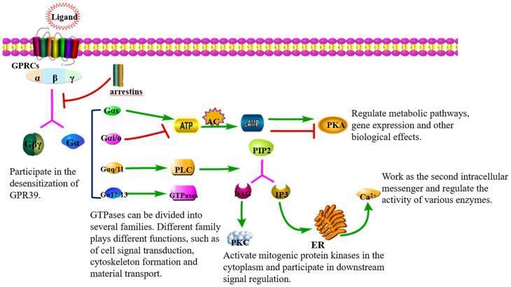
This pathophysiology diagram illustrates G-protein-coupled receptor (GPCR) signal transduction pathways across a plasma membrane. The schematic details four primary Gα subunit cascades. The Gαs pathway activates adenylate cyclase (AC) to convert ATP into cAMP, which stimulates Protein Kinase A (PKA). Conversely, Gαi/o inhibits AC, reducing cAMP and inhibiting PKA activity. The Gαq/11 pathway activates Phospholipase C (PLC), which cleaves PIP2 into diacylglycerol (DAG) and inositol triphosphate (IP3). DAG subsequently activates Protein Kinase C (PKC), while IP3 triggers calcium (Ca2+) release from the endoplasmic reticulum (ER) to serve as a second messenger. The Gα12/13 pathway is shown activating GTPases, involved in cytoskeleton and signal regulation. Additionally, the diagram depicts GPCR desensitization through arrestins and the dissociation of the Gβγ complex. The visual highlights the molecular mechanisms by which extracellular ligands regulate metabolic pathways, gene expression, and enzyme activity in human cellular physiology.
Role of Calcium as a Secondary Messenger
Introduction
1. Sources of Intracellular Calcium
- Extracellular influx: Ca²⁺ enters through voltage-gated or receptor-operated calcium channels in the plasma membrane.
- Intracellular release: Ca²⁺ is released from the endoplasmic reticulum (ER) and sarcoplasmic reticulum (SR) via two receptor-channels:
- IP3 receptor (IP3R): Opened by inositol trisphosphate (IP3), a second messenger derived from PIP2 hydrolysis.
- Ryanodine receptor (RyR): Opened by cytoplasmic Ca²⁺ itself, producing calcium-induced calcium release (CICR). - Principles of Neural Science (Kandel), 6th Ed., p. 355
2. The PIP2 - IP3 - Calcium Pathway
- A first messenger (hormone/neurotransmitter) binds a Gq-protein-coupled receptor on the cell surface.
- The activated Gq protein stimulates membrane-bound phospholipase C (PLC).
- PLC cleaves membrane phosphatidylinositol 4,5-bisphosphate (PIP2) into two products:
- IP3 (inositol trisphosphate) - water-soluble, diffuses to the ER
- DAG (diacylglycerol) - remains in the membrane
- IP3 binds its receptor on the ER membrane, opening a Ca²⁺ channel and releasing Ca²⁺ into the cytoplasm.
- The free Ca²⁺ now acts as a second messenger. - Guyton and Hall, p. 912

3. The Calcium-Calmodulin System
- Calmodulin is a small (148 amino acid), ubiquitous cytoplasmic protein with four calcium-binding sites.
- When 3 to 4 of these sites are occupied by Ca²⁺, calmodulin undergoes a conformational change, becoming activated.
- The Ca²⁺-calmodulin complex then binds to and activates or inhibits a variety of target enzymes and proteins. - Guyton and Hall, p. 912
| Target | Effect |
|---|---|
| CaM kinase (CaMK) | Phosphorylates serine/threonine residues on target proteins; autophosphorylation makes it persistently active even after Ca²⁺ falls |
| Myosin light chain kinase (MLCK) | Phosphorylates myosin - causes smooth muscle contraction |
| Phosphorylase kinase | Activates glycogen phosphorylase - promotes glycogenolysis |
| Calcineurin | A phosphatase that activates NFAT transcription factor - important in immune cell activation |
| Nitric oxide synthase (NOS) | Generates NO, a potent vasodilator |
- Basic Medical Biochemistry (6th Ed.), p. 948; Principles of Neural Science (Kandel), p. 355
4. Protein Kinase C (PKC) - The DAG Partner
- DAG activates Protein Kinase C (PKC) in cooperation with Ca²⁺.
- PKC phosphorylates numerous proteins, amplifying and diversifying the intracellular response.
- This dual arm of the PLC pathway (IP3 → Ca²⁺ and DAG → PKC) allows a single extracellular signal to trigger multiple intracellular effects simultaneously. - Guyton and Hall, p. 912
5. Physiological Actions Mediated by Calcium as a Second Messenger
| System | Action |
|---|---|
| Smooth muscle | Contraction via MLCK activation by Ca²⁺-calmodulin |
| Skeletal muscle | Ca²⁺ binds troponin C (structurally similar to calmodulin), causing contraction |
| Cardiac muscle | Ca²⁺ triggers CICR via ryanodine receptors in the SR |
| Secretory cells | Exocytosis of vesicles (e.g., neurotransmitters, hormones, enzymes) |
| Glycogen metabolism | Phosphorylase kinase activation by Ca²⁺/calmodulin → glycogenolysis; inhibition of glycogen synthase |
| Neurotransmission | CaM kinase autophosphorylation contributes to long-term potentiation (memory) |
| Immune cells | Calcineurin/NFAT pathway - T-cell activation; mast cell degranulation via IP3/Ca²⁺ |
| Gene expression | Ca²⁺-activated transcription factors regulate cell proliferation and differentiation |
6. Termination of the Calcium Signal
- SERCA pumps (Sarco/Endoplasmic Reticulum Ca²⁺-ATPase): pump Ca²⁺ back into the ER.
- Plasma membrane Ca²⁺-ATPase (PMCA): extrudes Ca²⁺ out of the cell.
- Na⁺/Ca²⁺ exchanger (NCX): removes Ca²⁺ in exchange for Na⁺, especially in cardiac muscle.
- Mitochondrial uptake: buffering of large Ca²⁺ loads.
Summary
Comparison: Troponin C in skeletal muscle and calmodulin are structurally similar proteins that both use Ca²⁺ binding to regulate contraction - underscoring how evolution has repurposed the same molecular mechanism in different cell types. - Guyton and Hall, p. 912