I have all the textbook content needed from the previous session. Let me now deliver the complete, detailed answer.
Note on terminology: Taenia versicolor is a tapeworm of cats — it is not a recognized human pathogen. You almost certainly mean Tinea versicolor (also called Pityriasis versicolor), the chronic superficial fungal skin infection caused by Malassezia spp. This is one of the most common dermatoses in young adults and can persist for years if untreated or inadequately managed. The full clinical picture is addressed below.
Tinea (Pityriasis) Versicolor — Comprehensive Review
Case Context: 22-year-old male, 8-year history
1. Definition & Overview
Tinea versicolor (preferred modern term: Pityriasis versicolor) is a chronic, recurrent superficial mycosis of the stratum corneum caused by the dimorphic, lipophilic yeast Malassezia spp. The organism is a commensal of normal human skin that converts from its harmless yeast phase to a pathogenic hyphal (mycelial) form under favourable conditions — producing characteristic pigmentary changes and fine scaling.
The word versicolor ("various colours") reflects the hallmark feature: lesions vary from hypopigmented to hyperpigmented to salmon-pink, sometimes all in the same patient.
- Andrews' Diseases of the Skin, p. 358
- Red Book 2021, p. 918
2. Etiology & Pathophysiology
Causative Organisms
Malassezia spp. are obligate lipophilic yeasts that require long-chain fatty acids to grow. The main species implicated:
| Species | Frequency |
|---|
| M. globosa | Most common |
| M. furfur | Classic isolate |
| M. restricta | Common |
| M. sympodialis, M. obtusa, M. slooffiae | Less common |
Pathogenic Conversion
On normal skin the organism exists in yeast form. Overgrowth of the hyphal form causes disease. Triggers for conversion:
- High temperature and humidity
- Excessive sweating (seborrhoeic/oily skin)
- High sebaceous gland activity (peaks in young adults — explains why a 22-year-old male is a classic host)
- Corticosteroid use (topical or systemic)
- Immunosuppression
- Malnutrition, Cushing syndrome
Tintinalli's Emergency Medicine, p. 1681; Red Book 2021, p. 918–919
Mechanism of Hypopigmentation
Malassezia metabolises skin surface lipids → produces dicarboxylic acids (notably azelaic acid), which competitively inhibit tyrosinase in melanocytes → reduced melanin synthesis + abnormal melanosome transfer to keratinocytes → hypopigmented macules.
In tanned or dark-skinned individuals, the fungal-infected skin fails to tan (fungal hyphae physically block UV-induced melanogenesis), making lesions more conspicuous in summer.
- Dermatology 2-Volume Set 5e, block 17
- Dermatology 2-Volume Set 5e, block 14
3. Epidemiology
- Occurs in all climates and age groups but overwhelmingly favors adolescents and young adults
- Prevalence as high as 50% in tropical/subtropical regions; ~2% in cool temperate climates
- Not contagious — the organism is part of normal skin flora
- Males = females in most series, though some studies show slight male predominance
- Recurrence rate 60–80% after stopping treatment — the single most important clinical fact for managing an 8-year history
- Seborrhoea (oily skin) is the strongest predisposing factor, explaining persistence throughout the teenage–young adult years
Red Book 2021, p. 918; Tintinalli's Emergency Medicine, p. 1681
4. Clinical Features
Morphology
- Primary lesion: Round-to-oval macule, a few mm to 1–3 cm, with very fine, dusty "furfuraceous" (bran-like) scale
- Coalescing centrally → large confluent patches in areas of heavy involvement
- Colour variants:
| Colour | Context |
|---|
| Hypopigmented/white | Most striking on dark or tanned skin; the dominant complaint in this patient |
| Brown/hyperpigmented | More visible on fair, untanned skin in winter |
| Salmon/pink | Mild inflammatory variant; may be confused with dermatitis |
| Trichrome (all three colours simultaneously) | Classic, highly diagnostic |
Distribution (Sites of Predilection)
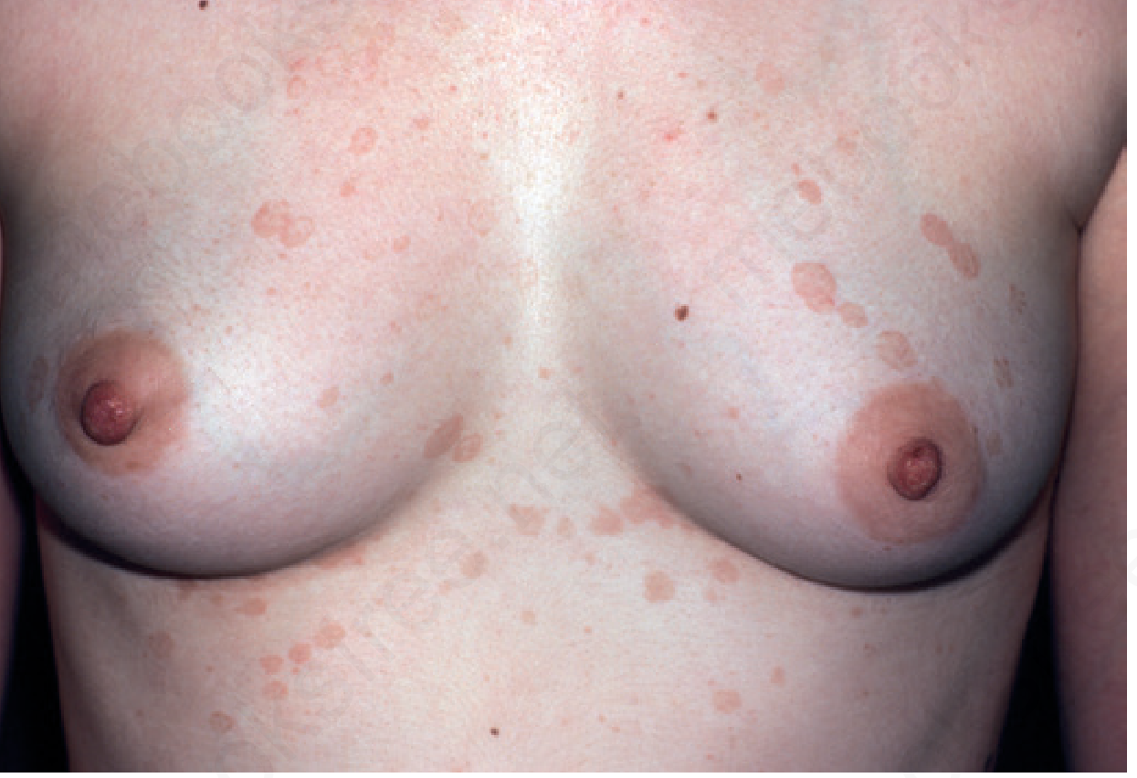
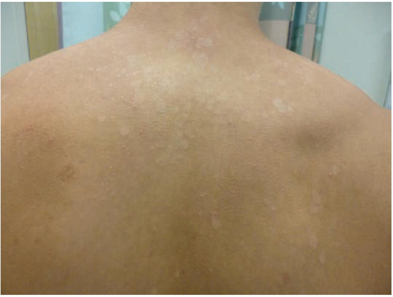
- Upper trunk (chest and back) — most common — governed by sebaceous gland density
- Shoulders, upper arms
- Sides of neck
- Abdomen and lower trunk
- Intertriginous areas (axillae, antecubital/popliteal fossae) — "inverse" tinea versicolor
- Face: mainly in children and immunocompromised adults
- Scalp, palms, soles (less common)
- Penile involvement reported; organism isolated in balanoposthitis
Tinea versicolor — hypopigmented coalescing macules on the anterior chest (Andrews' Diseases of the Skin)
Tinea versicolor — extensive hypopigmented involvement of the back (Tintinalli's Emergency Medicine)
Symptoms
- Usually asymptomatic — the main concern is cosmetic
- Mild pruritus may occur
- In this 22-year-old male with an 8-year history: the likely chief complaint is failure to tan normally in summer, or persistent discoloured patches despite intermittent treatments
Clinical Signs to Elicit
| Sign | Technique | Finding |
|---|
| Evoked scale sign | Stretch or rub the lesion | White, fuzzy scale confined to the lesion |
| Scratch sign | Gently scrape with slide/blade | Pale bran-like scale |
| Failure to tan | History | Lesions remain pale in summer |
5. Investigations & Diagnosis
Potassium Hydroxide (KOH) Preparation
Cornerstone of diagnosis. Scrape scale from lesion → mount in KOH → microscopy reveals:
"Spaghetti and meatballs" — short, stubby, curved hyphae ("spaghetti") + clusters of thick-walled yeast cells ("meatballs")
This combination of hyphae and spores is pathognomonic. Stains that enhance visualisation:
- Parker blue-black ink (1:1 with 20% KOH)
- 1% Chicago sky blue 6B with 8% KOH
- Gram stain
Wood's Lamp (UV-A, 365 nm)
- Lesions fluoresce yellow-green (due to porphyrins produced by the yeast)
- Accentuates pigmentary differences
- Identifies satellite lesions not visible to naked eye
Skin Biopsy (rarely needed)
- Thick basket-weave stratum corneum with hyphae and spores
- In the atrophic variant: effacement of rete ridges, subepidermal fibroplasia, pigment incontinence, elastolysis
- PAS or methenamine silver stains highlight organisms
Fungal Culture
Not routinely done — the organism is a commensal, so culture from skin surface is non-diagnostic. Culture requires lipid-enriched media (olive oil overlay). Only useful if Malassezia folliculitis/sepsis is suspected.
- Andrews' Diseases of the Skin, p. 358
- Red Book 2021, p. 919
6. Differential Diagnosis
| Condition | Distinguishing Features |
|---|
| Vitiligo | Complete depigmentation (not just hypo-), sharp borders, no scale, KOH negative |
| Pityriasis alba | Children, dry eczematous background, face predominance |
| Seborrhoeic dermatitis | Erythematous yellow-greasy scales, nasolabial folds/scalp/glabella |
| Pityriasis rosea | Herald patch, Christmas tree pattern, self-limiting 6–8 weeks |
| Pityriasis rubra pilaris | Follicular papules, orange hue, islands of sparing |
| Leprosy (tuberculoid) | Anhidrosis, loss of sensation, asymmetric, KOH negative |
| Confluent & reticulated papillomatosis (CARP) | Reticulated, warty texture, neck/trunk, fails antifungal |
| Secondary syphilis | Copper-coloured, indurated, adenopathy, positive serology |
| Postinflammatory hypopigmentation | History of prior inflammatory skin disease |
| Progressive macular hypomelanosis | Folliculocentric, no scale, KOH negative |
| Nummular eczema | Oozing, crusted, pruritic, responds to steroids |
The 8-year duration without systemic features and the young male demographic make vitiligo and leprosy the two most clinically important differentials to exclude.
7. Management
Why This Patient Poses a Challenge
An 8-year history in a 22-year-old signifies:
- Disease onset at ~14 years — peak sebaceous activity
- Likely inadequate prior treatment or failure to use prophylaxis
- High seborrhoea predisposition — needs maintenance therapy
- Pigmentation will not normalise immediately even after mycological cure — must counsel the patient
Topical Therapy (First-line)
| Agent | Regimen | Notes |
|---|
| Selenium sulfide 2.5% lotion/shampoo | Apply daily × 7 days, wash off after 5–10 min; OR single overnight application monthly | Most cost-effective; sulfur odour reduces compliance; covers scalp reservoir |
| Ketoconazole 2% shampoo | Daily × 1 week, leave 3–5 min | Highly effective; FDA discourages oral form for skin |
| Zinc pyrithione shampoo/soap | Daily treatment + monthly for prophylaxis | Well tolerated, cost-effective, easy to use in shower |
| Clotrimazole 1% cream | Twice daily × 2–3 weeks | Standard azole option |
| Miconazole, econazole, oxiconazole | Twice daily × 2–3 weeks | Equivalent efficacy |
| Ciclopirox olamine | Twice daily | Also effective for Malassezia folliculitis |
| Terbinafine cream 1% | Twice daily (superior to once daily) | Note: oral terbinafine is ineffective for tinea versicolor |
Shampoo formulations are easier to spread over extensive truncal involvement (as in this 8-year case) and improve compliance.
Systemic Therapy (for resistant, extensive, or recurrent disease — fits this case)
Given 8 years of disease with likely extensive involvement, oral therapy is appropriate:
| Drug | Regimen | Notes |
|---|
| Fluconazole (preferred) | 300–400 mg once weekly × 2–4 weeks | First-line oral agent; repeatable monthly for prophylaxis |
| Itraconazole | 200 mg/day × 7 days OR 400 mg single dose | Effective; exercise caution in hepatic disease |
| Ketoconazole (oral) | Avoid — FDA warns of liver toxicity, adrenal insufficiency, drug interactions; at least one fatality reported | Not recommended for uncomplicated skin infection |
| Oral terbinafine | Ineffective for tinea versicolor | Do not use systemically |
Important: Topical therapy is often equivalent or superior to systemic therapy. Oral agents have adverse effect profiles that must be considered. Monitor LFTs if multiple courses of oral azoles are given.
Prophylaxis (Critical for this patient)
Given the 60–80% recurrence rate, prophylactic therapy is mandatory:
| Prophylactic regimen | Frequency |
|---|
| Selenium sulfide shampoo/lotion | Weekly or monthly |
| Ketoconazole 2% or econazole shampoo | Monthly, overnight application |
| Fluconazole 400 mg | Monthly (oral) |
| Zinc pyrithione soap/shampoo | Weekly shower use |
| Bifonazole shampoo | Monthly, overnight |
- Andrews' Diseases of the Skin, p. 358
- Red Book 2021, p. 919–920
- Tintinalli's Emergency Medicine, p. 1681
Emerging/Alternative Therapies
- 5-aminolevulinic acid photodynamic therapy (PDT) has been reported effective in refractory cases
- Andrews' Diseases of the Skin, p. 358
8. Post-Treatment Pigmentation
One of the most important counselling points:
Hypopigmentation (and sometimes hyperpigmentation) persists for weeks to months after successful mycological cure. The absence of re-pigmentation immediately after treatment does not mean treatment failure.
Mechanisms:
- Damaged melanosomes take time to reconstitute
- Keratinocyte turnover must replace pigment-deficient cells
- UV exposure (sun or sunbed) can accelerate repigmentation once the fungus is eradicated
- Resolution of scale and negative KOH = treatment success — not return of normal skin colour
9. Malassezia Folliculitis (Important Complication to Know)
A related condition sometimes co-existing with or mistaken for tinea versicolor:
- Monomorphic dome-shaped follicular papules and tiny pustules
- Upper back, adjacent areas, face, scalp
- Pruritic; more common in transplant recipients and after antibiotic therapy (especially tetracyclines for acne suppress Propionibacterium acnes, allowing Malassezia overgrowth)
- Treat with fluconazole 400 mg single dose or itraconazole 200 mg/day × 7 days
10. Summary Table for the Case
| Feature | This Patient |
|---|
| Age/sex | 22M — peak risk group (high sebaceous activity) |
| Duration | 8 years — chronic, recurrent course typical |
| Cause | Malassezia globosa/furfur — commensal gone pathogenic |
| Diagnosis | KOH "spaghetti & meatballs" + Wood's lamp yellow-green fluorescence |
| Treatment | Topical azole/selenium sulfide + oral fluconazole for extensive/recurrent disease |
| Key counselling | Pigment normalisation takes months; prophylaxis mandatory; not contagious |
| Recurrence risk | 60–80% without maintenance |
| Do NOT use | Oral terbinafine (ineffective), oral ketoconazole (toxic) |
Sources:
- Andrews' Diseases of the Skin: Clinical Dermatology, p. 358
- Red Book 2021: Report of the Committee on Infectious Diseases, pp. 918–920
- Tintinalli's Emergency Medicine: A Comprehensive Study Guide, pp. 1680–1681
- Dermatology 2-Volume Set, 5th Edition, blocks 14 & 17