
Vitamin c chi tablet mi don gheu gheu shakto divsachya 500 mg ch 1 ahe vatt kahitari
- ✅ दैनंदिन गरज: प्रौढांना फक्त 65-90 mg/day लागते
- ✅ 500 mg हे जास्त असले तरी शरीर अतिरिक्त भाग लघवीतून बाहेर टाकते (water-soluble vitamin आहे)
- ⚠️ जेवणानंतर घ्या - रिकाम्या पोटी घेतल्यास पोटदुखी किंवा आम्लता होऊ शकते
- ⚠️ kidney stones चा इतिहास असल्यास जास्त dose टाळा
- ℹ️ Upper limit (UL): 2000 mg/day पर्यंत सुरक्षित मानले जाते

Aani he kashi gheu
📦 NEUROLA Forte म्हणजे काय?
| Vitamin | प्रत्येक टॅबलेटमध्ये |
|---|---|
| Vitamin B1 (Thiamine) | 1.0 mg |
| Vitamin B2 (Riboflavin) | 1.0 mg |
| Vitamin B6 (Pyridoxine) | 1.5 mg |
| Vitamin B12 | 2.2 mcg |
| Niacinamide (B3) | 15 mg |
| Calcium Pantothenate (B5) | 3 mg |
कसे घ्यावे?
"One tablet daily or as directed by the Dietician"
⚠️ महत्त्वाचे
- हे Health Supplement आहे, medicine नाही
- Pregnant किंवा breastfeeding असाल तर डॉक्टरांना विचारा
- थंड, कोरड्या जागी ठेवा - उन्हापासून दूर
He got hon sathi ghetoy mi
🧠 B-Complex कशासाठी फायदेशीर आहे?
⚡ Energy & थकवा कमी करणे
- B1, B2, B3 हे जेवणातून energy बनवतात
- रोज थकवा वाटत असेल तर हे मदत करते
🧠 Nervous System (मज्जासंस्था)
- B6, B12 मज्जातंतूंच्या आरोग्यासाठी
- हात-पाय सुन्न पडणे, मुंग्या येणे कमी होते
💆 Stress & मन
- B5 (Calcium Pantothenate) stress hormones नियंत्रित करतो
- मूड चांगला राहतो
💇 केस, त्वचा, नखे
- B2, B7 केस गळणे आणि त्वचेसाठी चांगले
🩸 Blood (रक्त)
- B12 लाल रक्तपेशी बनवण्यास मदत करते
- अशक्तपणा (Anemia) टाळतो
तुम्ही कोणत्या कारणासाठी घेत आहात?
💊 एक टॅबलेट सकाळी जेवणानंतर - हे पुरेसे आहे. जास्त घेऊ नका.
Gor honya sathi boltoy ani ho malaa te p. Varsicoluor hota paha to gelay fakt tya dagan sathi upyukt ahe na
थेट उत्तर:
🔴 NEUROLA Forte (B-Complex) - गोरे होण्यासाठी?
🟡 Pityriasis Versicolor चे डाग उरले असतील तर?
| काय | कशासाठी |
|---|---|
| Vitamin C (तुमची 500mg) | ✅ हो! Melanin कमी करते, skin tone सुधारते |
| Sunscreen रोज लावणे | ✅ डाग लवकर जातात |
| Moisturizer | ✅ त्वचा healthy राहते |
| B-Complex (NEUROLA) | ❌ त्वचेचा रंग बदलत नाही थेट |
सारांश:
- NEUROLA Forte - energy, nerves साठी चांगले, पण गोरेपणासाठी नाही
- व्हिटॅमिन C 500mg - हे मात्र डाग कमी करण्यासाठी थोडे उपयुक्त आहे
- Pityriasis चे डाग 3-6 महिन्यांत आपोआप जातात सूर्यप्रकाशाने
⚠️ "गोरे होण्यासाठी" कोणतेही supplement घेणे टाळा - त्यापेक्षा Sunscreen + Vitamin C हे जास्त effective आणि safe आहे! 😊
Pityriasis Versicolor चे कारण आणि उपचार
pityriasis versicolor skin lesions hypopigmented patches
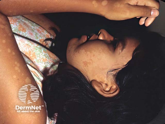
This dermatology clinical photograph depicts hypopigmented patches consistent with pityriasis versicolor (tinea versicolor) involving sun-exposed skin on the face and forearm of a patient labeled PV-patient1. The lesions appear as flat, well-demarcated, hypoesthetic macules with subtle surface scale. Facial patches are lighter than surrounding skin, while forearm lesions show a similar pigmentary disturbance. The appearance is characteristic for PV, caused by superficial Malassezia overgrowth that metabolizes lipids, producing pigmentary changes in the stratum corneum. Sun exposure can accentuate hypopigmentation, and Wood’s lamp examination may reveal fluorescence supportive of diagnosis. Clinically, recognize it as a benign, treatable dermatosis that should be differentiated from vitiligo, pityriasis alba, and post-inflammatory hypopigmentation. Management typically includes topical antifungal therapy (ketoconazole, itraconazole, terbinafine) or selenium sulfide, with potential recurrence. This image serves educational purposes in dermatology training, remote assessment, and AI-driven lesion recognition, illustrating characteristic distribution on the face and arms, the macular morphology, and the essential differential diagnoses in pigmentary disorders.
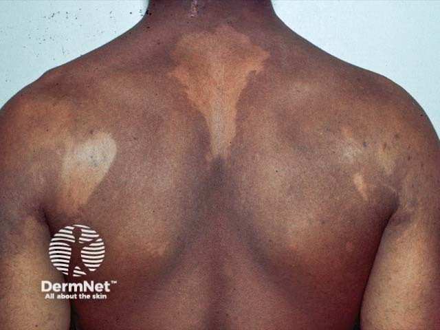
Clinical photography of the back showing large hypopigmented patches consistent with pityriasis versicolor (tinea versicolor). The dorsal trunk demonstrates irregular, well-demarcated macules with reduced pigment relative to surrounding skin and slight surface scaling. Lesions are primarily on the upper back and shoulders, sometimes extending centrally, with a predominantly hypo pigmented appearance in darker skin tones and mixed hypo/hyperpigmentation in lighter tones. The patches may appear less conspicuous in sun-exposed areas and more noticeable in cooler seasons. On close inspection, there is minimal erythema or itching; scale, when present, is fine and compelling under dermoscopy or magnification. The imaging modality is clinical photography, without contrast, intended for documentation and educational reference. The pattern is truncal, symmetric or patchy, often affecting the upper back, shoulders and central chest in typical presentations. In practice, differential diagnoses include pityriasis alba, post-inflammatory hypopigmentation, chronic eczema variants, and hypopigmented tinea corporis. Clinically, pityriasis versicolor is caused by Malassezia species; diagnosis is supported by KOH showing spaghetti-and-meatballs and Wood's lamp fluorescence. Management involves topical azoles or oral antifungals in extensive disease. This image supports dermatology training, telemedicine consultations, and research into pigmentary disorders, fungal colonization, and treatment response. It serves as a reference for pattern recognition globally.
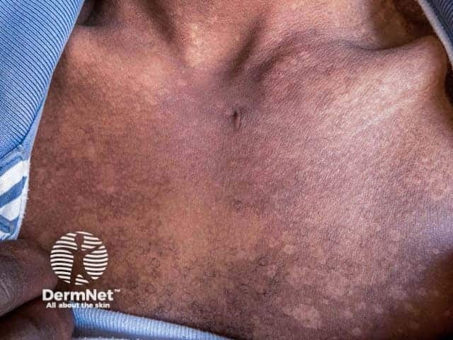
Clinical photography of the anterior trunk in a patient with pityriasis versicolor presenting in the hypopigmented stage. High-resolution, color-balanced digital image captured under standard visible light to document surface skin changes on the chest and upper abdomen with an anterior view. The lesions appear as numerous flat, ill‑defined macules with a pale to slightly hypopigmented hue relative to surrounding skin. The patches are variably coalescent, with fine scale evident at the margins and mild asynchronous tan response in adjacent areas. The distribution favors the central chest and proximal trunk, presenting as asymptomatic patches without erosion or crusting. Dermatoscopic features are not available; the surface is smooth, with subtle textural changes rather than prominent edema or vascular markings. Clinically, pityriasis versicolor is a superficial Malassezia yeast infection that disrupts melanin production, producing hypopigmented or hyperpigmented macules depending on skin type. The image illustrates hypopigmented lesions characteristic of the disease, aiding differential diagnosis from vitiligo, post-inflammatory hypopigmentation, and pityriasis alba. This image supports clinical correlation with Wood’s lamp examination, which often reveals copper-orange fluorescence in affected skin, and with mycological confirmation via KOH preparation showing spaghetti-and-meatballs. Potential clinical applications include education, dermatology training, and research on pigmentary disorders and superficial fungal infections.
🍄 Pityriasis Versicolor (Tinea Versicolor)
कारण काय? (Etiology)
- 🌡️ उष्ण व दमट हवामान (भारतात खूप common)
- 💦 जास्त घाम येणे
- 🧴 तेलकट त्वचा (sebaceous glands जास्त active)
- 😷 रोगप्रतिकारशक्ती कमी असणे
- 🧒 तरुण वयात (Adolescents मध्ये जास्त)
⚠️ हे सांसर्गिक (Contagious) नाही - दुसऱ्याला लागत नाही!
लक्षणे कशी दिसतात?
- पांढरे, तपकिरी, गुलाबी किंवा हलके डाग - एकाच माणसात वेगवेगळ्या रंगाचे असू शकतात
- उन्हाळ्यात डाग फिकट दिसतात (tan होत नाहीत), हिवाळ्यात जास्त गडद दिसतात - म्हणून नाव "Versicolor" (रंग बदलणारे)
- छाती, पाठ, खांदे, मान - हे भाग जास्त प्रभावित
- खाज कमी असते किंवा नसते
- त्वचा ताणल्यास पांढरी पूड दिसते (Evoked scale sign)
निदान कसे होते?
| चाचणी | काय दिसते |
|---|---|
| KOH Preparation | "Spaghetti & Meatballs" - लहान धागे + गोल yeast |
| Wood's Lamp | पिवळ्या-हिरव्या रंगाचा फ्लुरोसन्स |
| Clinical | वैशिष्ट्यपूर्ण डागांवरून |
उपचार (Treatment)
💊 टॉपिकल (त्वचेवर लावायचे) - पहिली निवड:
| औषध | कसे वापरावे |
|---|---|
| Ketoconazole shampoo/cream | रात्री लावा, सकाळी धुवा - 2-3 आठवडे |
| Selenium Sulfide shampoo | 5-10 मिनिटे ठेवा, धुवा - 3-7 दिवस |
| Clotrimazole cream | दिवसातून 2 वेळा - 2-3 आठवडे |
| Terbinafine / Ciclopirox | डॉक्टरांच्या सल्ल्याने |
💊 तोंडी औषधे (जास्त पसरलेल्या cases मध्ये):
- Fluconazole 300mg - एकच dose, आठवड्यातून एकदा × 2
- Itraconazole 200mg × 7 दिवस
- ⚠️ Oral Ketoconazole - liver side effects मुळे टाळावे
⚠️ डाग उरतातच का? (तुमचा प्रश्न!)
बुरशी मेली तरी डाग महिनोंमहिने राहतात कारण Melanin (त्वचेचा रंग) परत यायला वेळ लागतो.
- ☀️ सूर्यप्रकाश - melanin regeneration ला मदत करतो
- 🧴 Sunscreen रोज लावा - skin uniform होते
- Vitamin C - antioxidant, melanin synthesis सुधारते
- 3-6 महिने धीर धरा - डाग आपोआप जातील!
पुन्हा होणे (Recurrence) टाळण्यासाठी:
- दर महिन्याला 1-2 वेळा Ketoconazole shampoo अंघोळीत वापरा
- घाम आल्यावर लगेच अंघोळ करा
- Oily moisturizers टाळा प्रभावित भागावर
- उन्हाळ्यात preventive dose घेऊ शकता (डॉक्टरांच्या सल्ल्याने)