
18 yr old pt presented with eczema type appearance around nose on scalp , chest and behind ears After stretching and rubbing it showed whitish flakes what is diagnosis and treatment
seborrhoeic dermatitis face scalp whitish flakes
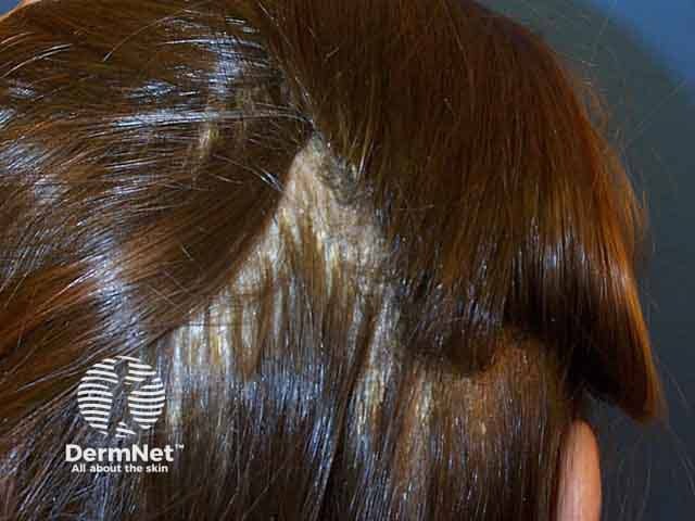
Comprehensive Description: This is a high-resolution clinical photograph of the scalp taken in vivo, using standard dermatology photography techniques, showing a lateral (side) view of the hair-bearing skin. The image demonstrates extensive seborrhoeic dermatitis with overlapping greasy scales adherent to erythematous plaques over the scalp, most prominently along the temporal and vertex regions. The scales appear yellowish-white, oily in appearance, and coalesce to form layered flakes that extend into the hair margins. Mild diffuse erythema and scaling are present without visible purpura or crusting. Hair remains present, with no obvious scarring alopecia. The morphology is consistent with inflammatory dermatosis of the scalp; differential diagnoses include psoriasis capitis, tinea capitis, and atopic dermatitis, though the greasy character with fine lamellar scaling favors seborrheic dermatitis. Clinically, this image is relevant for dermatology education, trichology assessment, and differential diagnosis practice, as well as for evaluating treatment response to antifungal shampoos (ketoconazole, head-and-shoulders zinc pyrithione), corticosteroid, or calcineurin inhibitor regimens. It can be used in teaching modules on scalp dermatoses, in digital archives for image-guided diagnosis, and for research into Malassezia-associated inflammation, inflammatory skin disorders, and patient counseling on scalp hygiene. This descriptor supports retrieval in education, clinical case review, and dermatology datasets globally.
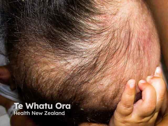
This is a high-resolution clinical photograph of an infant scalp demonstrating seborrhoeic dermatitis with yellow greasy scales preferentially located on the frontal/temporoparietal scalp and sparing the face. The image captures the characteristic matting and flaking scales adherent to hair shafts, with mild erythema and diffuse scale accumulation at the crown and temple regions. The lesion is non-vesicular, non-exudative, and appears symmetrical across the scanned scalp area. The findings are consistent with cradle cap, a common, benign dermatologic condition of infancy believed to reflect sebaceous gland activity and infant skin immaturity. Differential considerations include tinea capitis if alopecia or perifollicular scaling is prominent, atopic dermatitis with more diffuse erythema, or psoriasis with well-demarcated plaques though these are less typical in early life. Clinically correlated, this pattern typically responds to routine infant scalp hygiene and gentle, non-irritating cleansing; medicated soaps or antifungal shampoos may be used under pediatric guidance if scales persist. This image is valuable for educational reference in pediatric dermatology, dermoscopy correlation not shown, and radiology cross-disciplinary discussions about differential diagnoses of scalp scaling. Potential applications: medical education, image-based differential diagnosis training, clinical case discussions, and telemedicine triage for infants presenting with cradle cap.
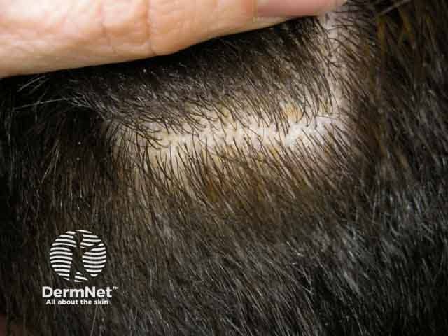
Clinical photograph of the scalp demonstrates seborrhoeic dermatitis characterized by honey-coloured, greasy scales adherent to hair shafts with mild surrounding erythema. Involvement is patchy to diffuse over hair-bearing skin, most pronounced at the vertex and temple regions, with scales accumulating along follicular openings. The yellowish scales are waxy and easily removable, yet can be persistent; pruritus is commonly reported in patients. Hair shafts appear preserved and no frank alopecia is evident in this image. This presentation aligns with the typical clinical phenotype of seborrhoeic dermatitis, a common inflammatory scalp dermatosis linked to sebaceous gland activity and colonization by Malassezia species. The visual features help distinguish it from psoriasis (silvery scales, well-demarcated plaques) and tinea capitis (hair loss, broken hairs, erythematous patches). Diagnostic significance lies in recognizing the greasy, yellow scales on the scalp as a hallmark, guiding topical therapies such as selenium sulfide, zinc pyrithione, ketoconazole antifungal shampoos, and low‑potency corticosteroids when appropriate. Clinically, the image is relevant for dermatology education, differential diagnosis exercises, and treatment response documentation in adult and pediatric patients. The image supports hands-on recognition of seborrhoeic dermatitis patterns in scalp dermatology curricula and serves as a reference for teledermatology consultations.
seborrheic dermatitis treatment
Diagnosis: Seborrhoeic Dermatitis
- 18-year-old (adult form, chronic course)
- Eczema-like erythema in sebaceous gland-rich zones: nasolabial folds/around nose, scalp, chest, retroauricular (behind ears)
- Whitish/yellowish bran-like scales revealed on stretching/rubbing - the pathognomonic sign
- Symmetric distribution
Pathogenesis (Why it happens)
- Sebum overproduction - sebaceous gland activity is high in adolescence/young adults
- Malassezia yeast (formerly Pityrosporum ovale/M. furfur) - a lipophilic commensal yeast that proliferates in sebaceous areas and triggers inflammation
- Individual susceptibility - impaired epidermal barrier, immune response
Differential Diagnosis
| Condition | Distinguishing Features |
|---|---|
| Psoriasis | Silvery-white scales, well-demarcated plaques, Auspitz sign, extensor surfaces |
| Atopic dermatitis | Flexural distribution, intense itch, personal/family atopy history |
| Rosacea | Flushing, papulopustules, telangiectasia, no greasy scales |
| Tinea capitis | Hair loss, broken hairs, KOH positive, mainly children |
| SLE (butterfly rash) | Photodistribution, spares nasolabial folds, systemic features |
Treatment
Scalp
- First-line: Ketoconazole 2% shampoo - apply 2-3x/week, leave on 5 minutes, rinse. Use for 4 weeks, then maintenance 1x/week (75-90% response rate in double-blind trials).
- Alternatives: Ciclopirox olamine shampoo, selenium sulfide shampoo, zinc pyrithione shampoo, coal tar shampoo
- For inflammation/pruritus: add a low-to-mid potency topical corticosteroid lotion (e.g. betamethasone valerate) short-term
Face and Behind Ears
- Ketoconazole 2% cream - apply twice daily for 2-4 weeks, then taper to maintenance
- Ciclopirox olamine 1% cream - equally effective, anti-inflammatory and antifungal
- Low-potency topical corticosteroid (e.g. hydrocortisone 1%) - short-term use only for the face (risk of skin atrophy/rosacea with prolonged use)
- Topical calcineurin inhibitors (tacrolimus 0.1%, pimecrolimus 1%) - steroid-sparing option, safe for long-term facial use
Chest/Body
- Ketoconazole cream or ciclopirox cream as above
- Mild emollients to support skin barrier
Maintenance (important - SD is chronic/relapsing)
Systemic (severe/resistant cases only)
- Oral fluconazole 300mg once weekly for 2-4 weeks
- Oral itraconazole - second option
Patient Counseling Points
- This is a chronic, relapsing condition - not curable, but manageable
- Avoid hot water, harsh soaps, and excessive scrubbing (worsens inflammation)
- Sun exposure in moderation may help (summer improvement is common)
- Stress, fatigue, and febrile illnesses can trigger flares
- Scales on dark clothing are a cosmetic concern - regular use of medicated shampoos prevents this
Create a treatment protocol document (scalp vs face medication regimens)
ls -la /tmp/workspace/ 2>/dev/null || mkdir -p /tmp/workspace && echo "created"
mkdir -p /tmp/workspace/seborrhoeic-protocol && python3 << 'EOF' from docx import Document from docx.shared import Pt, Inches, RGBColor from docx.enum.text import WD_ALIGN_PARAGRAPH from docx.oxml.ns import qn from docx.oxml import OxmlElement import copy doc = Document() # ── Page margins ────────────────────────────────────────────── section = doc.sections[0] section.top_margin = Inches(1.0) section.bottom_margin = Inches(1.0) section.left_margin = Inches(1.2) section.right_margin = Inches(1.2) # ── Base font ───────────────────────────────────────────────── normal = doc.styles["Normal"] normal.font.name = "Calibri" normal.font.size = Pt(11) # ── Helper: shade a table row ───────────────────────────────── def shade_cell(cell, hex_color): tc = cell._tc tcPr = tc.get_or_add_tcPr() shd = OxmlElement("w:shd") shd.set(qn("w:val"), "clear") shd.set(qn("w:color"), "auto") shd.set(qn("w:fill"), hex_color) tcPr.append(shd) def set_cell_border(cell, **kwargs): """Add borders to a cell.""" tc = cell._tc tcPr = tc.get_or_add_tcPr() tcBorders = OxmlElement("w:tcBorders") for edge in ("top","left","bottom","right","insideH","insideV"): tag = OxmlElement(f"w:{'insideH' if edge=='insideH' else 'insideV' if edge=='insideV' else edge}") tag.set(qn("w:val"), "single") tag.set(qn("w:sz"), "4") tag.set(qn("w:space"),"0") tag.set(qn("w:color"),"4472C4") tcBorders.append(tag) tcPr.append(tcBorders) def add_bold_run(para, text, size=None, color=None): run = para.add_run(text) run.bold = True if size: run.font.size = Pt(size) if color: run.font.color.rgb = RGBColor(*color) return run def header_row(table, texts, bg="1F4E79"): row = table.rows[0] for i, txt in enumerate(texts): cell = row.cells[i] shade_cell(cell, bg) p = cell.paragraphs[0] p.alignment = WD_ALIGN_PARAGRAPH.CENTER run = p.add_run(txt) run.bold = True run.font.color.rgb = RGBColor(0xFF, 0xFF, 0xFF) run.font.size = Pt(10) def add_data_row(table, values, bg=None): row = table.add_row() for i, val in enumerate(values): cell = row.cells[i] if bg: shade_cell(cell, bg) p = cell.paragraphs[0] p.alignment = WD_ALIGN_PARAGRAPH.LEFT p.add_run(val).font.size = Pt(10) return row # ═══════════════════════════════════════════════════════════════ # HEADER BLOCK # ═══════════════════════════════════════════════════════════════ title_para = doc.add_paragraph() title_para.alignment = WD_ALIGN_PARAGRAPH.CENTER r = title_para.add_run("SEBORRHOEIC DERMATITIS") r.bold = True; r.font.size = Pt(20); r.font.color.rgb = RGBColor(0x1F,0x4E,0x79) sub_para = doc.add_paragraph() sub_para.alignment = WD_ALIGN_PARAGRAPH.CENTER r2 = sub_para.add_run("Clinical Treatment Protocol") r2.bold = True; r2.font.size = Pt(14); r2.font.color.rgb = RGBColor(0x2E,0x74,0xB5) divider = doc.add_paragraph() divider.alignment = WD_ALIGN_PARAGRAPH.CENTER r3 = divider.add_run("─" * 70) r3.font.color.rgb = RGBColor(0x2E,0x74,0xB5) meta = doc.add_paragraph() meta.alignment = WD_ALIGN_PARAGRAPH.CENTER m = meta.add_run("Prepared for: Clinical Practice | Patient Age: Adolescent / Adult | Date: July 2026") m.italic = True; m.font.size = Pt(9); m.font.color.rgb = RGBColor(0x59,0x59,0x59) doc.add_paragraph() # ═══════════════════════════════════════════════════════════════ # DIAGNOSIS OVERVIEW BOX # ═══════════════════════════════════════════════════════════════ h = doc.add_heading("1. DIAGNOSIS OVERVIEW", level=1) h.runs[0].font.color.rgb = RGBColor(0x1F,0x4E,0x79) dx_table = doc.add_table(rows=1, cols=2) dx_table.style = "Table Grid" dx_table.columns[0].width = Inches(2.2) dx_table.columns[1].width = Inches(4.5) header_row(dx_table, ["Parameter", "Details"]) rows_data = [ ("Condition", "Seborrhoeic Dermatitis (ICD-10: L21)"), ("Causative Factor","Malassezia spp. yeast (M. furfur / M. globosa) + sebum overproduction"), ("Age Group", "Adolescents & adults (peak: 2nd-6th decade); Males > Females"), ("Key Sign", "Yellowish/whitish bran-like scales on erythematous base; revealed on stretching"), ("Distribution", "Scalp, nasolabial folds, eyebrows, retroauricular, chest (presternal)"), ("Course", "Chronic, relapsing — maintenance therapy is required"), ("Red Flags", "Severe/resistant SD in young pt → screen for HIV, immunosuppression"), ] for i, (k, v) in enumerate(rows_data): bg = "EBF3FB" if i % 2 == 0 else "FFFFFF" row = dx_table.add_row() shade_cell(row.cells[0], "D6E4F0") row.cells[0].paragraphs[0].add_run(k).bold = True row.cells[0].paragraphs[0].runs[0].font.size = Pt(10) shade_cell(row.cells[1], bg) row.cells[1].paragraphs[0].add_run(v).font.size = Pt(10) doc.add_paragraph() # ═══════════════════════════════════════════════════════════════ # SECTION 2 — SCALP REGIMEN # ═══════════════════════════════════════════════════════════════ h2 = doc.add_heading("2. SCALP TREATMENT REGIMEN", level=1) h2.runs[0].font.color.rgb = RGBColor(0x1F,0x4E,0x79) # Subheading sh = doc.add_paragraph() r = sh.add_run("A. Acute Phase (Weeks 1-4)") r.bold = True; r.font.size = Pt(12); r.font.color.rgb = RGBColor(0x2E,0x74,0xB5) scalp_acute = doc.add_table(rows=1, cols=5) scalp_acute.style = "Table Grid" header_row(scalp_acute, ["Drug", "Formulation", "Dosing", "Duration", "Notes"], bg="2E74B5") scalp_acute_data = [ ("Ketoconazole 2%", "Shampoo (1st line)", "2-3x/week\nLeave on 5 min", "4 weeks", "75-90% response rate; antifungal"), ("Ciclopirox olamine 1%","Shampoo (1st line)", "2-3x/week", "4 weeks", "Antifungal + anti-inflammatory"), ("Selenium Sulfide 2.5%","Shampoo (alt)", "2x/week", "4 weeks", "Effective; cosmetically less acceptable"), ("Zinc Pyrithione 1-2%","Shampoo (OTC alt)", "2-3x/week", "4 weeks", "Widely available; milder"), ("Coal Tar 2-5%", "Shampoo (alt)", "2-3x/week", "4 weeks", "Anti-proliferative; use if scales thick"), ("Betamethasone\nValerate 0.1%","Scalp lotion/solution","Once daily\n(evening)", "2-3 weeks", "For inflammation & itch; taper after control"), ("Clobetasol\nPropionate 0.05%","Scalp solution","Once daily", "Max 2 weeks", "High-potency; for severe scalp only"), ] for i, row_data in enumerate(scalp_acute_data): bg = "EBF3FB" if i % 2 == 0 else "FFFFFF" add_data_row(scalp_acute, row_data, bg=bg) doc.add_paragraph() sh2 = doc.add_paragraph() r = sh2.add_run("B. Maintenance Phase (After Week 4 — ongoing)") r.bold = True; r.font.size = Pt(12); r.font.color.rgb = RGBColor(0x2E,0x74,0xB5) scalp_maint = doc.add_table(rows=1, cols=4) scalp_maint.style = "Table Grid" header_row(scalp_maint, ["Drug", "Formulation", "Frequency", "Notes"], bg="2E74B5") maint_data = [ ("Ketoconazole 2%", "Shampoo", "Once weekly", "Prevents Malassezia re-proliferation"), ("Ciclopirox olamine 1%","Shampoo", "Once weekly", "Good long-term tolerability"), ("Zinc Pyrithione 1-2%","Shampoo", "1-2x/week", "OTC; suitable for mild cases"), ("Selenium Sulfide 2.5%","Shampoo", "Once weekly", "Alternate if above not tolerated"), ] for i, row_data in enumerate(maint_data): bg = "EBF3FB" if i % 2 == 0 else "FFFFFF" add_data_row(scalp_maint, row_data, bg=bg) doc.add_paragraph() # ═══════════════════════════════════════════════════════════════ # SECTION 3 — FACE / RETROAURICULAR / CHEST REGIMEN # ═══════════════════════════════════════════════════════════════ h3 = doc.add_heading("3. FACE, RETROAURICULAR & CHEST REGIMEN", level=1) h3.runs[0].font.color.rgb = RGBColor(0x1F,0x4E,0x79) sh3 = doc.add_paragraph() r = sh3.add_run("A. Acute Phase (Weeks 1-4)") r.bold = True; r.font.size = Pt(12); r.font.color.rgb = RGBColor(0x2E,0x74,0xB5) face_acute = doc.add_table(rows=1, cols=5) face_acute.style = "Table Grid" header_row(face_acute, ["Drug", "Formulation", "Dosing", "Duration", "Notes"], bg="1F4E79") face_data = [ ("Ketoconazole 2%", "Cream (1st line)", "Twice daily", "2-4 weeks", "Apply to nasolabial, retroauricular, chest"), ("Ciclopirox olamine 1%", "Cream (1st line)", "Twice daily", "2-4 weeks", "Antifungal + anti-inflammatory; safe long-term"), ("Hydrocortisone 1%", "Cream (low-potency steroid)", "Once-twice daily", "Max 2 weeks", "Short-term only on face; risk of atrophy"), ("Desonide 0.05%", "Cream (low-potency steroid)", "Twice daily", "Max 2 weeks", "For marked erythema; do not use long-term"), ("Tacrolimus 0.1%", "Ointment (calcineurin inhib.)","Twice daily", "4-8 weeks", "Steroid-sparing; safe for long-term facial use"), ("Pimecrolimus 1%", "Cream (calcineurin inhib.)", "Twice daily", "4-8 weeks", "Well-tolerated on face; no atrophy risk"), ("Ketoconazole 2%\n+ HC 1%","Cream combo", "Twice daily", "2 weeks", "Combine antifungal + steroid for rapid control"), ] for i, row_data in enumerate(face_data): bg = "EBF3FB" if i % 2 == 0 else "FFFFFF" add_data_row(face_acute, row_data, bg=bg) doc.add_paragraph() sh4 = doc.add_paragraph() r = sh4.add_run("B. Face Maintenance Phase (After Week 4)") r.bold = True; r.font.size = Pt(12); r.font.color.rgb = RGBColor(0x2E,0x74,0xB5) face_maint = doc.add_table(rows=1, cols=4) face_maint.style = "Table Grid" header_row(face_maint, ["Drug", "Formulation", "Frequency", "Notes"], bg="1F4E79") face_maint_data = [ ("Ketoconazole 2%", "Cream", "2-3x/week", "Best evidence for maintenance"), ("Ciclopirox olamine 1%","Cream", "2-3x/week", "Safe for prolonged use"), ("Pimecrolimus 1%", "Cream", "2-3x/week", "Preferred if steroids caused adverse effects"), ("Tacrolimus 0.1%", "Ointment", "As needed", "On-demand use at first sign of flare"), ] for i, row_data in enumerate(face_maint_data): bg = "EBF3FB" if i % 2 == 0 else "FFFFFF" add_data_row(face_maint, row_data, bg=bg) doc.add_paragraph() # ═══════════════════════════════════════════════════════════════ # SECTION 4 — SYSTEMIC THERAPY # ═══════════════════════════════════════════════════════════════ h4 = doc.add_heading("4. SYSTEMIC THERAPY (Severe / Resistant Cases)", level=1) h4.runs[0].font.color.rgb = RGBColor(0x1F,0x4E,0x79) note_p = doc.add_paragraph() note_p.add_run("Reserved for cases refractory to topical therapy, extensive body involvement, or frequent rapid relapsers.").italic = True note_p.runs[0].font.size = Pt(10) note_p.runs[0].font.color.rgb = RGBColor(0x59,0x59,0x59) doc.add_paragraph() sys_table = doc.add_table(rows=1, cols=5) sys_table.style = "Table Grid" header_row(sys_table, ["Drug", "Dose", "Duration", "Evidence", "Notes"], bg="833C00") sys_data = [ ("Fluconazole", "300mg once weekly\n(or 50mg daily)", "2-4 weeks", "RCTs", "First-line oral; well-tolerated"), ("Itraconazole", "200mg daily", "1-2 weeks", "RCTs", "Alternative to fluconazole"), ("Ketoconazole\n(oral)", "200mg daily", "2-4 weeks", "Cochrane", "Avoid long-term; hepatotoxicity risk"), ("Terbinafine", "250mg daily", "4 weeks", "Limited", "Less active vs Malassezia; 3rd line"), ] for i, row_data in enumerate(sys_data): bg = "FFF2CC" if i % 2 == 0 else "FFFFFF" add_data_row(sys_table, row_data, bg=bg) doc.add_paragraph() # ═══════════════════════════════════════════════════════════════ # SECTION 5 — STEPWISE ALGORITHM # ═══════════════════════════════════════════════════════════════ h5 = doc.add_heading("5. STEPWISE TREATMENT ALGORITHM", level=1) h5.runs[0].font.color.rgb = RGBColor(0x1F,0x4E,0x79) steps = [ ("Step 1", "Mild SD (dandruff only, no erythema)", "Zinc pyrithione or selenium sulfide shampoo 2-3x/week. Gentle face wash. OTC ciclopirox or ketoconazole cream for face if needed."), ("Step 2", "Moderate SD (erythema + scaling, scalp + face)", "Ketoconazole 2% shampoo (scalp) + Ketoconazole 2% cream (face/retroauricular). Add low-potency topical steroid (HC 1%) for face short-term if inflamed."), ("Step 3", "Moderate SD not responding to Step 2", "Switch to ciclopirox olamine OR add topical calcineurin inhibitor (tacrolimus/pimecrolimus) for face. Consider mid-potency steroid for scalp (betamethasone valerate lotion) short-term."), ("Step 4", "Severe / Resistant / Frequent relapser", "Add oral fluconazole 300mg/week x 2-4 weeks. Investigate for HIV / immunosuppression. Consider dermatology referral."), ("Maintenance", "All patients after acute control", "Medicated shampoo once weekly (scalp). Ketoconazole or ciclopirox cream 2-3x/week (face). Long-term use of calcineurin inhibitors is safe for the face."), ] step_table = doc.add_table(rows=1, cols=3) step_table.style = "Table Grid" header_row(step_table, ["Step", "Indication", "Treatment"], bg="375623") for i, (step, ind, tx) in enumerate(steps): bg = "EBF3E8" if i % 2 == 0 else "FFFFFF" row = step_table.add_row() shade_cell(row.cells[0], "375623" if step.startswith("Step") else "1F4E79") p0 = row.cells[0].paragraphs[0] p0.alignment = WD_ALIGN_PARAGRAPH.CENTER r0 = p0.add_run(step) r0.bold = True; r0.font.color.rgb = RGBColor(0xFF,0xFF,0xFF); r0.font.size = Pt(10) shade_cell(row.cells[1], bg) row.cells[1].paragraphs[0].add_run(ind).font.size = Pt(10) row.cells[1].paragraphs[0].runs[0].bold = True shade_cell(row.cells[2], bg) row.cells[2].paragraphs[0].add_run(tx).font.size = Pt(10) doc.add_paragraph() # ═══════════════════════════════════════════════════════════════ # SECTION 6 — KEY CAUTIONS # ═══════════════════════════════════════════════════════════════ h6 = doc.add_heading("6. KEY CAUTIONS & MONITORING", level=1) h6.runs[0].font.color.rgb = RGBColor(0x1F,0x4E,0x79) cautions = [ ("Topical steroids on face", "Do NOT use for >2 weeks continuously — risk of skin atrophy, steroid rosacea, perioral dermatitis, and rebound flares."), ("High-potency steroids", "Avoid on facial SD. Limit scalp use of clobetasol to <2 weeks."), ("Oral ketoconazole", "Hepatotoxic with prolonged use. LFTs if >2-4 weeks. Prefer fluconazole orally."), ("Maintenance compliance", "Patients must continue maintenance regimen — relapse occurs within 2-4 weeks of stopping (Malassezia proliferation rate)."), ("Flare triggers", "Sun exposure (UV may help), heat, stress, febrile illness, systemic corticosteroid taper."), ("HIV / Immunosuppression", "Severe, extensive, or treatment-resistant SD in a young patient warrants HIV testing."), ("Drug-induced SD", "Neuroleptics, atypical antipsychotics, tyrosine kinase inhibitors, dupilumab can precipitate SD-like eruptions."), ] caut_table = doc.add_table(rows=1, cols=2) caut_table.style = "Table Grid" header_row(caut_table, ["Concern", "Guidance"], bg="C00000") for i, (concern, guidance) in enumerate(cautions): bg = "FFF2F2" if i % 2 == 0 else "FFFFFF" row = caut_table.add_row() shade_cell(row.cells[0], "FADBD8" if i % 2 == 0 else "FDECEA") row.cells[0].paragraphs[0].add_run(concern).bold = True row.cells[0].paragraphs[0].runs[0].font.size = Pt(10) shade_cell(row.cells[1], bg) row.cells[1].paragraphs[0].add_run(guidance).font.size = Pt(10) doc.add_paragraph() # ═══════════════════════════════════════════════════════════════ # SECTION 7 — PATIENT COUNSELLING # ═══════════════════════════════════════════════════════════════ h7 = doc.add_heading("7. PATIENT COUNSELLING POINTS", level=1) h7.runs[0].font.color.rgb = RGBColor(0x1F,0x4E,0x79) points = [ "This is a chronic condition — it can be controlled but not cured. Maintenance therapy is essential.", "Use medicated shampoo regularly even when symptom-free to prevent relapse.", "Avoid hot water, harsh soaps, and vigorous scrubbing — these worsen inflammation.", "Stress, fatigue, and illness can trigger flares. Manage these factors.", "Scales on dark clothing are cosmetically bothersome — regular shampoo use prevents this.", "Mild sun exposure may help; excessive heat can worsen the condition.", "Do not stop topical steroids abruptly — always taper as directed.", "Return for review if no improvement in 4 weeks or if rash extends/worsens.", ] for pt in points: p = doc.add_paragraph(style="List Bullet") p.add_run(pt).font.size = Pt(10) p.paragraph_format.space_after = Pt(3) doc.add_paragraph() # ── Footer line ─────────────────────────────────────────────── footer_div = doc.add_paragraph() footer_div.alignment = WD_ALIGN_PARAGRAPH.CENTER r_fd = footer_div.add_run("─" * 70) r_fd.font.color.rgb = RGBColor(0x2E,0x74,0xB5) footer_txt = doc.add_paragraph() footer_txt.alignment = WD_ALIGN_PARAGRAPH.CENTER rf = footer_txt.add_run( "Sources: Dermatology 2-Volume Set 5e (Elsevier) | Fitzpatrick's Dermatology Vol 1-2 | " "Dermatology 2-Volume Set 5e, pp. 281-284" ) rf.italic = True; rf.font.size = Pt(8); rf.font.color.rgb = RGBColor(0x59,0x59,0x59) # ── Save ───────────────────────────────────────────────────── out = "/tmp/workspace/seborrhoeic-protocol/Seborrhoeic_Dermatitis_Treatment_Protocol.docx" doc.save(out) print(f"Saved: {out}") EOF
Seborrhoeic Dermatitis Treatment Protocol
Word Document · DOCX
| Section | Contents |
|---|---|
| 1. Diagnosis Overview | ICD-10, causative factors, age group, key sign, red flags table |
| 2. Scalp Regimen | Acute phase table (ketoconazole, ciclopirox, selenium sulfide, zinc pyrithione, coal tar, corticosteroids) + Maintenance phase table |
| 3. Face / Retroauricular / Chest Regimen | Acute phase table (creams, low-potency steroids, calcineurin inhibitors) + Maintenance table |
| 4. Systemic Therapy | Fluconazole, itraconazole, oral ketoconazole with doses and evidence grade |
| 5. Stepwise Algorithm | Step 1 (mild) through Step 4 (severe/resistant) + maintenance, colour-coded by severity |
| 6. Key Cautions & Monitoring | Topical steroid risks, hepatotoxicity warnings, flare triggers, HIV red flag |
| 7. Patient Counselling Points | 8 bullet-point instructions for the patient |
How it is different from psoriasis
seborrhoeic dermatitis vs psoriasis scalp comparison
Clinical photograph of the scalp demonstrates seborrhoeic dermatitis characterized by honey-coloured, greasy scales adherent to hair shafts with mild surrounding erythema. Involvement is patchy to diffuse over hair-bearing skin, most pronounced at the vertex and temple regions, with scales accumulating along follicular openings. The yellowish scales are waxy and easily removable, yet can be persistent; pruritus is commonly reported in patients. Hair shafts appear preserved and no frank alopecia is evident in this image. This presentation aligns with the typical clinical phenotype of seborrhoeic dermatitis, a common inflammatory scalp dermatosis linked to sebaceous gland activity and colonization by Malassezia species. The visual features help distinguish it from psoriasis (silvery scales, well-demarcated plaques) and tinea capitis (hair loss, broken hairs, erythematous patches). Diagnostic significance lies in recognizing the greasy, yellow scales on the scalp as a hallmark, guiding topical therapies such as selenium sulfide, zinc pyrithione, ketoconazole antifungal shampoos, and low‑potency corticosteroids when appropriate. Clinically, the image is relevant for dermatology education, differential diagnosis exercises, and treatment response documentation in adult and pediatric patients. The image supports hands-on recognition of seborrhoeic dermatitis patterns in scalp dermatology curricula and serves as a reference for teledermatology consultations.
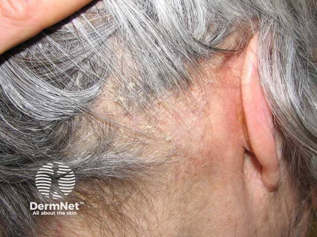
Clinical photography of the scalp demonstrating seborrhoeic dermatitis with thick adherent scales. The image presents a macroscopic view of the left temporal scalp and retroauricular region. Specimen type: human scalp skin. Imaging perspective: close-up, lateral scalp with ear visible. The scales are thick, greasy, yellowish, lamellate, coating the scalp surface and obscuring underlying hair shafts. Surrounding skin shows mild erythema and minimal crusting; hair density is preserved; no ulceration. This pattern is characteristic of seborrhoeic dermatitis: chronic relapsing inflammatory dermatosis associated with Malassezia yeast, sebaceous gland activity, and host inflammatory response. The lesion displays confluent plaques with adherent scale, diffuse but more prominent in chronically affected areas such as the temporal and retroauricular scalp. Diagnostic significance: photographic documentation aids clinical diagnosis, guides topical antifungal or anti-seborrheic therapy, and serves as baseline for response assessment. Differential considerations include psoriasis capitis (more silvery scale and well-demarcated plaques), tinea capitis (alopecia, hair shedding, broken hairs), and contact dermatitis. Clinical correlation includes pruritus, oiliness, and seasonal flares. This image is useful for education, dermatology training, and image-based search queries for seborrheic dermatitis of the scalp. Images like this support differential diagnosis, treatment planning, and educational demonstrations for medical students, residents, and clinicians in clinics.
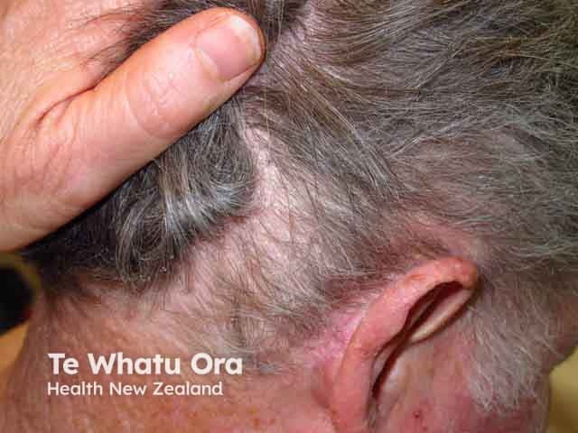
Clinical photography of the left retroauricular skin region demonstrates classic features of seborrhoeic dermatitis. The image shows erythematous, plaque-like patches with greasy, yellow-white scaling along the postauricular sulcus and adjacent scalp margins. The surrounding skin is mildly inflamed, with fine desquamation and mild edema; hair-bearing skin in the temporal area appears unaffected or variably involved. No secondary bacterial infection or crusting is evident. The appearance is symmetrical with typical sebaceous-rich distribution, reflecting fungal lipid degradation products and inflammatory response to Malassezia species. The lesion margins are relatively well defined, and the coloration ranges from pink to salmon with golden-scale overtones. Pruritus may be present but is not clearly depicted in the still image. This presentation can extend to the behind-ear fold, scalp margins, and forehead in a seborrheic distribution. Clinically, this condition is benign but relapsing, often coexisting with scalp dermatitis or dandruff. Differential diagnoses include tinea (dermatophyte) infections, psoriasis in seborrheic areas, contact dermatitis, and less likely atopic dermatitis. Management typically involves topical antifungals (ketoconazole, ciclopirox), anti-inflammatory agents (low-potency corticosteroids or calcineurin inhibitors), and regular hygiene to reduce sebaceous load. Documentation of this retroauricular dermatitis supports diagnosis and guides targeted therapy, monitoring response and relapse potential in dermatology practice.
Comprehensive Description: This is a high-resolution clinical photograph of the scalp taken in vivo, using standard dermatology photography techniques, showing a lateral (side) view of the hair-bearing skin. The image demonstrates extensive seborrhoeic dermatitis with overlapping greasy scales adherent to erythematous plaques over the scalp, most prominently along the temporal and vertex regions. The scales appear yellowish-white, oily in appearance, and coalesce to form layered flakes that extend into the hair margins. Mild diffuse erythema and scaling are present without visible purpura or crusting. Hair remains present, with no obvious scarring alopecia. The morphology is consistent with inflammatory dermatosis of the scalp; differential diagnoses include psoriasis capitis, tinea capitis, and atopic dermatitis, though the greasy character with fine lamellar scaling favors seborrheic dermatitis. Clinically, this image is relevant for dermatology education, trichology assessment, and differential diagnosis practice, as well as for evaluating treatment response to antifungal shampoos (ketoconazole, head-and-shoulders zinc pyrithione), corticosteroid, or calcineurin inhibitor regimens. It can be used in teaching modules on scalp dermatoses, in digital archives for image-guided diagnosis, and for research into Malassezia-associated inflammation, inflammatory skin disorders, and patient counseling on scalp hygiene. This descriptor supports retrieval in education, clinical case review, and dermatology datasets globally.
scalp psoriasis silvery scales well demarcated plaques
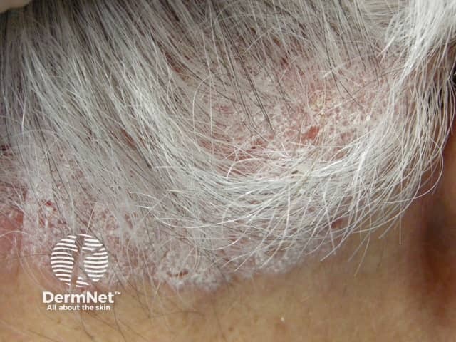
A high-resolution clinical photograph of the scalp shows classic plaque psoriasis with prominent silvery scales and erythematous, well‑demarcated plaques involving the hair-bearing scalp. The image demonstrates diffuse to patchy involvement, concentrated along the frontal and parietal regions with extension toward the occipital area and hairline. Adherent scale covers the plaques, producing a flaky, powdery surface in places and occasional fissuring at the scalp margins. Surrounding skin shows mild erythema without overt pustules, secondary infection, or exudate in this view. The lesion morphology is characteristic of plaque-type psoriasis (psoriasis vulgaris) rather than seborrheic dermatitis or tinea capitis, which may show more greasy scales or alopecia, respectively. This image is valuable for clinical education and research on psoriasis comorbidity with smoking: tobacco use is associated with increased disease severity, earlier onset, and poorer therapeutic response; smoking cessation can improve disease control. Therapeutic implications include topical regimens (shampoos with salicylic acid, coal tar, or corticosteroids) and systemic options for extensive disease in smokers. The photograph is suitable for dermatology training, patient counseling, and studies on lifestyle factors influencing psoriasis expression and management. It also illustrates differential considerations with seborrheic dermatitis, lichen simplex chronicus, and fungal infections in the scalp. Useful for exam questions.
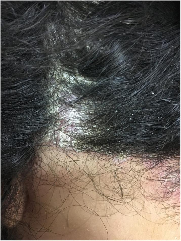
This clinical photograph shows a close-up view of the human scalp and posterior hairline, demonstrating well-demarcated erythematous plaques characteristic of scalp psoriasis. The lesions are covered with thick, silvery-white micaceous scales that adhere to both the underlying inflamed skin and the proximal hair shafts. The distribution is patchy, with prominent plaques extending beyond the frontal and temporal hair-bearing areas onto the adjacent non-terminal hair skin. The surrounding dark hair remains intact but contains scattered fine, flaky white debris (secondary scaling). The visible borders of the primary plaque are distinct and irregular. This image is an educational example of inflammatory dermatoses of the scalp, specifically illustrating the classic morphology and scaling pattern of psoriasis and pseudotinea amiantacea.
Seborrhoeic Dermatitis vs Psoriasis
Note: These two can overlap in some patients - the term "Sebopsoriasis" (or "seborrhiasis") is used when features of both coexist simultaneously.
Side-by-Side Comparison
| Feature | Seborrhoeic Dermatitis | Psoriasis |
|---|---|---|
| Scale colour | Yellowish, greasy, oily ("bran-like") | Silvery-white, dry, micaceous (peel in layers) |
| Scale texture | Soft, easily rubbed off, greasy | Thick, lamellar, adherent centrally, looser at periphery |
| Plaque borders | Poorly defined, blends into normal skin | Well-demarcated, sharp borders |
| Plaque thickness | Thin-moderate | Thick ("heaped up") |
| Auspitz sign | Absent | Present - pinpoint bleeding on scale removal (due to dilated dermal capillaries) |
| Koebner phenomenon | Absent | Present - psoriasis appears at sites of trauma/injury |
| Itch | Usually prominent | Mild to moderate (less itchy than SD) |
| Scalp appearance | Diffuse, poorly defined scaling; greasy hair | Discrete thick plaques; often extends beyond the hairline onto forehead skin |
| Nasolabial folds | Classically involved (hallmark site) | Spared or minimally involved |
| Extensor surfaces | Not involved | Elbows, knees - classic psoriasis sites |
| Nails | Normal | Nail pitting, onycholysis, oil drop sign |
| Joints | No joint involvement | Psoriatic arthritis in ~30% |
| Distribution | Sebaceous areas (scalp, face, chest, flexures) | Extensor surfaces, scalp, sacrum, umbilicus, nails |
| Pathogenesis | Malassezia yeast + sebum overproduction | Autoimmune T-cell mediated; IL-17/IL-23 axis; keratinocyte hyperproliferation |
| Family history | Not typically genetic | Strong genetic predisposition (HLA-Cw6) |
| Triggers | Stress, heat, fatigue, immunosuppression | Streptococcal infection, medications (beta-blockers, lithium, NSAIDs), stress, trauma |
| Histology | Mild spongiosis, superficial perivascular infiltrate, Malassezia organisms | Acanthosis, parakeratosis, munro microabscesses, elongated rete ridges, dilated capillaries |
| Response to antifungals | Excellent (ketoconazole is first-line) | Poor (antifungals do not help) |
The Key Clinical Tricks
- SD: yellow, greasy - like butter smeared on skin
- Psoriasis: silver-white, dry - like wax or mica flakes
- Remove a psoriasis scale and you see tiny bleeding points (dilated dermal papillary capillaries exposed)
- Absent in SD
- Psoriasis plaques cross the hairline onto the forehead - the "corona psoriatica"
- SD stays near the hairline, more diffuse
- Find elbows/knee plaques → psoriasis
- Find nasolabial/chest involvement with greasy scale → SD
- Find nail pitting → psoriasis
- Severe itch favours SD
- Psoriasis is often surprisingly non-itchy despite dramatic appearance
Clinical Images
Treatment Difference (Summary)
| SD | Psoriasis | |
|---|---|---|
| 1st line topical | Antifungals (ketoconazole, ciclopirox) | Topical corticosteroids + Vitamin D analogues (calcipotriol) |
| Shampoos | Ketoconazole, selenium sulfide, zinc pyrithione | Coal tar, salicylic acid (keratolytic), corticosteroid |
| Systemic | Oral fluconazole (severe only) | Methotrexate, cyclosporine, biologics (anti-TNF, anti-IL17, anti-IL23) |
| Biologics | Not used | Mainstay for moderate-severe disease |