
Explain me bornes of spinal cord
Reading File
The spinal cord extends from the foramen magnum (lower border of the medulla oblongata) to approximately L1-L2 in adults (conus medullaris), with a variable lower end. Let me pull the full details from the medical library.
Searching sources...
Finding Sources
Finding Sources
Reading File
Reading File
Reading File
Loading Image
Here is a detailed explanation of the bornes (boundaries/extent) of the spinal cord, based on Gray's Anatomy for Students and Bradley and Daroff's Neurology in Clinical Practice:
Boundaries (Bornes) of the Spinal Cord
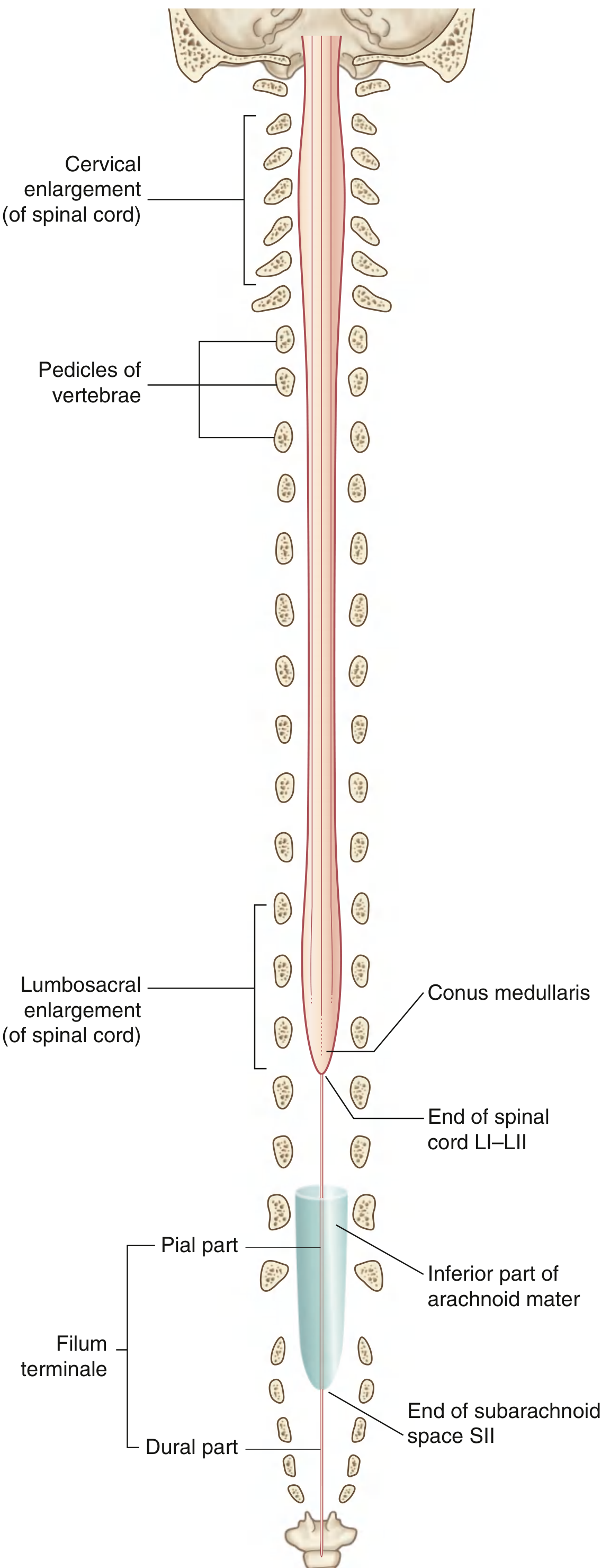
Upper Bound (Superior Limit)
The spinal cord begins at the foramen magnum, where it is continuous with the medulla oblongata (the lower part of the brainstem). This is the craniovertebral junction. The cord occupies the superior two-thirds of the vertebral canal.
Lower Bound (Inferior Limit) - The Conus Medullaris
The spinal cord tapers inferiorly into a cone-shaped tip called the conus medullaris. Its exact level varies:
| Population | Normal Range | Typical Level |
|---|---|---|
| Adults | T12 - L2/L3 disc | L1-L2 disc |
| Neonates | Up to L4 | L3 |
- In adults, it most commonly ends at the level of the disc between L1 and L2, but can range from as high as T12 to as low as the L2-L3 disc.
- In neonates, the cord extends further down to around L3 (and sometimes L4), because the vertebral column grows faster than the spinal cord after birth.
Gray's Anatomy for Students - p. 119
What Continues Below the Conus?
Once the spinal cord ends, two structures continue downward:
-
Filum terminale - A thin filament of connective tissue that extends from the apex of the conus medullaris. It has two parts:
- Pial part (filum terminale internum): extends from the conus through the subarachnoid space to S2.
- Dural part (filum terminale externum / coccygeal ligament): continues from S2 to the coccyx, anchoring the cord.
-
Cauda equina - The bundle of lumbar and sacral nerve roots that descend below the conus (since the spinal cord ends at L1-L2, but the spinal column extends to S5, the nerve roots travel a long distance within the subarachnoid space before exiting through their respective foramina).
The Subarachnoid Space
The subarachnoid space (which contains CSF) ends at approximately S2. This is clinically important - lumbar puncture is performed below L2 (usually at L3-L4 or L4-L5) to avoid injuring the spinal cord while still being within the subarachnoid space.
Length and Diameter
- The spinal cord is not uniform in diameter. It has two fusiform swellings called enlargements:
- Cervical enlargement - at C5 to T1 level (for innervation of upper limbs)
- Lumbosacral enlargement - at L1 to S3 level (for innervation of lower limbs)
Clinical Relevance
- Lumbar puncture is performed at L3-L4 or L4-L5 to avoid puncturing the cord.
- Conus medullaris syndrome (injury at T12-L2): causes a mixed upper + lower motor neuron picture, saddle anesthesia, and bladder/bowel dysfunction.
- Cauda equina syndrome (injury below conus): pure lower motor neuron syndrome with flaccid weakness, areflexia, and variable sensory loss. (Bradley and Daroff's Neurology, p. 485)
Summary Table:
| Landmark | Level |
|---|---|
| Upper border | Foramen magnum (= medulla oblongata junction) |
| Lower border (adult) | L1-L2 disc (range: T12 to L2-L3) |
| Lower border (neonate) | L3 (range: up to L4) |
| End of subarachnoid space | S2 |
| End of filum terminale | Coccyx |
This is a shared conversation. Sign in to Orris to start your own chat.