

Make a introduction about this topic
neoplasms AND metastasis
National Cancer Institute definition neoplasia benign malignant carcinoma in situ metastasis
Introduction: Neoplasia and Tumour Metastasis
Explain indetail about neoplasia
neoplasms AND pathology
site:cancer.gov cancer biology cancer begins cells grow out of control definition neoplasia
-
Basic definitions and terminology
- Neoplasia, neoplasm, tumour, cancer
- Differences from hyperplasia, hypertrophy, metaplasia, and dysplasia
- Nomenclature of benign and malignant tumours
-
Benign versus malignant tumours
- Differentiation and anaplasia
- Rate of growth
- Local invasion
- Metastasis
- Effects on the patient
-
Cancer cell biology
- Hallmarks of cancer
- Sustained proliferation and avoidance of growth inhibition
- Evasion of apoptosis and immune destruction
- Angiogenesis, invasion, and metastatic capability
-
Molecular basis of cancer
- Oncogenes and proto-oncogenes
- Tumour-suppressor genes such as TP53 and RB
- DNA-repair genes
- Multistep carcinogenesis and clonal evolution
-
Precancerous changes
- Dysplasia
- Carcinoma in situ
- Progression to invasive carcinoma
-
Tumour progression and metastasis
- Local invasion
- Lymphatic, haematogenous, and transcoelomic spread
- Tumour-host interactions and angiogenesis
-
Clinical effects, grading, staging, and diagnosis
- Local and systemic effects
- Cancer cachexia and paraneoplastic syndromes
- Tumour grade and TNM stage
- Principles of pathological diagnosis
More about Neoplasia
neoplasia pathology
site:cancer.gov benign tumors malignant tumors cancer definition invasion metastasis
Neoplasia
- Benign - non-cancerous and usually localised.
- Malignant - cancerous, capable of invading surrounding tissues and spreading to distant organs.
1. Normal cell growth versus neoplastic growth
- Cell proliferation
- Differentiation into specialised cells
- Cellular ageing
- Programmed cell death, called apoptosis
2. Difference between neoplasia and other adaptive changes
| Condition | Meaning | Reversibility |
|---|---|---|
| Hyperplasia | Increase in the number of normal cells in response to a stimulus | Usually reversible |
| Hypertrophy | Increase in cell size | Usually reversible |
| Metaplasia | Replacement of one mature cell type by another mature cell type | Usually reversible |
| Dysplasia | Disordered growth and maturation of cells, often in epithelium | May be reversible, but can progress |
| Neoplasia | Autonomous, clonal new growth caused by genetic alterations | Generally irreversible |
3. Classification of neoplasms
A. Benign neoplasms
- Lipoma - benign tumour of fat tissue
- Fibroma - benign tumour of fibrous tissue
- Adenoma - benign tumour of glandular epithelium
- Papilloma - benign epithelial tumour producing finger-like projections
- Leiomyoma - benign tumour of smooth muscle, for example uterine fibroid
B. Malignant neoplasms
- Carcinoma - malignant tumour arising from epithelial cells
Examples: squamous cell carcinoma, adenocarcinoma. - Sarcoma - malignant tumour arising from connective or mesenchymal tissues
Examples: osteosarcoma, fibrosarcoma, liposarcoma. - Leukaemia - malignant tumour of blood-forming cells, usually in bone marrow.
- Lymphoma - malignant tumour of lymphoid tissue.
- Melanoma - malignant tumour of melanocytes.
4. Benign versus malignant neoplasms
- Differentiation and anaplasia
- Local invasion
- Metastasis
| Feature | Benign tumour | Malignant tumour |
|---|---|---|
| Differentiation | Well differentiated, resembles tissue of origin | Often poorly differentiated or anaplastic |
| Growth rate | Usually slow | Variable, often faster |
| Margins | Well circumscribed, sometimes encapsulated | Irregular, poorly circumscribed |
| Local invasion | Does not invade adjacent tissue | Invades and destroys adjacent tissues |
| Metastasis | Absent | May be present |
| Recurrence after removal | Uncommon if fully removed | More likely |
5. Differentiation and anaplasia
- A well-differentiated tumour closely resembles the parent tissue.
- A poorly differentiated tumour shows less resemblance.
- Anaplasia means lack of differentiation and is a characteristic feature of many malignant tumours.
Microscopic features of anaplasia
- Pleomorphism - variation in size and shape of cells and nuclei
- Hyperchromatic nuclei - darkly stained nuclei due to excess DNA
- High nucleus-to-cytoplasm ratio
- Large, irregular nucleoli
- Numerous or abnormal mitoses
- Loss of normal tissue architecture
- Tumour giant cells
6. Dysplasia and carcinoma in situ
- Loss of uniformity of cells
- Cellular pleomorphism
- Enlarged, hyperchromatic nuclei
- Increased mitotic activity
- Disturbed arrangement and maturation of cells
- Cervical carcinoma in situ
- Ductal carcinoma in situ of the breast
- Squamous cell carcinoma in situ of skin
7. Molecular basis of neoplasia
A. Proto-oncogenes and oncogenes
- RAS
- MYC
- ERBB2/HER2
- BCR-ABL
B. Tumour-suppressor genes
- TP53 - responds to DNA damage by arresting the cell cycle or causing apoptosis
- RB - regulates progression from G1 to S phase of the cell cycle
- APC - important in colorectal tumour development
- BRCA1 and BRCA2 - involved in DNA repair
C. DNA-repair genes
- BRCA1/BRCA2
- Mismatch-repair genes such as MLH1 and MSH2
8. Hallmarks of cancer
- Sustained proliferative signalling
- Resistance to growth-inhibitory signals
- Evasion of apoptosis
- Limitless replicative capacity
- Induction of angiogenesis
- Invasion and metastasis
- Reprogramming of energy metabolism
- Evasion of immune destruction
- Genomic instability
- Tumour-promoting inflammation
9. Tumour growth and angiogenesis
10. Tumour invasion and metastasis
- Lymphatic spread - common in carcinomas
Example: breast carcinoma spreading to axillary lymph nodes. - Haematogenous spread - common in sarcomas, though carcinomas can also spread this way
Common sites: liver, lungs, bone, and brain. - Transcoelomic seeding - spread across body cavities
Example: ovarian carcinoma spreading throughout the peritoneal cavity.
11. Causes and risk factors for neoplasia
- Tobacco use
- Alcohol
- Ultraviolet and ionising radiation
- Obesity and dietary factors
- Chronic inflammation
- Occupational carcinogens, such as asbestos and benzene
- Oncogenic infections:
- Human papillomavirus - cervical and anogenital cancers
- Hepatitis B and C viruses - hepatocellular carcinoma
- Epstein-Barr virus - some lymphomas and nasopharyngeal carcinoma
- Helicobacter pylori - gastric adenocarcinoma and lymphoma
- Inherited mutations, for example BRCA mutations or familial adenomatous polyposis
Key points
- Neoplasia is autonomous, clonal, abnormal cellular proliferation.
- A neoplasm may be benign or malignant.
- Malignancy is identified mainly by anaplasia, local invasion, and metastasis.
- Carcinoma in situ is a full-thickness epithelial abnormality without basement membrane invasion.
- Cancer results from accumulated genetic alterations affecting oncogenes, tumour-suppressor genes, and DNA-repair genes.
- Metastasis is the major cause of morbidity and death in many cancers.
Explain nomenclature
neoplasms nomenclature pathology
site:cancer.gov cancer terms carcinoma sarcoma adenoma papilloma nomenclature
Nomenclature of Neoplasms
- The cell or tissue of origin
- Whether the tumour is benign or malignant
- Sometimes, its microscopic pattern or gross appearance
1. Naming benign tumours
A. Benign tumours of mesenchymal origin
Name of tissue/cell + “oma” = benign tumour
| Tissue of origin | Benign tumour | Meaning |
|---|---|---|
| Fibrous tissue | Fibroma | Benign tumour of fibroblasts |
| Fat | Lipoma | Benign tumour of adipose tissue |
| Cartilage | Chondroma | Benign tumour of cartilage |
| Bone | Osteoma | Benign tumour of bone |
| Smooth muscle | Leiomyoma | Benign smooth-muscle tumour |
| Skeletal muscle | Rhabdomyoma | Benign skeletal-muscle tumour |
| Blood vessels | Hemangioma | Benign vascular tumour |
| Lymphatic vessels | Lymphangioma | Benign lymphatic-vessel tumour |
- A lipoma is a benign mass made of mature fat cells.
- A uterine leiomyoma, also called a fibroid, is a benign tumour of smooth muscle in the uterus.
B. Benign epithelial tumours
1. Adenoma
- Thyroid adenoma
- Adrenal adenoma
- Colonic adenoma
- Pituitary adenoma
2. Papilloma
- Squamous papilloma of skin or oral cavity
- Urothelial papilloma of the urinary bladder
3. Cystadenoma
- Ovarian serous cystadenoma
- Ovarian mucinous cystadenoma
4. Polyp
- Non-neoplastic, for example an inflammatory nasal polyp
- Benign neoplastic, for example an adenomatous colonic polyp
- Malignant, in some cases
2. Naming malignant tumours
A. Carcinoma
| Type | Description | Example |
|---|---|---|
| Squamous cell carcinoma | Malignant tumour of squamous epithelium | Cancer of skin, cervix, oral cavity, oesophagus |
| Adenocarcinoma | Malignant gland-forming epithelial tumour | Cancer of colon, prostate, breast, stomach |
| Urothelial carcinoma | Malignant tumour of transitional urothelium | Urinary bladder carcinoma |
| Basal cell carcinoma | Malignant tumour of basal epidermal cells | Basal cell carcinoma of skin |
A malignant gland-forming epithelial tumour in the colon is called colonic adenocarcinoma.
B. Sarcoma
Name of tissue/cell + “sarcoma” = malignant tumour
| Tissue of origin | Malignant tumour |
|---|---|
| Fibrous tissue | Fibrosarcoma |
| Fat | Liposarcoma |
| Cartilage | Chondrosarcoma |
| Bone | Osteosarcoma |
| Smooth muscle | Leiomyosarcoma |
| Skeletal muscle | Rhabdomyosarcoma |
| Blood vessels | Angiosarcoma |
An osteosarcoma is a malignant tumour that produces bone or osteoid.
3. Tumours with special names
A. Malignant tumours ending in “-oma”
| Tumour | Nature |
|---|---|
| Melanoma | Malignant tumour of melanocytes |
| Lymphoma | Malignant tumour of lymphoid cells |
| Leukaemia | Malignant neoplasm of haematopoietic cells, usually involving marrow and blood |
| Mesothelioma | Usually malignant tumour of mesothelial cells |
| Hepatoma | Traditional term for hepatocellular carcinoma, a malignant liver tumour |
| Seminoma | Malignant germ-cell tumour of testis |
| Glioma | A tumour of glial cells, may be benign or malignant depending on type and grade |
B. Tumours named according to their cell type
| Tumour | Cell/tissue of origin |
|---|---|
| Melanoma | Melanocytes |
| Lymphoma | Lymphocytes and lymphoid cells |
| Leukaemia | Bone marrow blood-forming cells |
| Mesothelioma | Mesothelial lining of pleura, peritoneum, or pericardium |
| Germ-cell tumour | Germ cells of gonads or extragonadal sites |
| Neuroblastoma | Primitive sympathetic nervous-system cells |
| Retinoblastoma | Primitive retinal cells |
| Wilms tumour (nephroblastoma) | Primitive kidney tissue in children |
4. Mixed tumours
Pleomorphic adenoma
- It contains epithelial and myoepithelial tumour cells.
- The tumour cells may produce a myxoid, cartilaginous, or fibrous-looking stroma.
- Despite its mixed appearance, it usually arises from a single clone of cells.
Carcinosarcoma
- Carcinomatous component: malignant epithelial tissue
- Sarcomatous component: malignant mesenchymal-looking tissue
5. Hamartoma and choristoma
Hamartoma
Choristoma
6. Important terminology
| Term | Meaning |
|---|---|
| Neoplasm | Autonomous abnormal new growth of cells |
| Tumour | A mass caused by abnormal cell proliferation; often used synonymously with neoplasm |
| Benign | Localised tumour that does not invade or metastasise |
| Malignant | Tumour capable of invasion and metastasis |
| Carcinoma | Malignant epithelial tumour |
| Sarcoma | Malignant mesenchymal tumour |
| Adenoma | Benign epithelial tumour of glandular origin or forming glands |
| Papilloma | Benign epithelial tumour with finger-like projections |
| Polyp | Mucosal projection, which may or may not be neoplastic |
| Carcinoma in situ | Full-thickness epithelial malignancy without invasion through the basement membrane |
Easy summary
-
Benign mesenchymal tumour: tissue name + oma
Example: fat → lipoma -
Malignant mesenchymal tumour: tissue name + sarcoma
Example: fat → liposarcoma -
Malignant epithelial tumour: carcinoma
Example: glandular epithelium → adenocarcinoma -
Do not assume that all tumours ending in “-oma” are benign.
Examples of malignant “-oma” tumours include melanoma, lymphoma, mesothelioma, and seminoma.
Explain normal or cancer cells
cancer cells normal cells
site:cancer.gov differences between cancer cells and normal cells
Normal Cells and Cancer Cells
1. Normal cells
A. Controlled cell division
- During growth and development
- To replace worn-out cells
- During wound healing
- In response to appropriate growth factors or hormones
B. Differentiation
- Red blood cells transport oxygen.
- Neurons conduct electrical impulses.
- Hepatocytes carry out metabolic and detoxification functions.
- Muscle cells contract.
- Glandular epithelial cells produce secretions.
C. Response to growth-regulating signals
- Divide
- Stop dividing
- Differentiate
- Repair DNA
- Undergo programmed cell death
D. Contact inhibition and anchorage dependence
E. Limited life span
F. Orderly cell death
2. How normal cells become cancer cells
| Gene group | Normal role | Change in cancer |
|---|---|---|
| Proto-oncogenes | Promote normal controlled cell growth | Become oncogenes that drive excessive growth |
| Tumour-suppressor genes | Inhibit cell division, repair damage, or induce apoptosis | Are lost or inactivated |
| DNA-repair genes | Correct DNA errors | Become defective, causing mutations to accumulate |
| Apoptosis-regulating genes | Remove damaged cells | Allow abnormal cells to survive |
- RAS mutations can continuously stimulate cell proliferation.
- Loss of TP53 prevents proper DNA-damage response and apoptosis.
- Loss of RB removes an important cell-cycle brake.
- BRCA1/BRCA2 defects impair DNA repair.
3. Characteristics of cancer cells
A. Uncontrolled proliferation
Cancer cells: divide continuously or excessively.
B. Resistance to growth inhibition
C. Evasion of apoptosis
- Losing TP53 function
- Increasing anti-apoptotic proteins such as BCL-2
- Decreasing pro-apoptotic signals
D. Replicative immortality
E. Reduced differentiation and anaplasia
- Pleomorphism: variation in cell and nuclear size and shape
- Enlarged, dark-staining nuclei: hyperchromasia
- High nuclear-to-cytoplasmic ratio
- Prominent nucleoli
- Numerous or abnormal mitotic figures
- Loss of normal tissue architecture
F. Altered metabolism
G. Angiogenesis
H. Invasion and metastasis
- Detach from neighbouring cells
- Break down surrounding extracellular matrix and basement membrane
- Invade nearby tissues
- Enter lymphatic vessels or blood vessels
- Travel to distant organs
- Form secondary tumours, called metastases
I. Immune evasion
- Reducing tumour-antigen visibility
- Creating an immunosuppressive microenvironment
- Expressing proteins such as PD-L1 that inhibit T-cell activity
4. Normal cells versus cancer cells
| Feature | Normal cells | Cancer cells |
|---|---|---|
| Growth | Controlled and regulated | Excessive and relatively autonomous |
| Response to growth signals | Divide only when stimulated appropriately | May grow without external stimulation |
| Response to stop signals | Stop dividing when required | Often ignore growth-inhibitory signals |
| Apoptosis | Die when damaged or no longer needed | Resist apoptosis |
| Differentiation | Well differentiated and specialised | May be poorly differentiated or anaplastic |
| Shape and nucleus | Uniform size and shape | Pleomorphism, hyperchromasia, abnormal nuclei |
| Contact inhibition | Present | Often lost |
| Anchorage dependence | Usually required | May be lost |
| Replicative capacity | Limited | Often unlimited due to telomerase activation |
| Metabolism | Balanced energy production | Increased glucose uptake and altered metabolism |
| Angiogenesis | Occurs only when physiologically needed | Stimulates new vessel formation |
| Invasion | Remain within normal boundaries | May invade neighbouring tissues |
| Metastasis | Never metastasise | May spread through lymph, blood, or body cavities |
| Genetic stability | Usually maintained by repair mechanisms | Often genetically unstable |
5. Progression from normal cell to cancer
- Hyperplasia: increased number of cells, but cells look normal.
- Dysplasia: disordered growth with abnormal cell appearance and arrangement.
- Carcinoma in situ: severe dysplasia involving the full epithelial thickness, but the basement membrane is intact.
- Invasive carcinoma: tumour cells penetrate the basement membrane and invade surrounding tissue.
- Metastatic carcinoma: cells establish secondary deposits at distant sites.
6. Hallmarks of cancer cells
- Sustained proliferative signalling
- Evasion of growth suppressors
- Resistance to cell death
- Replicative immortality
- Induction of angiogenesis
- Invasion and metastasis
- Reprogrammed cellular metabolism
- Avoidance of immune destruction
- Genome instability and mutation
- Tumour-promoting inflammation
Key takeaway
Diagram of normal and abnormal cells
educational diagram comparing normal cells with cancer cells showing uncontrolled cell growth
NCI diagram normal cells versus cancer cells uncontrolled growth
Educational medical diagram comparing normal cells and cancer cells side by side. Left panel: neatly arranged, uniform epithelial cells with small regular nuclei, intact basement membrane, labels: controlled division, normal differentiation, apoptosis of damaged cells. Right panel: crowded disorganized pleomorphic cancer cells with enlarged dark irregular nuclei, many mitoses, basement membrane breached with cells invading connective tissue, labels: uncontrolled growth, loss of differentiation, invasion. Include a clear title: Normal Cells vs Cancer Cells.
Diagram: Normal Cells vs Cancer Cells
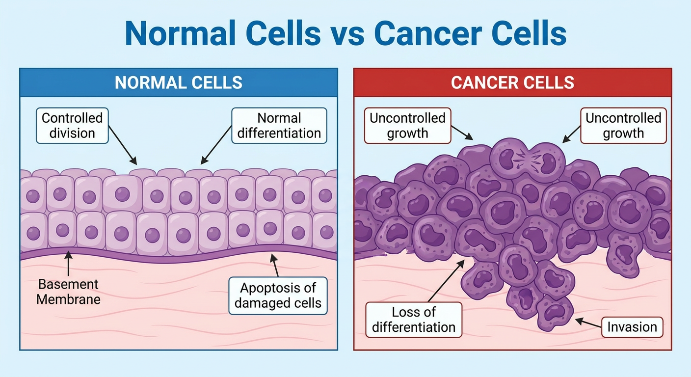
- Normal cells: uniform in size and shape, arranged in an orderly pattern, have regular nuclei, divide only when needed, and remain above the basement membrane.
- Cancer cells: variable in size and shape, crowded and disorganised, possess enlarged dark irregular nuclei, divide excessively, and can breach the basement membrane to invade surrounding tissue.
Explain benign and malignant tumours
neoplasms pathology
site:cancer.gov benign tumors malignant tumors invade metastasis
educational pathology diagram comparison benign tumor malignant tumor invasion metastasis
Benign and Malignant Tumours
- Benign tumours are non-cancerous. They remain localised and do not invade or metastasise.
- Malignant tumours are cancers. They invade surrounding tissues and can spread to distant sites, forming secondary tumours called metastases.
1. Benign tumours
Features
- Cells are usually well differentiated and resemble the normal cells from which they developed.
- Growth is generally slow, though exceptions occur.
- The tumour is often well-circumscribed and may be enclosed by a fibrous capsule.
- It expands by pushing on adjacent tissue rather than infiltrating it.
- It does not invade surrounding tissue.
- It never metastasises.
- Complete surgical removal is usually curative.
Examples
| Tissue | Benign tumour |
|---|---|
| Fat | Lipoma |
| Fibrous tissue | Fibroma |
| Cartilage | Chondroma |
| Smooth muscle | Leiomyoma |
| Glandular epithelium | Adenoma |
| Blood vessels | Hemangioma |
Can benign tumours be dangerous?
- Compression: a benign brain tumour can compress vital brain centres.
- Obstruction: a colonic polyp or bronchial adenoma may block a passage.
- Hormone production: a pituitary adenoma may produce excess hormones.
- Bleeding, ulceration, or rupture: depending on its site and size.
2. Malignant tumours
Features
- Cells can be well differentiated, poorly differentiated, or undifferentiated.
- Growth may be rapid, but growth rate alone does not prove malignancy.
- The mass is often irregular and poorly circumscribed.
- It infiltrates and destroys nearby normal tissue.
- It can recur after incomplete removal.
- It may spread through lymphatics, blood vessels, or body cavities.
- It can cause severe local damage, systemic illness, and death.
Examples
| Tissue of origin | Malignant tumour |
|---|---|
| Squamous epithelium | Squamous cell carcinoma |
| Glandular epithelium | Adenocarcinoma |
| Bone | Osteosarcoma |
| Cartilage | Chondrosarcoma |
| Fat | Liposarcoma |
| Smooth muscle | Leiomyosarcoma |
| Blood-forming cells | Leukaemia |
| Lymphoid tissue | Lymphoma |
3. Major differences
| Feature | Benign tumour | Malignant tumour |
|---|---|---|
| Nature | Non-cancerous | Cancerous |
| Differentiation | Usually well differentiated | May be poorly differentiated or anaplastic |
| Cell appearance | Uniform, similar to normal cells | Pleomorphism, enlarged dark nuclei, abnormal mitoses may occur |
| Rate of growth | Usually slow | Variable, often faster |
| Margins | Well circumscribed, often encapsulated | Ill-defined and infiltrative |
| Local invasion | Absent | Present |
| Metastasis | Never occurs | May occur |
| Recurrence | Uncommon after complete removal | More common, especially if incompletely removed |
| Effects on patient | Usually local pressure or hormone effects | Local destruction, metastasis, cachexia, paraneoplastic effects |
4. Differentiation and anaplasia
- Benign tumours are generally well differentiated.
- Example: a lipoma contains cells closely resembling normal fat cells.
- Malignant tumours may be well differentiated, but aggressive cancers are often poorly differentiated.
Features of anaplasia
- Pleomorphism: variation in cellular and nuclear size and shape
- Hyperchromasia: dark-staining nuclei due to increased DNA
- High nuclear-to-cytoplasmic ratio
- Large, prominent nucleoli
- Numerous or atypical mitotic figures
- Loss of normal tissue architecture
- Tumour giant cells
5. Growth pattern and local invasion
Benign tumour growth
Malignant tumour growth
- Damages normal organs
- Causes pain, bleeding, obstruction, and organ failure
- Makes complete surgical removal difficult
- Provides access to lymphatic and blood vessels
6. Metastasis
Main pathways
-
Lymphatic spread
- More common in carcinomas.
- Example: breast carcinoma spreading to axillary lymph nodes.
-
Haematogenous spread
- Common in sarcomas, but also occurs in carcinomas.
- Tumour cells enter blood vessels and commonly spread to the liver, lungs, bones, or brain.
-
Transcoelomic seeding
- Tumour cells spread across a body cavity.
- Example: ovarian carcinoma spreading throughout the peritoneal cavity.
7. Why malignant tumours are life-threatening
- Destruction of vital organs
- Obstruction of airways, intestines, bile ducts, or urinary tract
- Haemorrhage
- Infection due to tissue breakdown or immunosuppression
- Metastasis to organs such as the brain, liver, lungs, or bone marrow
- Severe weight loss and muscle wasting, called cancer cachexia
- Paraneoplastic syndromes, such as inappropriate hormone production or clotting disorders
Simple memory point
Malignant = invasive, destructive, and potentially metastatic.