
Pathophysiology of tuberculosis
tuberculosis pathophysiology granuloma lung
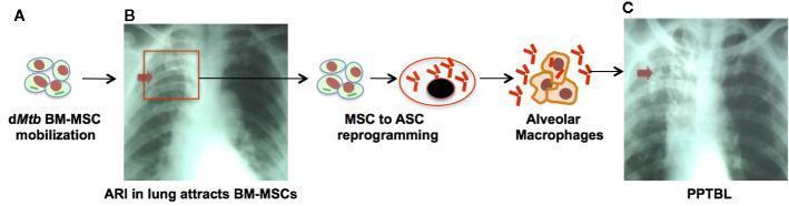
This pathophysiology diagram and clinical imaging sequence illustrate the development of Post-Primary Tuberculosis of the Lung (PPTBL) involving bone marrow-derived mesenchymal stem cells (BM-MSCs). Panel A depicts the mobilization of dormant Mycobacterium tuberculosis (dMtb)-harboring BM-MSCs, triggered by acute respiratory tract infection (ARI). Panel B features a chest X-ray of a subclinical phase, showing a focal pneumonia-like exudative opacity in the right lung apex (indicated by a red arrow). The diagram shows the subsequent reprogramming of MSCs into 'altruistic stem cells' (ASCs) which facilitate Mtb replication and infection of alveolar macrophages. Panel C presents a clinical PPTBL chest X-ray showing disease progression; the red arrow highlights more prominent, extensive opacities and cavitary lesions in the right upper lobe. The visual summarizes the transition from stem cell-mediated dMtb latency to active clinical disease characterized by host immune response, granuloma formation, and pulmonary cavitation. This material is designed for medical education focusing on immunology, pulmonology, and infectious disease mechanisms.
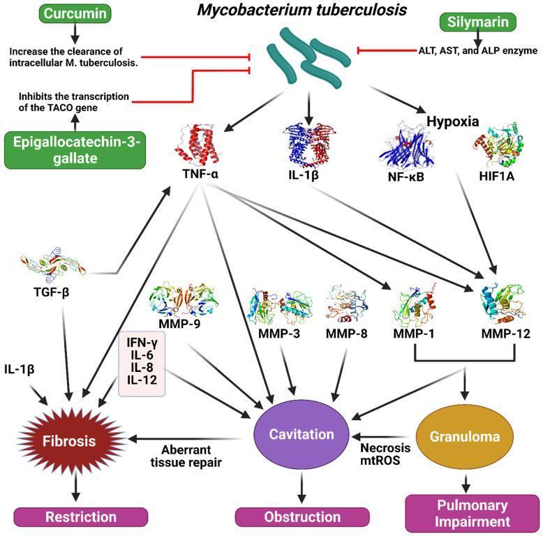
This pathophysiology diagram illustrates the signaling pathways and molecular mechanisms of Mycobacterium tuberculosis (M. tuberculosis) infection and the modulatory roles of specific polyphenols. The flowchart shows M. tuberculosis stimulating a cascade of inflammatory mediators including TNF-α, IL-1β, NF-κB, and HIF1A (hypoxia-inducible factor). These mediators further activate various Matrix Metalloproteinases (MMP-1, 3, 8, 9, 12) and TGF-β. The downstream clinical consequences are categorized into three major pathological states: Fibrosis (associated with pulmonary restriction), Cavitation (associated with obstruction), and Granuloma formation (leading to pulmonary impairment). The diagram also integrates therapeutic modulation by showing Curcumin increasing intracellular M. tuberculosis clearance, Epigallocatechin-3-gallate inhibiting TACO gene transcription, and Silymarin inhibiting liver enzymes (ALT, AST, ALP) often elevated during treatment. Secondary cytokines like IFN-γ, IL-6, IL-8, and IL-12 are shown contributing to the aberrant tissue repair and necrosis/mtROS processes that drive chronic lung damage. This diagram is designed for advanced medical education in pulmonology, infectious disease, and immunology.
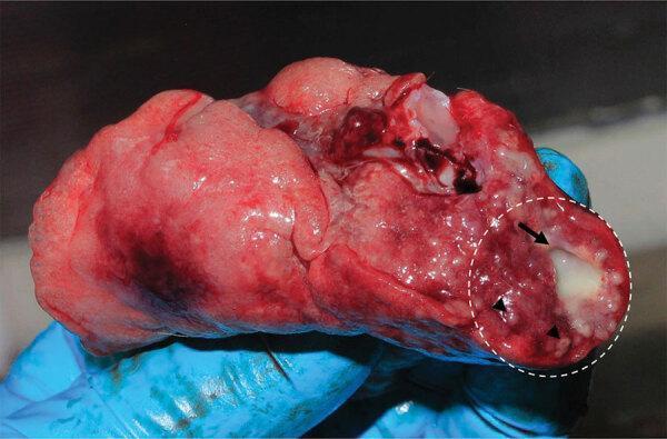
This clinical photograph displays a gross pathological specimen of lung tissue, demonstrating characteristic manifestations of a Mycobacterium tuberculosis infection. The lung parenchyma appears primarily reddish-pink with focal areas of dark red congestion and hemorrhage. A dashed white circle highlights a significant region of granulomatous inflammation. Within this region, a prominent, opaque, whitish-tan mass (indicated by a black arrow) represents a large granuloma, likely containing caseous necrosis. Surrounding this primary lesion, multiple smaller, milliary-sized white nodules (indicated by arrowheads) are scattered throughout the parenchyma, representing smaller granulomas. These visual findings are hallmark macroscopic signs of pulmonary tuberculosis, illustrating the diffuse and localized inflammatory response of the immune system to the bacilli. The specimen demonstrates the typical morphology of granulomatous disease in a primate model, used here for zoonotic research and diagnostic validation.
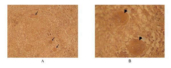
This diagnostic image consists of two light microscopy panels (A and B) showing immunohistochemical (immunoperoxidase) staining of human lung tissue from a tuberculosis (TB) patient. Panel A, at 160x magnification, displays the architectural context of a pulmonary granuloma, characterized by a central necrotic core and surrounding inflammatory cells. Arrows highlight sparsely distributed, circular regions of positive (brown) staining. Panel B provides a higher magnification (240x) view of the same tissue, revealing the detailed morphology of these positive staining areas. These are identified as multinucleated Langhans giant cells, exhibiting horseshoe-shaped arrangements of nuclei and concentrated immunoperoxidase signal within the cytoplasm. The staining utilizes a phage-displayed single-chain T-cell receptor (scTCR) specific for the Ac2SGL:CD1b complex, a lipid antigen presentation marker relevant to Mycobacterium tuberculosis infection. This image demonstrates the localization of specific lipid-antigen-presenting cells within the granulomatous microenvironment of human TB, emphasizing the clinical significance of CD1b-restricted immune responses.
tuberculosis pathogenesis granuloma immune response
tuberculosis Ghon complex primary infection chest X-ray
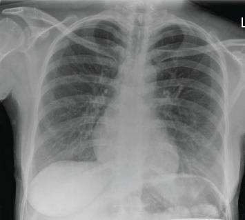
This diagnostic image is a posterior-anterior (PA) chest X-ray of an adult patient. The thoracic cage, mediastinum, and cardiac silhouette appear within normal limits for size and contour. The primary findings are localized to the left lung base, which exhibits mild peribronchial thickening and subtle linear opacities consistent with minimal scarring. There is a notable absence of definitive focal consolidation, pulmonary masses, or pleural effusions. Furthermore, no visual evidence of a Ghon complex, calcified granulomas, or hilar/mediastinal lymphadenopathy is observed, which are key negatives in the screening for tuberculosis. The costophrenic angles are sharp, and the diaphragmatic domes are well-defined. This radiograph serves as an educational example of subtle, non-specific pulmonary findings in the context of clinical infectious disease investigation.
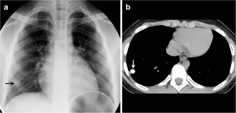
This dual-modality clinical imaging set illustrates a Ghon focus in a 7-year-old pediatric patient, characteristic of primary tuberculous infection. Panel A is an anteroposterior (AP) chest radiograph showing a subtle, well-defined, solitary nodular opacity in the peripheral right lower lung field (indicated by a black arrow). The surrounding pulmonary parenchyma, hilar structures, and cardiomediastinal silhouette appear unremarkable, without evident lymphadenopathy or pleural effusion. Panel B displays a non-contrast axial chest CT image at the level of the lower lobes, confirming the presence of a small, hyperdense, calcified pulmonary nodule in the right lower lobe (white arrow). The high attenuation value on CT indicates a healed or dormant granulomatous process. This visual finding is a classic representation of a Ghon focus, a localized area of parenchymal inflammation that has undergone caseating necrosis and subsequent calcification. This imaging is highly relevant for pediatric pulmonology and infectious disease education, demonstrating the typical radiological appearance of latent tuberculosis and the primary complex.
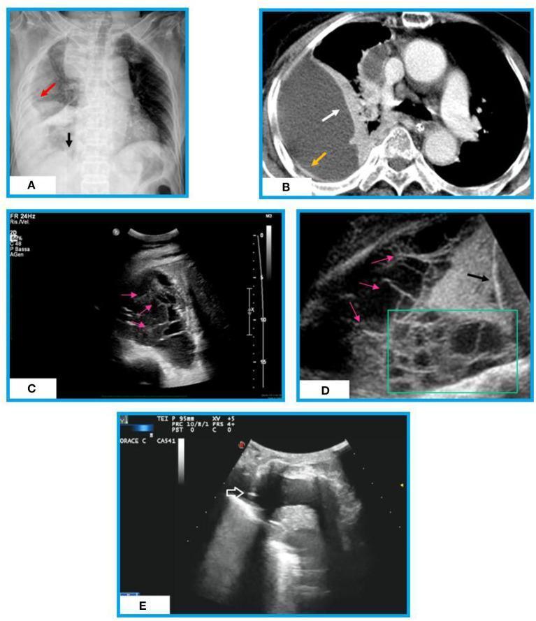
A multi-modal comparison of complex pleural effusion in a patient with post-primary tuberculosis across three imaging modalities. (A) Chest X-ray (CXR) depicts a non-gravity-dependent right pulmonary opacity with medial visualization of the diaphragm. (B) Contrast-enhanced axial CT of the chest shows a large, loculated right pleural collection with visceral pleural thickening (split-pleura sign) and adjacent compressive lung atelectasis; however, internal septations are poorly visualized. (C, D) Thoracic ultrasound (TUS) provides superior visualization of the effusion's internal architecture, showing multiple hyperechoic, linear, and mobile fibrin strands forming complex septations and loculations. (E) Real-time TUS-guided thoracentesis demonstrates a hyperechoic needle tip successfully placed within an anechoic fluid pocket. This educational series illustrates the diagnostic strengths of various modalities: CXR and CT for global assessment and pleural morphology, and TUS for identifying fibrin-rich, complex internal structures and facilitating safe procedural guidance in organized exudative effusions.
Pathophysiology of Tuberculosis
1. The Organism
2. Transmission
3. Primary Infection - Three Stages
Stage 1: Initial Macrophage Infection (Days 1-7)
- Inhaled bacilli land in the alveoli (typically the lower or middle zones, which receive the most airflow)
- Alveolar macrophages ingest the bacilli via phagocytosis
- MTB survives intracellularly by blocking phagosome-lysosome fusion, preventing acidification and enzymatic killing
- The bacilli replicate unchecked within macrophages
Stage 2: Silent Bacterial Multiplication (Days 7-21)
- Infected macrophages carry bacilli to regional hilar lymph nodes
- The patient remains asymptomatic
- Bacteria continue multiplying within macrophages
- Bacilli may enter the bloodstream (primary bacteremia) and seed distant organs (apices of lungs, kidneys, vertebrae, meninges) - these are seeds for later reactivation
Stage 3: Cell-Mediated Immunity and Granuloma Formation (Week 3 onward)
- CD4+ helper T cells (Th1 type) are activated and produce IFN-γ
- IFN-γ activates macrophages, which acquire enhanced bactericidal capacity (via nitric oxide, reactive oxygen species, and autophagy)
- Delayed-type hypersensitivity (DTH) develops - this is what makes the tuberculin skin test turn positive
- Activated macrophages die in large numbers → granuloma formation begins
4. The Granuloma - Core Pathological Lesion
| Cell Type | Role |
|---|---|
| Epithelioid macrophages | Fused, activated macrophages with abundant cytoplasm; main bacterial killers |
| Langhans giant cells | Multinucleated giant cells (fused macrophages, nuclei at periphery in horseshoe pattern); hallmark of TB |
| CD4+ T lymphocytes | Surround the macrophage core; provide IFN-γ |
| CD8+ T cells | Cytotoxic function at the periphery |
| B lymphocytes | Outer mantle; antibody production |
| Fibroblasts | Outer fibrous capsule |
- Low oxygen tension within the granuloma inhibits macrophage function and bacterial growth
- Macrophage death produces a central zone of caseous (cheese-like) necrosis - a coagulative necrosis unique to TB
- The necrotic center is relatively hostile to bacterial replication (low pH, low O₂, fatty acids toxic to MTB)
5. The Ghon Complex and Primary TB
- The Ghon focus = a small peripheral parenchymal granuloma, usually in the middle zone of the lung (where inhaled particles most commonly land)
- The Ghon complex = Ghon focus + enlarged draining hilar/mediastinal lymph nodes (from lymphatic spread)
- In most immunocompetent individuals (>90%), primary infection is controlled here. The Ghon focus heals by fibrosis and calcification, leaving a calcified nodule as the only trace of infection
- The patient is now in the latent TB phase: TST/IGRA positive, no symptoms, no active disease
6. Latent Tuberculosis
- Bacilli are not eliminated but are contained within dormant granulomas
- Bacteria can persist for decades in a metabolically inactive state
- Defined by: positive TST (tuberculin skin test) or IGRA (QuantiFERON-Gold or T-SPOT.TB) without clinical or radiographic evidence of disease
- IFN-γ remains a critical ongoing mediator maintaining granuloma integrity
- Lymph node lymphatic endothelial cells (LECs) can harbor replicating MTB, serving as a reservoir
7. Reactivation (Post-Primary) Tuberculosis
Trigger
- HIV infection (most important risk factor globally)
- Diabetes mellitus, malnutrition, alcoholism
- Immunosuppressive drugs (especially TNF-α inhibitors)
- Age-related immune decline
- Silicosis, renal failure, cancer
Mechanism of Reactivation
- Hydrolytic enzymes (metalloproteinases, including MMP-1, -3, -8, -9, -12) liquefy the caseous center
- Liquefied caseum = an ideal growth medium for MTB (rich nutrients, aerobic once cavity forms)
- Exponential bacterial replication → granuloma ruptures into bronchial tree → cavity formation
- Cavities create aerosol transmission capability → the patient becomes infectious
Why the Upper Lobes?
- Reactivation TB preferentially involves the apical and posterior segments of the upper lobes and the superior segments of the lower lobes
- Reason: highest oxygen tension in the body, favoring aerobic MTB growth; poor lymphatic drainage reduces immune surveillance
8. Tissue Destruction and Cavitation
- Edema, hemorrhage, and mononuclear cell infiltration spread
- Secondary infection with other bacteria, fungi, or yeasts can occur within cavities, accelerating tissue destruction
- Erosion of bronchial blood vessels → hemoptysis
- Cavity walls become fibrotic and thick over time
- Inner pyogenic layer (PMNs, necrotic debris)
- Middle granulomatous layer (epithelioid cells, Langhans cells)
- Outer fibrous capsule
9. Immune Evasion Mechanisms of MTB
| Mechanism | Effect |
|---|---|
| Block phagosome-lysosome fusion | Prevents acidification and enzymatic digestion |
| Inhibit reactive oxygen/nitrogen intermediates | Evades oxidative killing |
| Modulate apoptosis | Inhibits macrophage apoptosis early to preserve the intracellular niche |
| Alter antigen presentation | Downregulates MHC class II expression |
| TACO gene upregulation | Tryptophan aspartate-containing coat protein - further prevents phagosome maturation |
| Lipid-rich cell wall | Resists complement and antimicrobial peptides |
10. Dissemination Patterns
| Spread Type | Mechanism | Clinical Outcome |
|---|---|---|
| Lymphangitic | Via lymphatics to hilar nodes | Ghon complex, lymphadenopathy |
| Hematogenous | Via bloodstream | Miliary TB (uniform 1-2mm granulomas throughout lungs, liver, spleen, bone marrow) |
| Contiguous | Direct extension | Pleural TB (pleurisy), pericarditis, peritonitis |
| Endobronchial | Cavity rupture into bronchi | Laryngeal TB, tracheobronchial stenosis |
11. Inflammatory Mediators - Summary
- IFN-γ (from CD4+ T cells): macrophage activation, nitric oxide induction, autophagy - critical for containment
- TNF-α: granuloma formation and maintenance (TNF inhibitors dramatically increase reactivation risk)
- IL-12: drives Th1 differentiation from naive T cells
- IL-10: anti-inflammatory; excess IL-10 impairs granuloma function and promotes dissemination
- MMPs (1,3,8,9,12): matrix metalloproteinases drive cavity formation and tissue destruction
12. Extrapulmonary TB
- Miliary (disseminated) TB - hematogenous spread to multiple organs
- Serosal TB - pleural, pericardial, peritoneal, or meningeal involvement; TB meningitis is the most dangerous (permanent neurological damage, diagnosis difficult)
- Solid organ TB - lymph nodes (scrofula), bone (Pott's disease - vertebral TB), adrenal glands (formerly the leading cause of Addison disease), genitourinary TB
- Endobronchial TB can cause tracheobronchial stenosis in up to 90% of cases despite adequate treatment
Recent Evidence
Summary Flowchart
Inhalation of MTB droplet nuclei
        ↓
Alveolar macrophage phagocytosis
        ↓
MTB blocks phagolysosome fusion → survives intracellularly
        ↓
Bacteremia → seeding of apices, lymph nodes, other organs
        ↓
CD4+ Th1 activation → IFN-γ → macrophage activation
        ↓
Granuloma formation + caseous necrosis = PRIMARY TB (Ghon complex)
        ↓
       / \
      /   \
Controlled  Uncontrolled (immunodeficiency)
    ↓              ↓
Latent TB      Progressive primary or miliary TB
    ↓
Reactivation (waning immunity, MMP-mediated liquefaction)
    ↓
Upper lobe cavitary disease → POST-PRIMARY TB
    ↓
Infectious → transmission continues