

IMG_20260816_004422.jpg
Review
analyse_media
The image shows a clinical outpatient note. Let me read it carefully from the visual and provide a thorough review.
Clinical Note Review
Transcription
Patient: Male/Female, slip at around evening, c/o pain, twisting of Right foot and swelling over Right foot
BP: 120/80 mmHg
Date: 2/8/26 (August 2, 2026)
Dept: Ortho OPD (likely Orthopedic OPD)
C/O: Twist of Right foot
- Patient came for follow-up, "4 days, no other complaints"
O/E (On Examination):
- Swelling / fullness Right foot lateral compartment
- Tenderness over 5th metatarsal base
- ROM: Painful
- No external injury
X-Ray:
- Shows Right 5th metatarsal base - undisplaced (fracture, implied)
Advice:
- Below Knee Slab x 1 week
- Tab. PCM 500 mg 1-1-1 (TDS) x 3 days
- Tab. Serratiopeptidase 10 mg 1-0-1 (BD) x 3 days
- Tab. Pantoprazole 40 mg 1-0-1 (BD)
- Tab. Ibuprofen/Combiflam 400 mg 1-0-1 (BD)
- R/W (Review With) pin → OPD review on Thursday 6/8/26 (or next Thursday)
Clinical Review
Diagnosis
Undisplaced fracture of the base of the 5th metatarsal (Zone 1 - Avulsion fracture) - this is a classic inversion ankle injury where the peroneus brevis tendon avulses the base of the 5th metatarsal. The mechanism ("twist of right foot") and X-ray finding are consistent.
Management Assessment
| Component | Given | Appropriate? |
|---|---|---|
| Immobilization | Below-knee slab x 1 week | Appropriate for initial management of undisplaced Zone 1 fracture. Most guidelines accept POP slab or walking boot for 3-6 weeks. |
| Analgesic | PCM 500mg TDS x 3 days | Appropriate first-line analgesia |
| NSAID | Ibuprofen/Combiflam BD | Appropriate for pain and inflammation; note: some evidence NSAIDs may delay bone healing with prolonged use - short-term is acceptable |
| Enzyme | Serratiopeptidase 10mg BD | Commonly used in Indian orthopedic practice for reducing swelling/inflammation post-fracture; evidence is limited but widely practiced |
| GI cover | Pantoprazole 40mg BD | Appropriate gastroprotection with NSAIDs |
| Follow-up | Review in ~4 days (Thursday) | Appropriate - short interval to reassess, check slab, and confirm alignment |
Points to Note / Suggestions
-
Slab duration - 1 week is likely just the initial review interval, not total immobilization. At follow-up, the slab should be converted to a below-knee cast or walking boot for a total of 4-6 weeks from injury for Zone 1 fractures.
-
Weight-bearing status not clearly documented - for undisplaced Zone 1 fractures, weight-bearing as tolerated in a slab/boot is generally acceptable and should be advised.
-
Crutches - not mentioned; advisable for comfort especially if non-weight-bearing.
-
NSAIDs + PCM + Combiflam - Combiflam already contains both ibuprofen and paracetamol. If Combiflam is prescribed, separate PCM 500mg is potentially redundant and could lead to paracetamol double-dosing. Clarify whether both are intended.
-
Serratiopeptidase 10mg - the standard adult dose is 10 mg TDS (not BD); however BD dosing is also used. Minor point.
-
BP 120/80 - normal, no concerns.
-
Injury date context - complaint appears to be from an earlier visit; the OPD review note is dated 2/8/26, and follow-up scheduled for 6/8/26 (Thursday) - a 4-day interval which is appropriate for a fresh fracture review.
Summary
This is a well-documented, appropriate management plan for an undisplaced 5th metatarsal base avulsion fracture. The main concern is the potential paracetamol double-dose if both PCM 500mg and Combiflam (which contains paracetamol) are prescribed concurrently - this should be clarified. Weight-bearing instructions and total immobilization duration should be explicitly documented at follow-up.
After 2 weeks the slab removed but still have pain
For an undisplaced 5th metatarsal base fracture, 2 weeks of immobilization is too short - standard healing time is 4-6 weeks. Persistent pain at this stage is expected and the slab was likely removed prematurely.
Searching sources...
Reading File
Searching PubMed
5th metatarsal base fracture healing time management
Finding Sources
Reading Article
PMID: 40553553
Reading File
Reading File
Here is a complete clinical review of the situation:
Persistent Pain at 2 Weeks - 5th Metatarsal Base Fracture
Why Pain Persists: The Slab Was Removed Too Early
The slab was removed at 2 weeks, but standard evidence-based healing time for an undisplaced 5th metatarsal base (Zone 1 avulsion) fracture is:
- Conservative immobilization: 4-6 weeks minimum
- Full clinical healing (return to normal activity): 6-8 weeks
As per the Textbook of Family Medicine (9e): "Short-leg casting and immobilizer boots can lead to adequate healing in 6 to 8 weeks in most cases." Removing immobilization at 2 weeks leaves the fracture in the early callus stage - bone union is incomplete and weight-bearing pain is expected.
What to Do Now
1. Re-assess with X-Ray
Get a repeat X-ray (AP, lateral, oblique views) of the right foot to assess:
- Callus formation - is healing progressing?
- Any displacement that may have occurred
- Rule out a Zone 2 (Jones fracture) which is in a watershed vascular zone with high risk of nonunion and delayed healing
2. Re-immobilize
- Re-apply below-knee slab or walking boot for another 3-4 weeks (to complete a total of 5-6 weeks from the date of injury)
- The patient should ideally be partial/non-weight-bearing or use a walking boot that offloads the fracture site
3. Crutches
If not already advised, prescribe crutches for ambulation support
4. Medications
Continue or restart:
- Analgesic - PCM 500mg TDS as needed
- Calcium + Vitamin D3 supplementation (if not already given) - supports bone healing
- Avoid prolonged NSAIDs (>2 weeks) as they may impair fracture healing
5. Physiotherapy
Once healing confirmed at 6 weeks X-ray, start:
- Gentle ROM exercises
- Peroneal muscle strengthening
- Proprioception training
Red Flags - When to Escalate
Refer to orthopedic surgeon if:
- X-ray shows fracture displacement or non-union at 6-8 weeks
- Pain is increasing rather than decreasing
- X-ray suggests this is a Zone 2 (Jones fracture) - these have a high nonunion rate and often require intramedullary screw fixation, especially in active patients (per Campbell's Operative Orthopaedics 15th Ed, 2026)
Summary Table
| Issue | Current Status | Recommendation |
|---|---|---|
| Immobilization duration | 2 weeks only (too short) | Re-immobilize for 3-4 more weeks |
| Healing stage at 2 weeks | Early callus only | Bone NOT clinically united yet |
| Pain cause | Premature mobilization | Expected - protect the fracture |
| X-ray | Repeat now | Confirm no displacement or zone reclassification |
| Bone support | Not mentioned | Add Calcium + Vit D3 |
| Return to full activity | Not yet | After 6-week X-ray confirms union |
Pain at 2 weeks after a 5th metatarsal base fracture with only 2 weeks of slab is entirely expected - this is not a complication yet, but continued unprotected weight-bearing risks displacement and delayed union. Re-immobilize promptly.
Sever conditions that may cause
The user is asking about severe conditions that may cause persistent pain after a 5th metatarsal fracture - likely referring to complications or serious underlying conditions. Let me search the textbooks for a thorough answer.
Searching sources...
Finding Sources
Finding Sources
Finding Sources
Finding Sources
Reading File
Finding Sources
Finding Sources
Reading File
Finding Sources
Loading Image
Here is a thorough breakdown of all serious conditions that can cause persistent pain in this scenario:
Severe / Serious Conditions That May Cause Persistent Pain After 5th Metatarsal Base Fracture
First - Understand the Zone (Critical)
The most important question is: which zone is the fracture in?
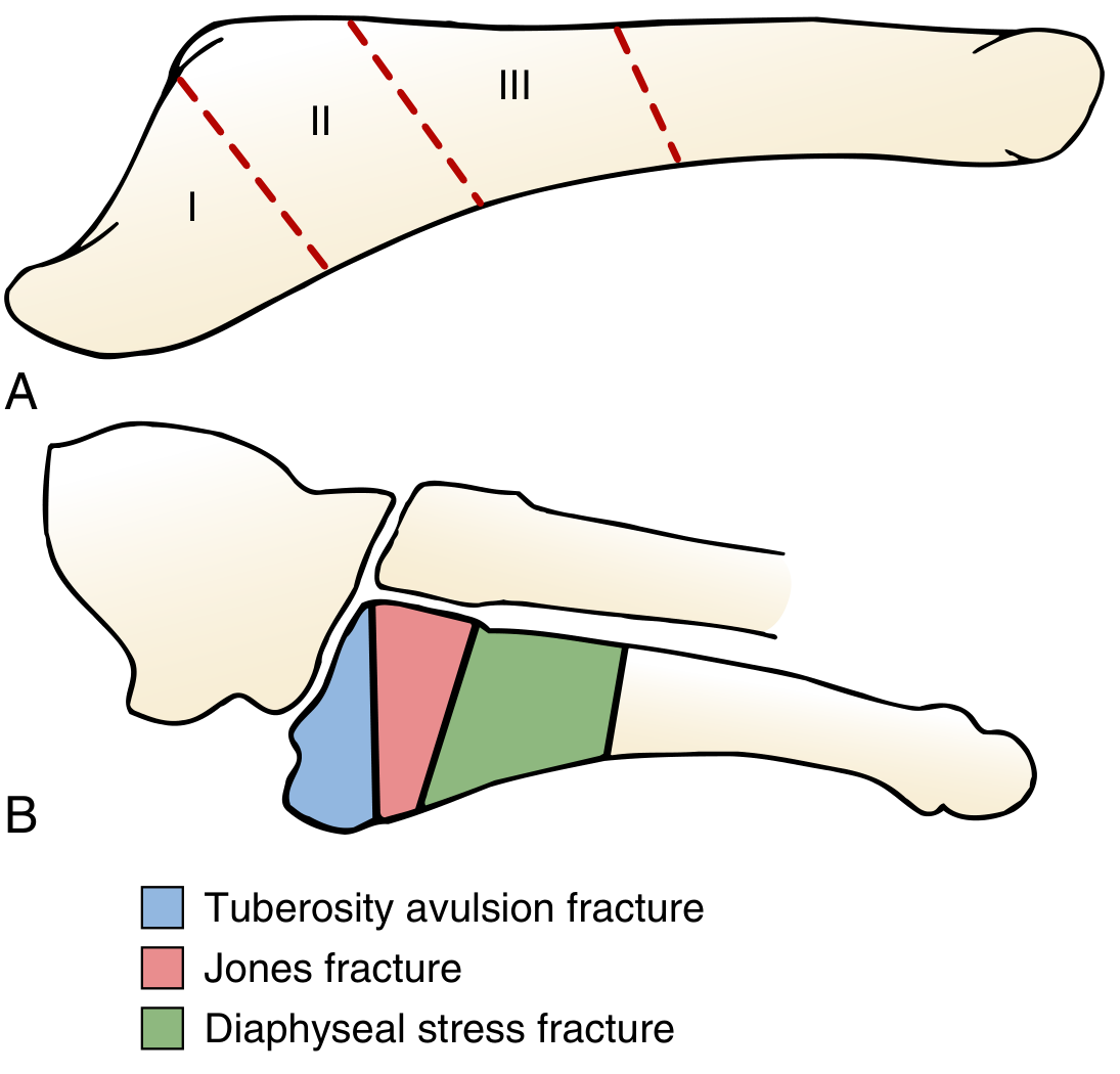
| Zone | Location | Name | Healing Risk |
|---|---|---|---|
| Zone 1 (blue) | Tuberosity - most proximal | Avulsion fracture (pseudo-Jones) | Good blood supply, heals well |
| Zone 2 (pink) | Metaphyseal-diaphyseal junction | Jones fracture | 15-25% nonunion risk |
| Zone 3 (green) | Proximal diaphysis | Diaphyseal stress fracture | Highest nonunion risk, slow healing |
1. Misclassification - This May Be a Jones Fracture (Zone 2)
The most serious and most commonly missed cause of persistent pain.
- The original X-ray was read as "undisplaced 5th metatarsal base fracture" but if the fracture line actually extends into the metaphyseal-diaphyseal junction (Zone 2), it is a true Jones fracture
- Jones fractures occupy a vascular watershed zone with poor blood supply - they carry a 15-25% nonunion rate with conservative management alone
- At 2 weeks with continued pain after slab removal, a Zone 2 fracture should be actively excluded
- Action: Repeat oblique X-ray or MRI to confirm zone classification
2. Nonunion / Delayed Union
- Nonunion = fracture fails to heal after sufficient time (typically >3 months with no radiographic progression)
- Delayed union = slower than expected healing, still possible but taking longer
- Risk factors in this patient:
- Premature slab removal at 2 weeks
- Early weight-bearing without protection
- Possible Zone 2/3 fracture
- Undetected metabolic issues (diabetes, osteoporosis, Vitamin D deficiency)
- X-ray finding: sclerosis at fracture ends, persistent fracture line, no callus bridging
- Treatment if confirmed: Intramedullary screw fixation ± bone grafting (per Miller's Review of Orthopaedics 9th Ed)
3. Fracture Displacement (Secondary)
- An originally undisplaced fracture can displace secondarily if the patient bears full weight prematurely without adequate immobilization
- Displaced fractures involving the 5th metatarsal-cuboid articular surface require ORIF (open reduction internal fixation)
- Tenting of the skin is an indication for emergency fixation
- Action: Urgent repeat X-ray
4. Refracture
- Returning to activity before radiographic union is confirmed sharply increases refracture risk
- Zone 3 stress fractures carry a 33% refracture risk with nonoperative treatment
- Clinically presents as sudden worsening of pain or new pop during weight-bearing
5. Complex Regional Pain Syndrome (CRPS) / Reflex Sympathetic Dystrophy
- A neuro-vascular pain disorder triggered by trauma/fracture
- Presents with burning, disproportionate pain, allodynia (pain to light touch), swelling, skin color/temperature changes, and hypersensitivity
- More common after inadequately managed or prolonged immobilized injuries
- Diagnosis: Clinical (Budapest criteria); bone scan or MRI may help
- Treatment: Early mobilization, physiotherapy, nerve blocks, medications (amitriptyline, gabapentin)
6. Underlying Metabolic Bone Disease
Conditions that severely impair fracture healing and cause prolonged pain:
| Condition | Mechanism | Clue |
|---|---|---|
| Diabetes mellitus | Impaired bone microvascular supply, neuropathy | History, HbA1c |
| Vitamin D deficiency | Impairs mineralization of callus | Serum 25-OH Vit D |
| Osteoporosis | Poor bone quality, slow healing | DEXA scan, history |
| Hyperparathyroidism | Increased bone resorption | Serum Ca, PTH |
| Female athlete triad | Anorexia + amenorrhea + low bone density | History in young females |
Per Miller's Review of Orthopaedics 9th Ed: "Evaluation for metabolic bone disease in these patients (especially if insidious onset) - assessment of Vitamin D level with treatment if needed."
7. Cavovarus Foot Deformity (Underlying Biomechanical Cause)
- A high-arched (cavus) foot with varus alignment places excess lateral loading on the 5th metatarsal
- This can cause persistent pain, stress fractures, and recurrent fractures
- Per Miller's Review: "Presence of a varus foot deformity is not uncommon in the patient with a Zone 3 fracture, and concomitant lateral closed-wedge calcaneal osteotomy should be considered to prevent recurrence"
- Action: Assess foot arch; may need custom orthotics or corrective surgery
8. Peroneus Brevis Tendon Injury
- The same inversion mechanism that fractures the 5th metatarsal base can cause a longitudinal split tear of the peroneus brevis tendon
- This is frequently missed on plain X-ray
- Causes persistent lateral foot/ankle pain even after fracture heals
- Diagnosis: MRI of ankle
- Treatment: Physiotherapy, or surgical repair if severe
9. Lisfranc Injury (Missed Associated Injury)
- A more severe inversion injury can cause tarsometatarsal (Lisfranc) ligament disruption alongside the 5th metatarsal fracture
- If missed and untreated, leads to chronic midfoot pain and progressive flat foot deformity
- Action: Check X-ray for widening of 1st-2nd metatarsal base gap (>2mm suspicious)
Summary - Priority Action Plan
Pain at 2 weeks + premature slab removal
         ↓
1. Repeat X-ray (AP + oblique) → Confirm zone, check displacement, callus
2. If Zone 2/3 confirmed → Orthopedic surgeon referral for screw fixation
3. Bloods: FBS/HbA1c, Calcium, Vit D, PTH
4. MRI if X-ray normal but pain persists → Rule out peroneus brevis tear, CRPS, occult fracture
5. Re-immobilize immediately (walking boot/slab) until imaging reviewed
The two most urgent concerns are fracture zone misclassification (Jones fracture) and secondary displacement from premature mobilization - both require imaging today.
Now almost 3 week and still have pain, but it is bearable and she is walking using normally but still paining
Finding Sources
Searching PubMed
fifth metatarsal avulsion fracture conservative treatment outcome weight bearing
This is a reassuring update. Let me put it in context of the timeline and what the evidence supports.
Assessment: This Is Consistent With Normal Healing
Trajectory so far:
- Week 0: Injury, undisplaced 5th metatarsal base fracture
- Week 1-2: Below-knee slab
- Week 2: Slab removed, pain on mobilization (expected, too early functionally)
- Week ~3: Pain now bearable, walking with normal gait — improving
This is exactly the pattern expected for an undisplaced Zone 1 avulsion fracture healing well. Mild residual ache with normal ambulation at 3 weeks is a good sign, not a concern — it reflects ongoing callus maturation while function returns ahead of full radiographic union, which is typical. The systematic review of fifth metatarsal base fracture treatment supports that most Zone 1 avulsion fractures do well with symptomatic/functional management and progressive return to weight-bearing over 4-6 weeks (Herterich et al., 2021).
What This Means Practically
- No red flags present — no worsening pain, no new deformity, no inability to bear weight, no night pain described. Bearable pain with normal gait is the expected pattern of a fracture transitioning from protected to full function.
- Continue current course: gentle activity as tolerated, avoid high-impact loading (running, jumping, twisting) until ~6 weeks.
- No need to reimmobilize unless pain suddenly worsens or gait changes.
Watch For (Red Flags Requiring Re-evaluation)
- Pain worsening rather than plateauing/improving
- New swelling, warmth, or redness (infection or CRPS)
- Sudden sharp pain or inability to bear weight (possible refracture/displacement)
- Pain lasting unchanged beyond 6-8 weeks (possible nonunion, especially if this turns out to be a Jones-zone fracture rather than a simple avulsion)
Recommended Follow-Up
- Clinical review with the treating doctor around 6 weeks from injury to confirm resolution and consider a repeat X-ray if pain hasn't fully resolved by then.
- Continue Calcium + Vitamin D if not already on it, and avoid prolonged NSAID use now that acute inflammation has settled.
- Gradual return to full activity (sports, prolonged standing) only after she is pain-free.
Overall, this update is reassuring — bearable pain with normal walking at 3 weeks fits a normal healing trajectory for this type of fracture. No urgent action needed; just continue monitoring toward the 6-week mark.
This is a shared conversation. Sign in to Orris to start your own chat.