
Red spots on skin
When you press on the spots, do they turn pale/white (blanch) or stay red? · What best describes the spots and any other symptoms?
red spots on skin causes when to see a doctor
petechiae and purpura on skin
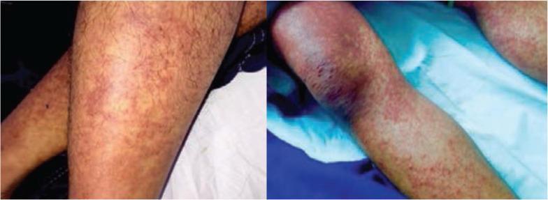
Two clinical photographs depict the lower limbs of a patient featuring an extensive cutaneous eruption. The rash is characterized by diffuse, reddish-purple macules and small papules that vary in morphology across different anatomical sites. On the shins and calves, the lesions appear as discrete, pinpoint petechiae and maculopapular elements distributed in a somewhat scattered fashion. In contrast, the region surrounding the knee shows higher lesion density with confluent erythematous patches and deep purpuric areas, suggesting palpable purpura or intense vascular inflammation. The underlying skin exhibits mild mottling and generalized erythema. This visual presentation is highly characteristic of systemic vasculitis (such as IgA vasculitis/Henoch-Schönlein purpura) or other hematologic conditions presenting with dependent purpura. These images serve as an educational resource for identifying vascular skin lesions, distinguishing between petechiae and confluent purpuric patches, and observing the characteristic dependent distribution of such dermatological manifestations.
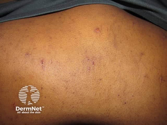
Clinical photography of cutaneous lesions on the back (posterior trunk). The image captures multiple tiny non-blanching petechiae and purpuric macules scattered across the thoracic–lumbar region. Lesions are discrete, punctate to 2–4 mm, flat, with a violaceous to brownish hue and no evident central necrosis or blistering. Some clusters appear confluent, forming larger patchy areas along relaxed skin folds. The surrounding skin shows mild textural variation but no significant edema. The overall pattern is asymptomatic in appearance, with lesions primarily in exposed or pressure-prone zones, suggesting superficial dermal hemorrhage without epidermal disruption. This appearance corresponds to purpura/petechiae rather than wheals or vesicles. Pathophysiologically, extravasation of red blood cells into the dermis produces the characteristic color change; lesion size and distribution can reflect microvascular fragility, thrombocytopenia, vasculitis, or coagulopathy. Clinically, the finding prompts correlation with systemic signs (fever, mucosal bleeding, fatigue) and complete blood count, coagulation studies, and review for medications or infectious etiologies. Differential diagnoses include benign frictional petechiae, senile purpura, drug-induced purpura, and inflammatory vasculitis. This image is valuable for dermatology education, triage training, and research on cutaneous hemorrhagic patterns; it supports teaching about diagnostic workup, documentation, and pattern recognition in pigmentary and vascular skin disorders. Educational, clinical, and research.
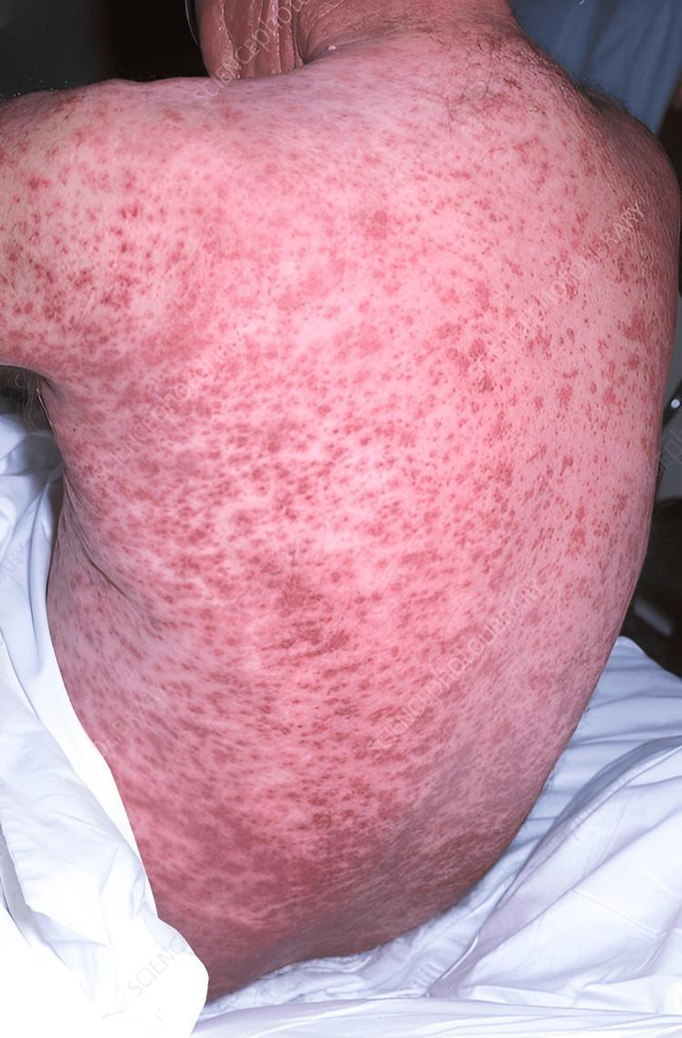
This clinical photograph displays the posterior torso of a patient exhibiting a widespread, confluent purpuric rash. The skin is densely populated with numerous non-blanching hemorrhagic lesions that vary in morphology. Smaller, pinpoint red lesions consistent with petechiae are visible, particularly on the upper back and shoulders. These lesions coalesce in the mid-to-lower back regions into larger, irregular macules and patches of deep red to purple discoloration, characteristic of purpura. The underlying background skin shows a generalized erythematous or sallow pinkish tone, suggesting systemic inflammation or vascular involvement. The extensive and symmetrical distribution across the back is clinically significant for evaluating systemic conditions such as severe malnutrition (e.g., Vitamin C deficiency/scurvy), vasculitis, or hematological disorders. This visual is intended for medical students and clinicians to recognize the cutaneous manifestations of systemic disease and differentiate between petechiae and palpable or non-palpable purpura.
Red Spots on Skin: What They Could Mean
1. Spots that blanch (fade) with pressure
- Hives/urticaria - raised, itchy welts, often from an allergic reaction, food, medication, or heat/cold
- Contact dermatitis / allergic reaction - itchy red rash after touching an irritant (poison ivy, new soap, etc.)
- Heat rash (miliaria) - small red bumps in sweaty areas
- Insect bites/stings - localized red bumps, often itchy or with a central punctum
- Viral exanthem - flat/raised red spots with fever, common in children (e.g., roseola, measles-like rashes)
- Rosacea/acne - chronic facial redness or inflamed bumps
- Cherry angiomas - small, stable, benign red-purple dome-shaped growths from tiny blood vessels, common with age
2. Spots that do NOT blanch (stay red/purple under pressure)
- Petechiae: pinhead-sized (under 3 mm), flat, red/purple/brown, non-blanching dots. They occur in crops, often on the lower legs or other dependent areas, and "usually imply a disorder of platelets rather than of coagulation factors" - Andrews' Diseases of the Skin. Causes range from minor (vigorous coughing/vomiting, tight clothing, straining) to serious (low platelet count, infection/sepsis, leukemia, vasculitis).
- Purpura: larger (several mm to cm), non-blanching macules/plaques from deeper hemorrhage. According to Symptom to Diagnosis: An Evidence-Based Guide, purpura "are associated with a variety of life-threatening diseases, such as vasculitis and sepsis." Causes include thrombocytopenia (ITP, TTP, DIC), vasculitis (e.g., IgA vasculitis/Henoch-Schönlein purpura), medication reactions, meningococcemia, and clotting disorders.
- Ecchymoses (bruises): larger, geometric or irregular blue-purple patches from trauma or fragile vessels (aging, steroid use, vitamin C deficiency/scurvy, blood thinners).
Red flags - seek urgent medical care if the spots come with:
- Fever, especially with a rapidly spreading non-blanching rash (can signal meningococcemia - a medical emergency)
- Unexplained bruising, bleeding gums, blood in urine/stool
- Fatigue, weight loss, or easy bruising (possible blood disorder such as leukemia)
- Joint pain, abdominal pain, or blood in urine alongside the rash (possible vasculitis)
- Rapid spread, blistering, or the spots not fading within a week or two
- Recent new medication (possible drug-induced thrombocytopenia or hypersensitivity reaction)
What to do
- Press a spot with your finger: if it fades, it's likely a benign vascular/inflammatory cause; if it stays red/purple, it needs medical evaluation (CBC, coagulation studies).
- Note distribution (localized vs. widespread), whether it's itchy or painful, and any associated symptoms - this history is what a clinician uses to narrow the differential.
- Isolated, stable, blanching cherry angiomas or a known allergic rash that's improving usually don't need urgent care, but new, spreading, or non-blanching spots - especially with fever or bleeding - warrant prompt evaluation.