
Hypokalaemia path of physiology in a diagrammatic representation
Reading File
Finding Sources
Searching PubMed
hypokalemia AND physiology
Searching the Web
hypokalemia pathophysiology potassium shift membrane potential reputable medical reference
Generating Image
Educational medical flowchart explaining hypokalaemia pathophysiology: causes lead to low extracellular potassium, then show hyperpolarisation of skeletal muscle, cardiac muscle, and gastrointestinal smooth muscle; also show renal potassium wasting, intracellular shift, and reduced intake. Include downstream signs such as weakness, ileus, ECG changes and arrhythmias. Label arrows clearly.
Reading File
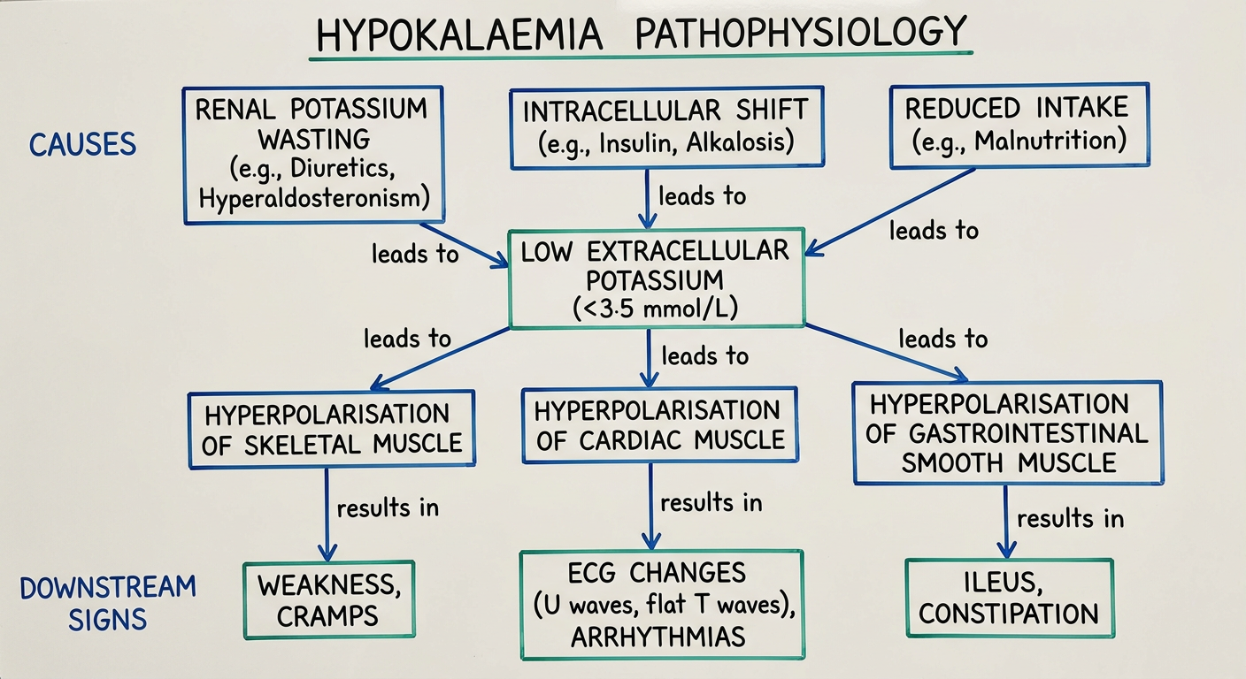
Hypokalaemia: pathophysiology flow diagram
                         HYPOKALAEMIA
                    Serum K+ < 3.5 mmol/L
                              │
       ┌──────────────────────┼────────────────────────┐
       │                      │                        │
       ▼                      ▼                        ▼
↓ Total-body potassium    Shift of K+ into cells   Reduced potassium intake
       │                      │
       │                      ├─ Insulin
       │                      ├─ β2-adrenergic stimulation
       │                      └─ Alkalosis
       │
       ├───────────────────┬──────────────────────┐
       ▼                   ▼                      ▼
Renal K+ loss          GI K+ loss              Skin loss
- Loop/thiazide         - Diarrhoea            - Excess sweating
  diuretics             - Vomiting*
- Hyperaldosteronism
- Renal tubular disease
- Hypomagnesaemia

*Vomiting mainly produces hypokalaemia indirectly:
 volume depletion → ↑ aldosterone → ↑ renal K+ secretion
                              │
                              ▼
                  ↓ Extracellular [K+]
                              │
                              ▼
      ↑ K+ gradient from inside the cell to extracellular fluid
                              │
                              ▼
      More negative resting membrane potential
                   (HYPERPOLARISATION)
                              │
              ┌───────────────┼────────────────┐
              ▼               ▼                ▼
      Skeletal muscle      Smooth muscle       Cardiac muscle
      ↓ excitability       ↓ motility          Abnormal repolarisation
              │               │                │
              ▼               ▼                ▼
      Weakness, cramps,    Constipation,      Flattened T waves
      fatigue, paralysis   paralytic ileus    ST depression
                                               Prominent U waves
                                               Prolonged QU interval
                                               Atrial/ventricular
                                               arrhythmias
Key physiological concept
Potassium is the main intracellular cation. About 98% of body potassium lies inside cells, so extracellular potassium concentration strongly influences the resting membrane potential. When extracellular K+ falls, excitable cells generally become hyperpolarised and are harder to activate. This produces muscle weakness and reduced gastrointestinal motility.
In cardiac myocytes, hypokalaemia also reduces potassium conductance and delays repolarisation, producing the typical ECG changes and increasing susceptibility to dangerous arrhythmias. Merck Manual overview supports renal and gastrointestinal loss as common mechanisms.
Tintinalli's Emergency Medicine, pp. 710-729.
Tintinalli's Emergency Medicine, pp. 710-729.
This is a shared conversation. Sign in to Orris to start your own chat.