
Facisl nerve 20 marks
facial nerve anatomy branches course diagram
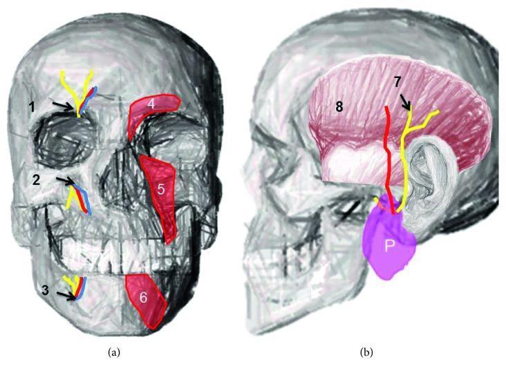
Anatomical diagrams of the human skull illustrating terminal branches of the trigeminal nerve and associated facial structures. Figure (a) is an anterior view highlighting the supraorbital notch (1), infraorbital foramen (2), and mental foramen (3), with accompanying neurovascular bundles shown in yellow (nerves) and blue/red (vessels). The diagram also depicts the overlying musculature: the corrugator supercilii (4), levator labii superioris (5), and depressor labii inferioris (6). Figure (b) provides a lateral view demonstrating the course of the auriculotemporal nerve (7) emerging from the parotid gland (P) and ascending across the temporal region. The temporalis muscle (8) is shown in pink, providing anatomical context for nerve distribution. This educational illustration demonstrates the relationship between bony landmarks, superficial facial muscles, and the sensory nerve exits essential for clinical procedures such as regional nerve blocks and the management of trigeminal neuralgias.
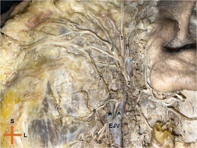
This clinical photograph displays a cadaveric dissection of the left extratemporal facial nerve (Cranial Nerve VII), highlighting its complex branching pattern and vascular relationships. The main trunk bifurcates into the temporofacial and cervicofacial divisions. The superior temporofacial division gives rise to the temporal and zygomatic branches, which course toward the upper face, and a buccal branch. The inferior cervicofacial division exhibits a rare anatomical variation where it bifurcates around the posterior facial vein; the upper branch passes superficially while the lower branch courses deep to the vein. Key anatomical landmarks include the retromandibular vein, the external jugular vein (EJV), and the masseter muscle. Labeled structures include the temporal (4), zygomatic (5), buccal (6), and marginal mandibular (8, 9) branches. This image is an essential educational resource for surgical anatomy, particularly for parotidectomy and maxillofacial procedures, demonstrating the critical proximity of neural branches to venous structures in the parotid region.
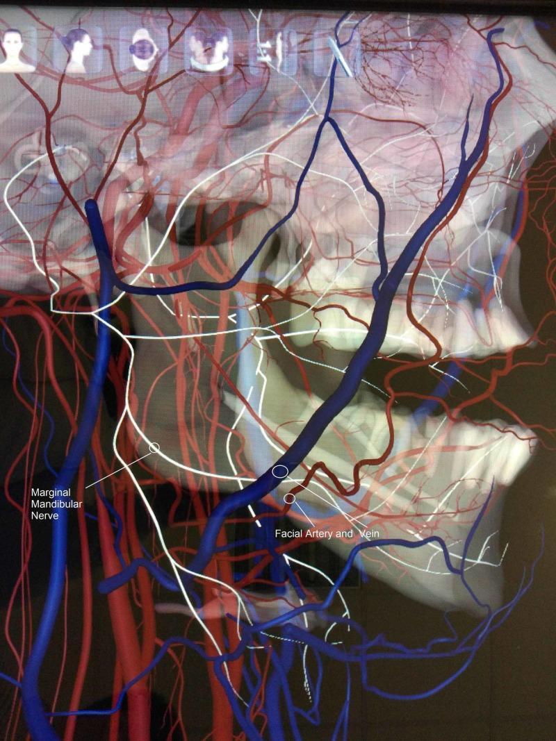
Anatomical diagram and 3D visualization illustrating a rare neurovascular anatomical variation in the maxillofacial region. This lateral view focuses on the right side of the mandible and cervical area, highlighting the spatial relationship between the marginal mandibular nerve (MMN), the facial artery, and the facial vein. The MMN is depicted as a blue tubular structure descending toward the lower border of the mandible. Crucially, the diagram demonstrates an atypical course where the MMN runs deep (medial) to the facial artery and vein (rendered in red), contrasting with the standard anatomical presentation where the nerve typically crosses superficial to these vessels. The visualization overlays these structures on a skeletal model of the skull and mandible, showing the nerve's proximity to the mandibular angle and body. This educational content is relevant for surgical anatomy, particularly in the context of neck dissections, submandibular gland excision, and rhytidectomy, where preserving the MMN is critical to prevent paralysis of the depressor muscles of the lower lip.
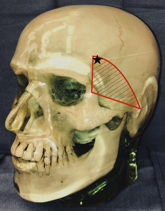
This anatomical diagram utilizes a lateral view of a human skull model to illustrate the surgical vulnerability of the temporal branch of the facial nerve. A red hatched triangular region is overlaid on the temporal fossa, bounded by the zygomatic arch inferiorly, the zygomatic process of the frontal bone anteriorly, and extending toward the superior temporal line. This area represents the danger zone where the temporal branch of the facial nerve is most superficial. A black star is marked within the superior-anterior portion of this shaded region, indicating the specific placement of a Mayfield head clamp pin that resulted in postoperative frontalis muscle palsy. The image serves as a clinical education tool for neurosurgical and orthopedic surgical positioning, highlighting the risk of iatrogenic nerve injury from external fixation devices when applied near the course of the facial nerve branches.
facial nerve segments intratemporal course stylomastoid foramen geniculate ganglion
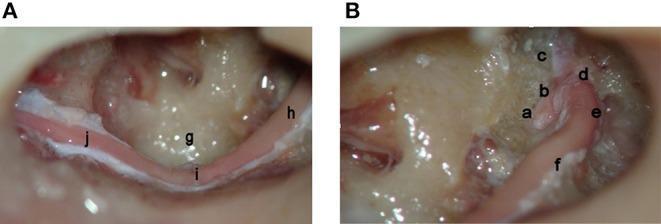
Two intraoperative endoscopic photographs (A and B) demonstrate a complete surgical decompression of the left facial nerve in a cadaveric specimen, tracing the nerve from the labyrinthine meatus to the stylomastoid foramen. Image B shows the proximal segments, including the meatal foramen (a), labyrinthine portion (b), greater superficial petrosal nerve (c), the first genu (d), the geniculate ganglion (e), and the proximal horizontal (tympanic) segment (f). Image A details the distal course, illustrating the continuation of the horizontal segment (h) as it passes the sinus tympani (g), reaching the second genu (i). From this point, the nerve transitions into its vertical (mastoid) segment (j), extending toward the stylomastoid foramen. This anatomical display highlights the relationship between the facial nerve and middle ear landmarks, such as the sinus tympani, and is relevant for otologic surgery, temporal bone fracture management, and facial nerve decompression procedures.
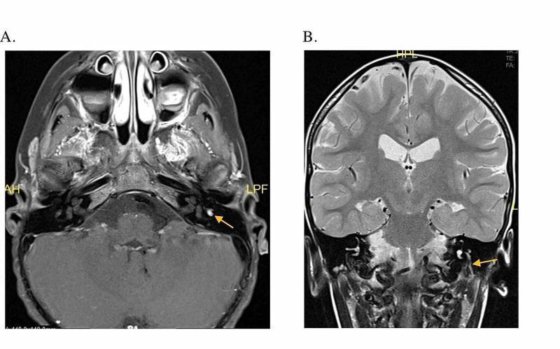
Diagnostic neuroimaging featuring two T1-weighted post-contrast MRI views focusing on the internal auditory canal and facial nerve (Cranial Nerve VII). Panel A (Axial view) demonstrates abnormal focal enhancement of the left facial nerve at the level of the geniculate ganglion, indicated by a yellow arrow, visible against the surrounding temporal bone and cerebellum. Panel B (Coronal view) shows linear enhancement and asymmetric enlargement of the descending mastoid segment of the left facial nerve, extending from the geniculate ganglion toward the stylomastoid foramen, also marked by a yellow arrow. Comparison with the contralateral right side reveals significant hyperintensity and thickening on the left. These findings are clinically significant in the diagnostic workup of facial nerve pathology, such as Bell's palsy or facial nerve schwannoma. General neuroanatomical structures including the lateral ventricles, temporal lobes, and brainstem are also visible.
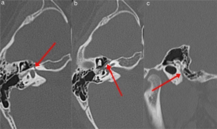
High-resolution axial computed tomography (CT) images of the left temporal bone, presented in three sections (A, B, and C) to demonstrate the course of the facial nerve (Fallopian) canal. Red arrows highlight different segments of the canal. Image A shows the labyrinthine segment, appearing as a narrow radiolucent channel within the dense petrous bone, traveling anterolaterally towards the geniculate ganglion. Image B depicts the tympanic (horizontal) segment, visualized as a thin lucency running along the medial wall of the tympanic cavity, inferior to the lateral semicircular canal. Image C illustrates the mastoid (descending) segment, where the canal assumes a vertical orientation within the mastoid portion of the temporal bone, posterior to the external auditory canal. These images are clinically relevant for evaluating anatomical variations or pathologies, such as Bell's palsy, by assessing the diameter and integrity of the bony canal across its various intratemporal segments.
Facial Nerve (Cranial Nerve VII) - 20 Marks
1. Introduction
2. Nuclei (Central Connections)
| Nucleus | Location | Fiber Type | Function |
|---|---|---|---|
| Motor nucleus of VII | Caudal pons | Special visceral efferent (SVE) | Facial expression, stapedius, stylohyoid, posterior digastric |
| Superior salivatory nucleus | Dorsal to motor nucleus, pons | General visceral efferent (GVE) | Parasympathetic to lacrimal, nasal, sublingual, submandibular glands |
| Nucleus solitarius (NTS) | Medulla | Special visceral afferent (SVA) | Taste from anterior 2/3 tongue |
3. Fiber Components (Five Fiber Types)
- Special visceral efferent (SVE) - motor to muscles of facial expression, stapedius, stylohyoid, posterior belly of digastric
- General visceral efferent (GVE) - preganglionic parasympathetic to lacrimal, nasal, submandibular, sublingual glands
- Special visceral afferent (SVA) - taste from anterior 2/3 of tongue, soft palate, tonsillar fossa
- General somatic afferent (GSA) - cutaneous sensation from EAC, concha, mastoid skin (small area)
- General visceral afferent (GVA) - afferents from nasal/pharyngeal/palatal mucosa
4. Course and Segments
Segment 1 - Intracranial (Cisternal) Segment (17-24 mm)
- Runs from the pons to the porus of the internal auditory canal (IAC)
- Traverses the cerebellopontine angle (CPA) with CN VIII
- The motor root and nervus intermedius (sensory/autonomic root) are separate here
Segment 2 - Meatal (Intracanalicular) Segment (8-10 mm)
- Runs within the IAC
- At the fundus of the IAC, occupies the anterosuperior quadrant (separated from CN VIII by Bill's bar above, and crista falciformis below)
- No branches arise here
Segment 3 - Labyrinthine Segment (4 mm) - SHORTEST
- Enters the fallopian canal (bony canal)
- Runs from the meatal foramen to the geniculate ganglion
- The narrowest and shortest intratemporal segment
- Most susceptible to ischemic compression during inflammation (e.g., Bell's palsy)
- Branch: Greater superficial petrosal nerve (GSPN) arises from the geniculate ganglion
Segment 4 - Tympanic (Horizontal) Segment (11-13 mm)
- From the geniculate ganglion to the second genu
- Runs along the medial wall of the tympanic cavity, superior to the oval window and cochleariform process
- No major branches arise here
- Susceptible to injury in middle ear surgery (most commonly dehiscent here)
Segment 5 - Mastoid (Vertical) Segment (13-20 mm)
- From the second genu (at the lateral semicircular canal) to the stylomastoid foramen
- Branches:
- Nerve to stapedius (upper mastoid segment)
- Chorda tympani (lower mastoid segment) - carries taste from anterior 2/3 tongue and parasympathetic fibers to submandibular/sublingual glands
Segment 6 - Extratemporal Segment
- Exits the skull at the stylomastoid foramen
- Before entering the parotid, gives off:
- Posterior auricular nerve (occipitalis, auricular muscles)
- Digastric branch (posterior belly of digastric)
- Stylohyoid branch (stylohyoid muscle)
- Enters the parotid gland and divides at the pes anserinus ("goose's foot") into:
| Branch | Muscles Innervated |
|---|---|
| Temporal | Frontalis, corrugator supercilii, upper orbicularis oculi |
| Zygomatic | Lower orbicularis oculi |
| Buccal | Buccinator, zygomaticus, nasalis, upper lip muscles |
| Marginal mandibular | Depressors of lower lip and chin |
| Cervical | Platysma |
5. Branches and Their Functions - Summary Table
| Branch | Site of Origin | Function |
|---|---|---|
| GSPN | Geniculate ganglion | Parasympathetic to lacrimal gland; taste from soft palate |
| Nerve to stapedius | Mastoid segment | Dampens ossicular vibration (protective reflex) |
| Chorda tympani | Mastoid segment | Taste anterior 2/3 tongue; parasympathetic to submandibular + sublingual glands |
| Posterior auricular | Near stylomastoid foramen | Occipitalis, auricular muscles |
| Digastric branch | Near stylomastoid foramen | Posterior belly of digastric |
| Stylohyoid branch | Near stylomastoid foramen | Stylohyoid |
| 5 terminal branches | In parotid (pes anserinus) | Muscles of facial expression, platysma |
6. Localizing Lesions of CN VII
| Level | Structures Affected | Clinical Features |
|---|---|---|
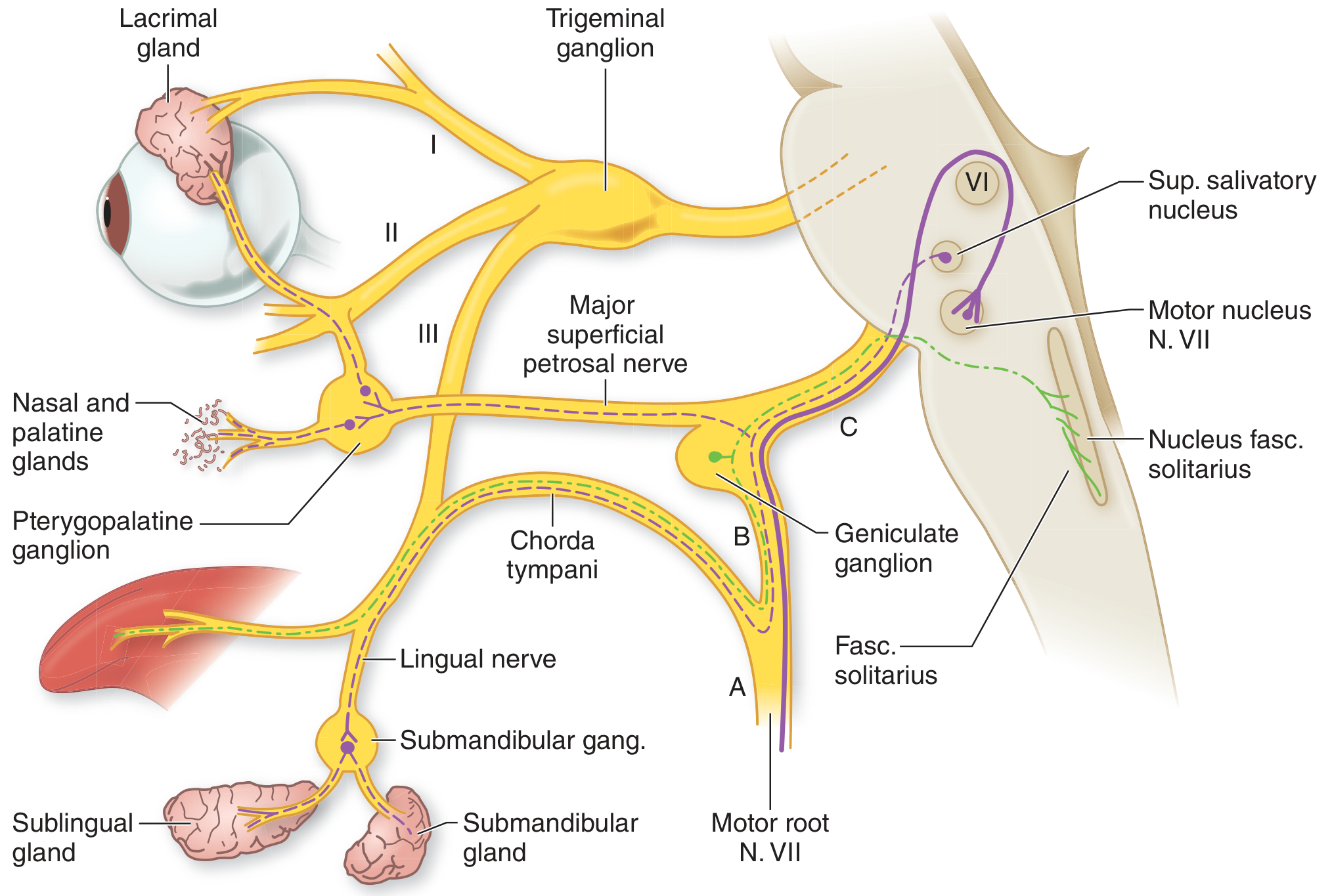
| A - Stylomastoid foramen | Motor root only | LMN facial palsy: all ipsilateral facial muscles. Taste intact, no hyperacusis, no lacrimation loss |
| B - Above chorda tympani, below geniculate | Motor + chorda tympani + stapedius | LMN palsy + loss of taste anterior 2/3 tongue + hyperacusis |
| C - Geniculate ganglion or proximal | Motor + chorda + stapedius + GSPN | All of the above + reduced lacrimation + possibly CN VIII involvement (deafness, vertigo) |
| UMN lesion | Corticobulbar fibers | Contralateral lower face palsy only; forehead spared (bilateral cortical representation) |
7. Grading of Facial Palsy - House-Brackmann Scale
| Grade | Description | Function |
|---|---|---|
| I | Normal | 100% function |
| II | Slight dysfunction | Subtle weakness; complete eye closure with effort |
| III | Moderate dysfunction | Obvious but not disfiguring difference; complete eye closure with effort |
| IV | Moderately severe | Obvious weakness; incomplete eye closure |
| V | Severe dysfunction | Barely perceptible motion |
| VI | Total paralysis | No movement |
8. Bell's Palsy (Idiopathic Facial Paralysis)
- Acute onset (<72 hrs) of unilateral LMN facial palsy
- Ipsilateral forehead weakness (distinguishes from UMN lesion)
- Lagophthalmos (incomplete eye closure) → risk of corneal exposure
- Loss of nasolabial fold, drooping corner of mouth
- Hyperacusis (stapedius involvement)
- Altered taste (chorda tympani involvement)
- Post-auricular pain may precede palsy (reactivation)
- Epiphora (excess tearing - lower lid ectropion + crocodile tears later in recovery)
- Clinical diagnosis; no routine imaging required
- MRI with contrast: linear enhancement at labyrinthine segment, geniculate ganglion, proximal tympanic segment (normal finding in Bell's palsy)
- EMG/nerve conduction if needed to assess severity
- Prednisolone 1 mg/kg/day (max 60 mg) for 10 days - start within 72 hours
- Antiviral (valacyclovir/acyclovir) - beneficial when added to steroids, especially in severe cases
- Eye care: Lubricating drops, night-time taping/patching to prevent corneal exposure
- Surgical decompression: reserved for severe cases with >90% degeneration on EMG + no recovery
9. Applied Anatomy and Clinical Correlations
- Parotidectomy: The most feared complication is facial nerve injury. The nerve is identified at the stylomastoid foramen or via the tragal pointer, tympanomastoid suture, or the digastric ridge. The intimate relationship of CN VII within the parotid makes surgical dissection challenging.
- Cholesteatoma/Middle ear surgery: The tympanic segment is most vulnerable, especially when the fallopian canal is dehiscent (found in up to 55% of temporal bones at this site).
- Temporal bone fracture: Transverse fractures (perpendicular to petrous axis) are more likely to injure the geniculate ganglion or labyrinthine segment; longitudinal fractures (parallel to petrous axis) more commonly cause delayed palsy from edema at the geniculate ganglion.
- Ramsay Hunt Syndrome: Varicella-zoster virus reactivation in the geniculate ganglion causes: facial palsy + herpetic vesicles on the ear (pinna, EAC) + sensorineural hearing loss + vertigo.
- Crocodile tears (gustatolacrimal reflex): Aberrant regeneration after CN VII injury where salivatory fibers grow into lacrimal pathways, causing tearing while eating.
- Synkinesis: Post-injury abnormal co-movements (e.g., eye blinking on smiling) due to misdirected axonal regeneration.
10. Blood Supply
- Labyrinthine artery (from AICA): supplies cisternal, meatal, and labyrinthine segments
- Petrosal artery (from middle meningeal): supplies geniculate ganglion and proximal tympanic segment
- Stylomastoid artery (from posterior auricular): supplies mastoid segment
- The labyrinthine segment is the most susceptible to ischemia due to end-arterial supply with no anastomoses.
Summary Diagram
- Shambaugh Surgery of the Ear (Segments, nuclei, fiber types)
- Adams and Victor's Principles of Neurology, 12th ed. (Lesion localization, Bell's palsy)
- K.J. Lee's Essential Otolaryngology (Segments, paralysis etiologies)
- Cummings Otolaryngology Head and Neck Surgery (Imaging, Bell's palsy, House-Brackmann grading)
- Scott-Brown's Otorhinolaryngology Head & Neck Surgery, Vol. 2 (Intratemporal anatomy, blood supply, grading)
- Localization in Clinical Neurology, 8e (Peripheral segments, clinical localization)