
python3 -c " import pdfplumber with pdfplumber.open('/home/daytona/workspace/attachments/d255bdb4-bcde-411d-8a9c-805d0cba30c9/PatientReport.pdf') as pdf: print(f'Pages: {len(pdf.pages)}') for i, page in enumerate(pdf.pages): print(f'--- PAGE {i+1} ---') text = page.extract_text() print(text) tables = page.extract_tables() if tables: for j, table in enumerate(tables): print(f'[Table {j+1}]') for row in table: print(row) "
"medial meniscus" AND "posterior horn" AND tear management
"anterior cruciate ligament" AND sprain AND "MRI" AND management
MRI Right Knee Joint - Patient Report Summary & Explanation
What the MRI Found
1. Grade III Posterior Horn Medial Meniscus Tear (Most Significant Finding)
- Grade I/II: Increased signal within the meniscus that does NOT reach the articular surface - represents internal degeneration, not a true tear
- Grade III: Increased (bright/hyperintense) signal that extends to the articular surface - this is a true tear
2. Grade I ACL Sprain (Partial Stretch, No Rupture)
| ACL Grade | MRI Finding | Clinical Significance |
|---|---|---|
| Grade I | Edema, thickening, no fiber discontinuity | Intact; conservative management |
| Grade II | Partial tear, some fibers disrupted | May need bracing/PT |
| Grade III | Complete rupture | Surgical reconstruction often needed |
3. Grade I MCL Sprain
4. Mild Knee Joint Effusion
5. Normal Findings (Reassuring)
- Lateral meniscus (anterior and posterior horns): Normal - no tear
- Anterior horn of medial meniscus: Normal
- PCL (posterior cruciate ligament): Normal course and signal
- Quadriceps tendon and patellar ligament: Normal
- Patella: Normal position
- Lateral collateral ligament (LCL): Normal, no tear
- Bone marrow (all bones): Normal signal - no bone bruise, fracture, or avascular necrosis
- Muscles and fat planes: Normal
Clinical Significance & What Happens Next
- Conservative (non-surgical): Physiotherapy, quadriceps strengthening, activity modification, anti-inflammatory medications, and possibly a knee brace. This is often tried first, especially for degenerative tears in middle-aged patients.
- Surgical (arthroscopic): If conservative treatment fails, arthroscopic partial meniscectomy (trimming the torn portion) or meniscal repair may be considered. Modern orthopedic practice increasingly favors tissue preservation over resection, especially in younger or active patients (Bailey & Love's Surgery, 28th Ed). The location and tear pattern influence repairability - peripheral tears with better blood supply have higher healing potential.
Summary Table
| Finding | Severity | Clinical Implication |
|---|---|---|
| Posterior horn medial meniscus tear | Grade III (true tear, reaches articular surface) | Orthopedic review; may need arthroscopy |
| ACL sprain | Grade I (edema, no fiber disruption) | Conservative - PT, bracing |
| MCL sprain | Grade I (periligamentous edema only) | Conservative - PT, bracing |
| Knee joint effusion | Mild | Resolves with treatment |
| Bones, other ligaments, tendons | Normal | No further action needed |
Note: This explanation is for educational purposes. Clinical decisions should be made by the treating physician with full clinical correlation, as the report itself advises.
Create physiotherapy protocol with mechanism of treatment
"meniscal tear" AND "rehabilitation" AND "physiotherapy"
"ACL sprain" AND "MCL" AND "conservative" AND "exercise"
"knee" AND "MCL" AND "collateral ligament" AND "rehabilitation" AND "exercise"
PMID: 41160820
PMID: 35996030
physiotherapy protocol Grade III medial meniscus tear Grade I ACL MCL sprain conservative 2024 2025
https://www.brighamandwomens.org/assets/BWH/patients-and-fam…
Physiotherapy Rehabilitation Protocol
Patient: Mrs. Rinku Jain | 48 years / Female
Diagnosis: Grade III Posterior Horn Medial Meniscus Tear + Grade I ACL Sprain + Grade I MCL Sprain + Mild Knee Effusion (Right Knee)
Guiding Principles & Evidence Base
Mechanism of Treatment
| Treatment | Physiological Mechanism |
|---|---|
| Cryotherapy (ice) | Reduces local blood flow via vasoconstriction; decreases prostaglandin-mediated inflammation; slows nerve conduction velocity, reducing pain perception (gate control) |
| Compression | Limits tissue edema by opposing capillary hydrostatic pressure; reduces hematoma formation |
| Elevation | Facilitates venous and lymphatic drainage via gravity; reduces hydrostatic pressure in peripheral vessels |
| TENS/Electrotherapy | Activates large Aβ fibers that inhibit pain signals at spinal cord (gate control theory); triggers endorphin release |
| Ultrasound therapy | Thermal: increases collagen extensibility, blood flow, and metabolic rate. Non-thermal (pulsed): promotes cell membrane permeability, accelerates tissue repair via cavitation |
| Isometric exercises | Produce muscle contraction without joint movement; maintain muscle bulk, stimulate mechanoreceptors, reduce quadriceps inhibition from effusion without meniscal loading |
| Open kinetic chain (OKC) exercises | Isolate specific muscle groups (quad/hamstring) in non-weight-bearing; minimize shear forces on the meniscus; used early when weight-bearing is painful |
| Closed kinetic chain (CKC) exercises | Simulate functional activities; distribute load across the entire lower limb; reduce anterior tibial shear; promote co-contraction of hamstrings and quads which protects both ACL and meniscus |
| Proprioception/neuromuscular training | Stimulates mechanoreceptors in the joint capsule, ligaments, and meniscus; restores afferent neural feedback to restore dynamic joint stability; compensates for ligament injury-related afferent loss |
| Hip strengthening | Gluteus medius/maximus control knee valgus and tibial rotation; reduces compressive and shear forces on medial compartment, directly protecting the healing meniscus and MCL |
| Manual therapy/joint mobilization | Reduces joint stiffness via neurophysiological pain inhibition; restores arthrokinematic glide; reduces capsular adhesions and improves synovial fluid distribution |
| Blood Flow Restriction (BFR) training | Applies partial venous occlusion to allow high metabolic stimulus at low resistance loads; achieves hypertrophic effect equivalent to heavy training without the compressive joint load - ideal for early-stage meniscus rehabilitation |
Phase-Based Protocol
PHASE 1: Acute / Inflammation Control
Precautions
- Avoid deep squatting, kneeling, or pivoting
- Avoid loaded knee flexion > 60° initially
- Use crutches if weight-bearing is painful
- Hinged knee brace: locked in extension for walking, unlocked for ROM exercises
Modalities (Physiological Mechanism)
| Modality | Parameters | Mechanism |
|---|---|---|
| Cryotherapy | Ice pack wrapped in towel, 15-20 min, 3-4x/day | Vasoconstriction reduces effusion; analgesic via nerve conduction slowing |
| Compression bandage | Tubigrip or crepe, figure-of-8 | Limits edema accumulation |
| Elevation | Leg elevated above heart level | Enhances venous/lymphatic return |
| TENS | 80-100 Hz (conventional), 30 min, 2x/day | Gate control pain inhibition |
| Pulsed therapeutic ultrasound | 1 MHz, 1.0-1.5 W/cm², 20% duty cycle, 5 min | Non-thermal: stimulates fibroblast proliferation and collagen synthesis in meniscus |
Therapeutic Exercises
| Exercise | Purpose / Mechanism |
|---|---|
| Isometric quadriceps sets (quad tightening, leg straight, 10 sec hold) | Reduces quadriceps inhibition from effusion; maintains muscle activation |
| Ankle pumps | Activates calf muscle pump; prevents DVT; reduces edema |
| Supine heel slides (gentle active knee flexion/extension) | Restores ROM; promotes synovial fluid nutrition of cartilage |
| Straight leg raises (SLR) - 4 planes: supine, sidelying (abduction/adduction), prone | Strengthens quad, hip flexors, abductors, extensors without knee joint loading |
| Isometric glute sets | Activates gluteals without lower limb loading |
| Supine bridging (hip extension) | Activates glutes and hamstrings in a safe, non-weight-bearing manner |
| Clamshells / Sidelying hip abduction | Activates gluteus medius; reduces valgus knee stress from the hip |
Weight Bearing
- Partial weight bearing with crutches as tolerated
- Progress to full weight bearing without pain (typically by end of Week 2)
PHASE 2: Subacute / Functional Loading
Criteria to Progress from Phase 1:
- Effusion largely resolved (minimal or no swelling)
- Full knee extension achieved
- Able to straight-leg raise without lag
- Pain < 3/10 at rest
Modalities
| Modality | Parameters | Mechanism |
|---|---|---|
| Moist heat / hydrotherapy (before exercise) | 15-20 min warm pack or pool | Increases tissue extensibility; reduces stiffness before loading |
| Ice (after exercise) | 15 min post-session | Controls post-exercise reactive inflammation |
| Neuromuscular electrical stimulation (NMES) | 50 Hz, 300-400 μs pulse width, 10 sec on/50 sec off | Facilitates quadriceps re-education via motor nerve stimulation |
| Continuous ultrasound (for MCL) | 1 MHz, 1.0 W/cm², 5-8 min | Thermal: increases collagen cross-linking during ligament healing |
Therapeutic Exercises
| Exercise | Sets/Reps | Mechanism |
|---|---|---|
| Stationary cycling (low resistance, seat high to limit flexion) | 15-20 min | Restores ROM; cardiovascular; low meniscal load; activates quadriceps/hamstrings through full arc |
| Mini squats (0-45° flexion, body weight) | 3x15 | Closed kinetic chain; promotes co-contraction; minimal meniscal load at shallow angles |
| Wall slides (0-60° knee flexion) | 3x15 | Graded loading of knee extensors in CKC; functional |
| Step-ups (forward and lateral) (10 cm step, progress height) | 3x10 each | Functional CKC; proprioceptive challenge; simulates stair climbing |
| Terminal knee extension with resistance band | 3x15 | Specifically targets VMO (vastus medialis oblique); reduces patellar tracking issues |
| Seated knee extension (partial arc, 90° to 40°) | 3x15 | Open kinetic chain; targets quadriceps in inner range |
| Prone hamstring curls (with or without ankle weight) | 3x15 | Hamstring strengthening; dynamic stabilizers of the knee, reducing tibiofemoral shear |
| Standing hip abduction/extension | 3x15 | Gluteal activation; controls knee valgus and tibial rotation |
| Single-leg standing (eyes open, then eyes closed) - 30 sec | 3 reps | Early proprioception; activates joint mechanoreceptors |
Gait Training
- Normalize walking pattern; wean off crutches
- Use hinged brace for walking if instability perceived
- Practice heel-toe walking; correct Trendelenburg if present
PHASE 3: Strengthening & Neuromuscular Control
Criteria to Progress from Phase 2:
- Full pain-free ROM (0-120° minimum)
- Full weight bearing without assistive device
- No significant effusion after exercise
- Single-leg stance > 30 seconds
Modalities
- Ice post-exercise as needed
- Sports massage / soft tissue mobilization to periligamentous structures (MCL region)
Therapeutic Exercises
| Exercise | Sets/Reps | Mechanism |
|---|---|---|
| Leg press (double then single leg, 30-90° arc) | 3x15, progressive load | Primary CKC quad + glute strengthening; arc limits peak meniscal stress |
| Goblet/box squat (to 90° once tolerated) | 3x12 | Full CKC functional lower limb strengthening |
| Romanian deadlift (RDL) / Single-leg RDL | 3x12 | Posterior chain (hamstrings, glutes); reduces anterior tibial shear (protective for ACL) |
| Lateral band walks / Monster walks | 3x20 steps | Hip abductor activation; reduces medial compartment loading |
| Hamstring Nordic curls (eccentric) | 3x8 | Eccentric hamstring strength; protects ACL by limiting tibial translation |
| Balance board / wobble board (bilateral then unilateral) | 3 x 1 min | Stimulates articular, ligamentous, and capsular mechanoreceptors; restores neuromuscular joint protection |
| Blood Flow Restriction (BFR) training (quad sets or leg press with cuff) | 4 sets: 30-15-15-15, 80% limb occlusion | High metabolic/hormonal response at low loads; muscle hypertrophy without joint overload - ideal for meniscus-protected strengthening (2024 EU-US Consensus, Grade B) |
| Treadmill walking (progressing speed) | 20 min | Gait normalization; cardiovascular; functional loading |
| Elliptical trainer | 20-30 min | Low-impact aerobic; simulates gait without impact; meniscus-friendly |
| Swimming (flutter kick only, no breaststroke frog kick) | 20-30 min | Non-weight-bearing aerobic; maintains fitness; hydrotherapy analgesic effect |
| Y-balance test training (reach in anterior, posteromedial, posterolateral directions) | 3x each direction | Dynamic proprioception; challenges neuromuscular control in 3 planes |
PHASE 4: Functional / Return to Activity
Criteria to Progress from Phase 3:
- Quadriceps strength > 80% of contralateral side (ideally tested with dynamometer or single-leg press)
- No pain or effusion during or after exercise
- Single-leg squat to 60° with good alignment (no valgus collapse)
- Single-leg hop test (timed or distance) > 85% limb symmetry index
Therapeutic Exercises
| Exercise | Purpose |
|---|---|
| Progressively loaded squats and lunges | Full functional lower limb strength |
| Step-down exercises (eccentric quad control) | Simulates descending stairs; high clinical relevance for this patient's daily function |
| Jogging on flat surface (if cleared by orthopaedician) | Gradual impact loading of the meniscus; tests healed capacity |
| Lateral agility drills (side shuffles, carioca) | Restores multi-directional control; challenges MCL and proprioception |
| Perturbation training (therapist-applied unexpected balance challenges) | Reflex neuromuscular response training; protects against re-injury during unexpected movements |
| Core stabilization (dead bugs, bird dogs, planks) | Lumbopelvic stability reduces abnormal forces transmitted to knee during functional activities |
Patient Education
- Avoid prolonged kneeling, deep squatting, and high-impact activities
- Recognize warning signs: swelling after activity = too much load, reduce and reassess
- Maintain a healthy weight: every 1 kg of body weight reduction = ~4 kg reduction in knee joint load
- Stair technique: lead with the stronger leg going up, and the operated/weaker leg going down
Complete Timeline Summary
| Phase | Weeks | Key Focus | Brace Use |
|---|---|---|---|
| 1 - Acute | 1-2 | Pain/effusion control, quad activation, passive ROM | Hinged brace, locked in extension for walking |
| 2 - Subacute | 3-6 | Full weight bearing, CKC initiation, gait normalization | Hinged brace, unlocked; wean off by Week 6 |
| 3 - Strengthening | 7-10 | Progressive resistance, neuromuscular training, BFR | Functional brace for demanding activities |
| 4 - Return to Activity | 11-16 | Functional movement, agility, patient education | Only for high-demand activity if needed |
Modality Reference Card
| Modality | Phase | Frequency | Duration | Goal |
|---|---|---|---|---|
| Cryotherapy | 1-2 (post-exercise throughout) | 3-4x/day Phase 1; after each session Phase 2+ | 15-20 min | Anti-inflammatory, analgesic |
| TENS | 1-2 | 2x/day | 30 min | Pain gate modulation |
| Pulsed ultrasound | 1-2 | 5x/week | 5 min | Tissue repair stimulation |
| Continuous ultrasound (MCL) | 2-3 | 3x/week | 5-8 min | Collagen remodeling |
| NMES | 2-3 | 3x/week | 15-20 min | Quadriceps re-education |
| Moist heat/hydrotherapy | 2 onward (pre-exercise) | Before sessions | 15 min | Tissue warming, flexibility |
| BFR training | 3-4 | 3x/week | Per protocol | Early strengthening with low joint load |
Red Flags - When to Refer Back to Orthopaedician
- Sudden increase in swelling or "locked" knee (inability to fully extend) - may indicate a displaced meniscal fragment
- Worsening pain despite 6-8 weeks of physiotherapy
- Mechanical symptoms: clicking, catching, or giving way
- No improvement in quadriceps strength after 8-10 weeks
- Patient desires return to high-demand sport or physical work
Note: This protocol is designed for conservative management of Mrs. Rinku Jain's specific injuries. It should be supervised by a registered physiotherapist and adjusted based on clinical response. Any progression should be symptom-guided, not purely time-based. Regular clinical correlation with the referring orthopaedician (Dr. Jagjit Singh) is advised.
I'm a physiotherapist i give patients a session and feel relief but the next day patients complains the same
"physiotherapy" AND "treatment effect" AND "pain recurrence" AND "home exercise"
why physiotherapy session gives relief but pain returns next day central sensitization neurophysiology explanation 2024
"pain neuroscience education" AND physiotherapy AND musculoskeletal outcomes
PMID: 33863860
Why Patients Feel Better in Session But Return in Pain the Next Day
The Core Problem: You're Treating the Alarm, Not the Wiring
The 3 Mechanisms Behind In-Session Relief
1. Gate Control (Immediate, Short-Lived)
2. Exercise-Induced Hypoalgesia (EIH)
3. Conditioned Pain Modulation (CPM)
Why the Pain Returns: Peripheral and Central Sensitization
Peripheral Sensitization
Central Sensitization (the bigger long-term problem)
- Pain from non-painful stimuli (allodynia)
- Amplified pain from mildly painful stimuli (hyperalgesia)
- Pain that spreads beyond the original injury site
- Pain that persists long after tissues have healed
The 5 Real Reasons Relief Doesn't Last (Clinical Checklist)
| Reason | What's Happening | What to Do |
|---|---|---|
| 1. No home exercise being done | Tissue adaptation requires daily mechanical loading; 2-3x/week clinic sessions are insufficient | Prescribe a specific, simple 10-15 min daily HEP; track compliance next session |
| 2. Central sensitization present | Nervous system is amplifying pain independent of tissue state | Add Pain Neuroscience Education (PNE) to every session; reframe pain as "volume knob" not "damage signal" |
| 3. Activity behaviour between sessions | Patient is either overdoing it (re-irritating tissue) or completely avoiding movement (deconditioning, fear-avoidance) | Teach activity pacing; set a daily step/movement target |
| 4. Sleep deprivation | Poor sleep independently amplifies central sensitization; cortisol dysregulation increases inflammatory markers | Ask about sleep quality; address sleep hygiene as part of treatment |
| 5. Psychosocial factors (yellow flags) | Fear of movement (kinesiophobia), catastrophizing, anxiety, depression all independently worsen pain outcomes | Screen with TSK (Tampa Scale of Kinesiophobia); address beliefs directly |
The Evidence-Based Solution: Shift Your Clinical Model
Add Pain Neuroscience Education (PNE) to Every Session
How to Explain it to Patients (3 minutes):
"The relief you feel in our session is real - your nervous system is being calmed down. But the nervous system is like a volume knob that has been turned up too high. Our job together is to slowly turn that knob down by gradually doing more movement, understanding what the pain means, and calming the alarm system. The pain returning overnight doesn't mean more damage has happened - it means the alarm is still sensitive. Every time you do your exercises at home, you're turning the knob down a tiny bit."
Restructure Your Treatment to Create Lasting Change
| Current Approach | Evidence-Based Adjustment |
|---|---|
| Passive modalities dominate the session | Use modalities only to reduce pain enough to enable active exercise - not as the primary treatment |
| Patient is passive recipient | Patient actively performs exercises in session and learns to self-manage |
| No home program, or patient not doing it | Prescribe 2-3 specific exercises (not a list of 10); demonstrate, have them do it in front of you; review at next session |
| No education about pain mechanisms | Spend 5-10 min every session on brief PNE; address fear-avoidance beliefs |
| Progress measured only by symptom relief | Measure function: how far they walk, how many steps, what they can do - not just pain VAS |
The Graded Exposure Principle
For Mrs. Rinku Jain Specifically
- Ongoing inflammatory mediators from the meniscal tear re-sensitizing peripheral nociceptors overnight
- Insufficient daily exercise - the quadriceps weakness and abnormal loading patterns resume the moment she returns to normal activity
- Kinesiophobia - fear of the knee giving way likely causes guarding, altered gait, and muscle tension, all of which worsen pain
The bottom line: You are already doing the right things in the session. The missing piece is what happens in the other 23 hours. Your role as a physiotherapist is to be a coach and educator, not just a hands-on provider. The more your patient understands their pain and does their daily exercises, the more your clinic sessions will produce lasting results instead of temporary ones.
Design a simple 15-min daily home exercise checklist for meniscus tear compliance tracking
ls /home/daytona/workspace/ && which wkhtmltopdf && which chromium-browser || which google-chrome || which chromium
mkdir -p /home/daytona/workspace/meniscus-checklist
~/meniscus-checklist/checklist.html
<!DOCTYPE html>
<html lang="en">
<head>
<meta charset="UTF-8"/>
<meta name="viewport" content="width=device-width, initial-scale=1.0"/>
<title>15-Min Home Exercise Checklist – Meniscus Tear</title>
<style>
@import url('https://fonts.googleapis.com/css2?family=Inter:wght@400;500;600;700;800&display=swap');
* { margin: 0; padding: 0; box-sizing: border-box; }
body {
font-family: 'Inter', Arial, sans-serif;
background: #f0f4f8;
padding: 20px;
color: #1a2332;
}
.page {
background: #ffffff;
max-width: 780px;
margin: 0 auto;
border-radius: 16px;
overflow: hidden;
box-shadow: 0 4px 32px rgba(0,0,0,0.10);
}
/* ── HEADER ── */
.header {
background: linear-gradient(135deg, #0f4c81 0%, #1a73c8 60%, #2196f3 100%);
padding: 28px 32px 22px;
position: relative;
}
.header-top {
display: flex;
justify-content: space-between;
align-items: flex-start;
}
.header h1 {
color: #ffffff;
font-size: 22px;
font-weight: 800;
letter-spacing: -0.3px;
line-height: 1.2;
}
.header h1 span {
display: block;
font-size: 13px;
font-weight: 500;
color: rgba(255,255,255,0.75);
margin-top: 4px;
letter-spacing: 0.5px;
text-transform: uppercase;
}
.badge {
background: rgba(255,255,255,0.18);
border: 1.5px solid rgba(255,255,255,0.35);
border-radius: 50px;
padding: 5px 14px;
color: #fff;
font-size: 12px;
font-weight: 600;
white-space: nowrap;
margin-top: 4px;
}
.patient-bar {
background: rgba(255,255,255,0.12);
border-radius: 8px;
padding: 10px 14px;
margin-top: 14px;
display: flex;
gap: 24px;
flex-wrap: wrap;
}
.patient-bar .field {
font-size: 11.5px;
color: rgba(255,255,255,0.85);
}
.patient-bar .field strong {
color: #fff;
font-weight: 700;
}
/* ── WEEK TRACKER ── */
.week-section {
padding: 18px 32px 14px;
background: #f7fafd;
border-bottom: 1px solid #e2eaf3;
}
.week-section h3 {
font-size: 11px;
text-transform: uppercase;
letter-spacing: 1px;
color: #6b8aad;
font-weight: 700;
margin-bottom: 10px;
}
.week-grid {
display: grid;
grid-template-columns: repeat(7, 1fr);
gap: 6px;
}
.day-box {
background: #fff;
border: 1.5px solid #c8daf0;
border-radius: 8px;
padding: 6px 4px;
text-align: center;
min-height: 54px;
}
.day-box .day-name {
font-size: 10px;
font-weight: 700;
color: #5580aa;
text-transform: uppercase;
letter-spacing: 0.5px;
}
.day-box .day-num {
font-size: 16px;
font-weight: 800;
color: #1a2332;
margin: 2px 0;
}
.day-box .check-circle {
width: 20px;
height: 20px;
border: 1.5px solid #b0c8e0;
border-radius: 50%;
margin: 2px auto 0;
background: #f0f6ff;
}
/* ── MAIN CONTENT ── */
.content {
padding: 20px 32px 24px;
}
.section-title {
font-size: 10.5px;
text-transform: uppercase;
letter-spacing: 1.2px;
font-weight: 800;
color: #0f4c81;
margin-bottom: 10px;
display: flex;
align-items: center;
gap: 8px;
}
.section-title::after {
content: '';
flex: 1;
height: 1.5px;
background: linear-gradient(90deg, #c2d9f0 0%, transparent 100%);
}
/* ── EXERCISE CARDS ── */
.exercise-list {
display: flex;
flex-direction: column;
gap: 7px;
margin-bottom: 18px;
}
.ex-card {
display: grid;
grid-template-columns: 28px 1fr auto 90px;
align-items: center;
gap: 10px;
background: #f7fbff;
border: 1.5px solid #d6e9fa;
border-radius: 10px;
padding: 9px 12px;
transition: border-color 0.2s;
}
.ex-card:hover { border-color: #1a73c8; }
.ex-num {
width: 26px;
height: 26px;
background: linear-gradient(135deg, #1a73c8, #0f4c81);
color: #fff;
font-size: 12px;
font-weight: 800;
border-radius: 50%;
display: flex;
align-items: center;
justify-content: center;
flex-shrink: 0;
}
.ex-info {}
.ex-name {
font-size: 13px;
font-weight: 700;
color: #1a2332;
line-height: 1.2;
}
.ex-detail {
font-size: 11px;
color: #5c7a99;
margin-top: 2px;
}
.ex-detail .reps {
background: #e3f0fd;
color: #1a73c8;
font-weight: 700;
padding: 1px 7px;
border-radius: 20px;
font-size: 10.5px;
margin-right: 5px;
}
.ex-detail .time-tag {
font-size: 10.5px;
color: #7a99b5;
}
.ex-pain {
text-align: center;
}
.pain-label {
font-size: 9px;
color: #8aabcc;
text-transform: uppercase;
letter-spacing: 0.5px;
font-weight: 600;
margin-bottom: 3px;
}
.pain-dots {
display: flex;
gap: 3px;
justify-content: center;
}
.pain-dots span {
width: 12px;
height: 12px;
border: 1.5px solid #b0c8e0;
border-radius: 50%;
display: inline-block;
background: #fff;
}
.ex-done {
display: flex;
flex-direction: column;
align-items: center;
gap: 3px;
}
.done-label {
font-size: 9px;
color: #8aabcc;
text-transform: uppercase;
letter-spacing: 0.5px;
font-weight: 600;
}
.done-box {
width: 28px;
height: 28px;
border: 2px solid #1a73c8;
border-radius: 6px;
background: #fff;
}
/* phase label */
.phase-label {
font-size: 10px;
font-weight: 700;
padding: 2px 9px;
border-radius: 20px;
margin-bottom: 8px;
display: inline-block;
}
.phase-1 { background: #fff3e0; color: #e65100; border: 1px solid #ffcc80; }
.phase-2 { background: #e8f5e9; color: #2e7d32; border: 1px solid #a5d6a7; }
/* ── ICE REMINDER ── */
.ice-bar {
background: linear-gradient(90deg, #e3f2fd 0%, #f0f8ff 100%);
border: 1.5px solid #90caf9;
border-radius: 10px;
padding: 10px 14px;
display: flex;
align-items: center;
gap: 12px;
margin-bottom: 18px;
}
.ice-icon {
font-size: 22px;
flex-shrink: 0;
}
.ice-text {
font-size: 11.5px;
color: #1565c0;
font-weight: 600;
flex: 1;
}
.ice-text span { font-weight: 400; color: #1976d2; }
.ice-check {
display: flex;
flex-direction: column;
align-items: center;
gap: 2px;
}
.ice-check-box {
width: 24px;
height: 24px;
border: 2px solid #1a73c8;
border-radius: 5px;
background: #fff;
}
.ice-check span {
font-size: 9px;
color: #8aabcc;
text-transform: uppercase;
font-weight: 600;
}
/* ── PAIN TRACKER ROW ── */
.pain-tracker {
background: #fff8f0;
border: 1.5px solid #ffcc80;
border-radius: 10px;
padding: 12px 14px;
margin-bottom: 18px;
}
.pain-tracker-title {
font-size: 10.5px;
font-weight: 800;
color: #e65100;
text-transform: uppercase;
letter-spacing: 0.8px;
margin-bottom: 8px;
}
.pain-scale {
display: flex;
align-items: center;
gap: 8px;
}
.pain-scale .label {
font-size: 10px;
color: #8d6e63;
font-weight: 600;
width: 50px;
flex-shrink: 0;
}
.pain-scale .label.right { text-align: right; }
.scale-boxes {
display: flex;
gap: 3px;
flex: 1;
}
.scale-boxes span {
flex: 1;
height: 22px;
border: 1.5px solid #ffcc80;
border-radius: 4px;
background: #fff;
display: flex;
align-items: center;
justify-content: center;
font-size: 10px;
color: #bcaaa4;
font-weight: 600;
}
.pain-note {
margin-top: 8px;
font-size: 11px;
color: #8d6e63;
}
.pain-note .note-line {
border-bottom: 1px solid #ffcc80;
margin-top: 4px;
height: 18px;
}
/* ── NOTES ── */
.notes-box {
background: #f9f9f9;
border: 1.5px solid #e0e0e0;
border-radius: 10px;
padding: 10px 14px;
margin-bottom: 16px;
}
.notes-box .notes-title {
font-size: 10.5px;
font-weight: 800;
color: #5c6b7c;
text-transform: uppercase;
letter-spacing: 0.8px;
margin-bottom: 7px;
}
.note-line {
border-bottom: 1px solid #e0e0e0;
height: 22px;
margin-bottom: 4px;
}
/* ── COMPLIANCE SUMMARY ── */
.compliance-row {
display: grid;
grid-template-columns: repeat(4, 1fr);
gap: 8px;
margin-bottom: 18px;
}
.comp-box {
background: #f7fafd;
border: 1.5px solid #d0e3f5;
border-radius: 10px;
padding: 10px 8px;
text-align: center;
}
.comp-box .comp-label {
font-size: 9.5px;
color: #6b8aad;
font-weight: 700;
text-transform: uppercase;
letter-spacing: 0.5px;
margin-bottom: 5px;
}
.comp-box .comp-value {
font-size: 22px;
font-weight: 800;
color: #1a2332;
}
.comp-box .comp-sub {
font-size: 9px;
color: #8aabcc;
margin-top: 2px;
}
/* ── WARNING BOX ── */
.warning-box {
background: #fff3f3;
border: 1.5px solid #ef9a9a;
border-radius: 10px;
padding: 10px 14px;
margin-bottom: 16px;
display: flex;
gap: 10px;
align-items: flex-start;
}
.warning-icon { font-size: 18px; flex-shrink: 0; margin-top: 1px; }
.warning-text { font-size: 11px; color: #b71c1c; }
.warning-text strong { font-size: 11.5px; display: block; margin-bottom: 3px; }
/* ── FOOTER ── */
.footer {
background: linear-gradient(135deg, #0f4c81, #1a73c8);
padding: 12px 32px;
display: flex;
justify-content: space-between;
align-items: center;
}
.footer p {
font-size: 10px;
color: rgba(255,255,255,0.7);
}
.footer strong {
color: #fff;
font-weight: 700;
}
/* ── PRINT STYLES ── */
@media print {
body { background: #fff; padding: 0; }
.page { box-shadow: none; border-radius: 0; }
}
</style>
</head>
<body>
<div class="page">
<!-- HEADER -->
<div class="header">
<div class="header-top">
<div>
<h1>
15-Min Daily Home Exercise Checklist
<span>Knee Rehabilitation · Meniscus Tear Program</span>
</h1>
</div>
<div class="badge">⏱ 15 Minutes</div>
</div>
<div class="patient-bar">
<div class="field"><strong>Patient:</strong> Mrs. Rinku Jain</div>
<div class="field"><strong>Age/Sex:</strong> 48 Yrs / Female</div>
<div class="field"><strong>Diagnosis:</strong> Gr. III Posterior Horn Medial Meniscus Tear + Gr. I ACL + MCL Sprain</div>
<div class="field"><strong>Week No.:</strong> ___________</div>
<div class="field"><strong>Date:</strong> ___________</div>
</div>
</div>
<!-- WEEK TRACKER -->
<div class="week-section">
<h3>Weekly Compliance Tracker — Tick each day you complete the full routine</h3>
<div class="week-grid">
<div class="day-box">
<div class="day-name">Mon</div>
<div class="day-num">__</div>
<div class="check-circle"></div>
</div>
<div class="day-box">
<div class="day-name">Tue</div>
<div class="day-num">__</div>
<div class="check-circle"></div>
</div>
<div class="day-box">
<div class="day-name">Wed</div>
<div class="day-num">__</div>
<div class="check-circle"></div>
</div>
<div class="day-box">
<div class="day-name">Thu</div>
<div class="day-num">__</div>
<div class="check-circle"></div>
</div>
<div class="day-box">
<div class="day-name">Fri</div>
<div class="day-num">__</div>
<div class="check-circle"></div>
</div>
<div class="day-box">
<div class="day-name">Sat</div>
<div class="day-num">__</div>
<div class="check-circle"></div>
</div>
<div class="day-box">
<div class="day-name">Sun</div>
<div class="day-num">__</div>
<div class="check-circle"></div>
</div>
</div>
</div>
<!-- MAIN CONTENT -->
<div class="content">
<!-- PRE-EXERCISE -->
<div class="section-title">Before You Start (1 min)</div>
<div class="exercise-list">
<div class="ex-card">
<div class="ex-num">✓</div>
<div class="ex-info">
<div class="ex-name">Prepare & Position</div>
<div class="ex-detail">
<span class="time-tag">Lie on a firm mat or bed. No pain-free = do NOT push through sharp pain. Mild ache is okay.</span>
</div>
</div>
<div></div>
<div class="ex-done">
<div class="done-label">Done</div>
<div class="done-box"></div>
</div>
</div>
</div>
<!-- PHASE 1 -->
<div class="phase-label phase-1">Phase 1 – Weeks 1–4 · Non-Weight Bearing Exercises</div>
<div class="section-title">Warm-Up Activation (4 min)</div>
<div class="exercise-list">
<div class="ex-card">
<div class="ex-num">1</div>
<div class="ex-info">
<div class="ex-name">Ankle Pumps</div>
<div class="ex-detail">
<span class="reps">30 reps</span>
<span class="time-tag">~1 min · Both feet · Up and down</span>
</div>
</div>
<div class="ex-pain">
<div class="pain-label">Pain 0–10</div>
<div class="pain-dots"><span></span><span></span><span></span></div>
</div>
<div class="ex-done">
<div class="done-label">Done</div>
<div class="done-box"></div>
</div>
</div>
<div class="ex-card">
<div class="ex-num">2</div>
<div class="ex-info">
<div class="ex-name">Isometric Quad Sets</div>
<div class="ex-detail">
<span class="reps">3 × 10 reps</span>
<span class="time-tag">~2 min · 10 sec hold · Tighten thigh, press knee down</span>
</div>
</div>
<div class="ex-pain">
<div class="pain-label">Pain 0–10</div>
<div class="pain-dots"><span></span><span></span><span></span></div>
</div>
<div class="ex-done">
<div class="done-label">Done</div>
<div class="done-box"></div>
</div>
</div>
<div class="ex-card">
<div class="ex-num">3</div>
<div class="ex-info">
<div class="ex-name">Supine Heel Slides</div>
<div class="ex-detail">
<span class="reps">2 × 15 reps</span>
<span class="time-tag">~1 min · Slide heel toward buttock, return slowly</span>
</div>
</div>
<div class="ex-pain">
<div class="pain-label">Pain 0–10</div>
<div class="pain-dots"><span></span><span></span><span></span></div>
</div>
<div class="ex-done">
<div class="done-label">Done</div>
<div class="done-box"></div>
</div>
</div>
</div>
<!-- STRENGTHENING -->
<div class="section-title">Strengthening Core (8 min)</div>
<div class="exercise-list">
<div class="ex-card">
<div class="ex-num">4</div>
<div class="ex-info">
<div class="ex-name">Straight Leg Raise (SLR)</div>
<div class="ex-detail">
<span class="reps">3 × 15 reps</span>
<span class="time-tag">~2 min · Tighten quad, raise leg 45°, hold 2 sec, lower slowly</span>
</div>
</div>
<div class="ex-pain">
<div class="pain-label">Pain 0–10</div>
<div class="pain-dots"><span></span><span></span><span></span></div>
</div>
<div class="ex-done">
<div class="done-label">Done</div>
<div class="done-box"></div>
</div>
</div>
<div class="ex-card">
<div class="ex-num">5</div>
<div class="ex-info">
<div class="ex-name">Supine Bridging (Glute Bridge)</div>
<div class="ex-detail">
<span class="reps">3 × 12 reps</span>
<span class="time-tag">~2 min · Feet flat, lift hips, hold 3 sec, lower slowly</span>
</div>
</div>
<div class="ex-pain">
<div class="pain-label">Pain 0–10</div>
<div class="pain-dots"><span></span><span></span><span></span></div>
</div>
<div class="ex-done">
<div class="done-label">Done</div>
<div class="done-box"></div>
</div>
</div>
<div class="ex-card">
<div class="ex-num">6</div>
<div class="ex-info">
<div class="ex-name">Sidelying Hip Abduction (Clamshell)</div>
<div class="ex-detail">
<span class="reps">3 × 15 reps</span>
<span class="time-tag">~2 min · Both sides · Knees bent, open like clamshell</span>
</div>
</div>
<div class="ex-pain">
<div class="pain-label">Pain 0–10</div>
<div class="pain-dots"><span></span><span></span><span></span></div>
</div>
<div class="ex-done">
<div class="done-label">Done</div>
<div class="done-box"></div>
</div>
</div>
<div class="ex-card">
<div class="ex-num">7</div>
<div class="ex-info">
<div class="ex-name">Prone Hamstring Curl</div>
<div class="ex-detail">
<span class="reps">2 × 15 reps</span>
<span class="time-tag">~2 min · Lie on stomach · Bend knee to 90° slowly, lower back</span>
</div>
</div>
<div class="ex-pain">
<div class="pain-label">Pain 0–10</div>
<div class="pain-dots"><span></span><span></span><span></span></div>
</div>
<div class="ex-done">
<div class="done-label">Done</div>
<div class="done-box"></div>
</div>
</div>
</div>
<!-- PHASE 2 UPGRADE -->
<div class="phase-label phase-2">Phase 2 – Weeks 5–8 · Add These When Weight-Bearing Is Comfortable</div>
<div class="section-title">Balance & Functional (2 min)</div>
<div class="exercise-list">
<div class="ex-card">
<div class="ex-num">8</div>
<div class="ex-info">
<div class="ex-name">Mini Wall Squat (0–45°)</div>
<div class="ex-detail">
<span class="reps">3 × 10 reps</span>
<span class="time-tag">~1 min · Back against wall · Slide down slowly, hold 5 sec</span>
</div>
</div>
<div class="ex-pain">
<div class="pain-label">Pain 0–10</div>
<div class="pain-dots"><span></span><span></span><span></span></div>
</div>
<div class="ex-done">
<div class="done-label">Done</div>
<div class="done-box"></div>
</div>
</div>
<div class="ex-card">
<div class="ex-num">9</div>
<div class="ex-info">
<div class="ex-name">Single-Leg Standing Balance</div>
<div class="ex-detail">
<span class="reps">3 × 30 sec</span>
<span class="time-tag">~1 min · Hold a wall if needed · Eyes open, then eyes closed</span>
</div>
</div>
<div class="ex-pain">
<div class="pain-label">Pain 0–10</div>
<div class="pain-dots"><span></span><span></span><span></span></div>
</div>
<div class="ex-done">
<div class="done-label">Done</div>
<div class="done-box"></div>
</div>
</div>
</div>
<!-- ICE REMINDER -->
<div class="ice-bar">
<div class="ice-icon">🧊</div>
<div class="ice-text">
Ice Pack After Exercise
<span>Wrap ice in cloth · Apply to knee for 15 minutes · Do NOT apply directly to skin</span>
</div>
<div class="ice-check">
<div class="ice-check-box"></div>
<span>Done</span>
</div>
</div>
<!-- PAIN TRACKER -->
<div class="pain-tracker">
<div class="pain-tracker-title">📊 Today's Pain Score (Circle or Write)</div>
<div class="pain-scale">
<div class="label">No Pain</div>
<div class="scale-boxes">
<span>0</span><span>1</span><span>2</span><span>3</span><span>4</span>
<span>5</span><span>6</span><span>7</span><span>8</span><span>9</span><span>10</span>
</div>
<div class="label right">Worst</div>
</div>
<div class="pain-note">
Describe any symptom: _______________________________________________
<div class="note-line"></div>
</div>
</div>
<!-- COMPLIANCE SUMMARY BOX -->
<div class="section-title">Weekly Summary (Fill at End of Week)</div>
<div class="compliance-row">
<div class="comp-box">
<div class="comp-label">Days Done</div>
<div class="comp-value">__ / 7</div>
<div class="comp-sub">Target: 7/7</div>
</div>
<div class="comp-box">
<div class="comp-label">Avg Pain</div>
<div class="comp-value">__ / 10</div>
<div class="comp-sub">Target: < 3</div>
</div>
<div class="comp-box">
<div class="comp-label">Exercises Skipped</div>
<div class="comp-value">__</div>
<div class="comp-sub">Note which ones</div>
</div>
<div class="comp-box">
<div class="comp-label">Swelling After?</div>
<div class="comp-value">Y / N</div>
<div class="comp-sub">Circle one</div>
</div>
</div>
<!-- WARNING BOX -->
<div class="warning-box">
<div class="warning-icon">⚠️</div>
<div class="warning-text">
<strong>Stop Exercise and Contact Your Physiotherapist If:</strong>
Sharp pain > 5/10 during any exercise · Knee swells significantly after session · Knee feels locked or unable to fully straighten · Giving-way episodes increase · Numbness or pins and needles in foot or leg
</div>
</div>
<!-- NOTES -->
<div class="notes-box">
<div class="notes-title">📝 Physiotherapist Notes / Progression Instructions</div>
<div class="note-line"></div>
<div class="note-line"></div>
<div class="note-line"></div>
</div>
</div><!-- /content -->
<!-- FOOTER -->
<div class="footer">
<p><strong>Physiotherapist:</strong> _________________________ | <strong>Contact:</strong> _________________ | <strong>Next Session:</strong> _________________</p>
<p>Orris · Evidence-Based Rehabilitation Protocol</p>
</div>
</div><!-- /page -->
</body>
</html>
chromium --headless --no-sandbox --disable-gpu \ --print-to-pdf=/home/daytona/workspace/meniscus-checklist/Meniscus_HEP_Checklist.pdf \ --print-to-pdf-no-header \ --no-pdf-header-footer \ file:///home/daytona/workspace/meniscus-checklist/checklist.html 2>&1 && echo "PDF OK"
Meniscus HEP Checklist
PDF Document · PDF
What the Sheet Contains
| # | Exercise | Time | Target |
|---|---|---|---|
| 1 | Ankle Pumps | 1 min | Edema reduction |
| 2 | Isometric Quad Sets | 2 min | Quad activation |
| 3 | Supine Heel Slides | 1 min | ROM |
| 4 | Straight Leg Raise | 2 min | Quad strength |
| 5 | Glute Bridge | 2 min | Posterior chain |
| 6 | Clamshell Hip Abduction | 2 min | Hip/valgus control |
| 7 | Prone Hamstring Curl | 2 min | Hamstring strength |
| 8 | Mini Wall Squat (Phase 2) | 1 min | CKC loading |
| 9 | Single-Leg Balance (Phase 2) | 1 min | Proprioception |
Patient feel intense pain at MCL AND LCL IN evening every day
patient MCL LCL both knee pain worse in evening causes differential diagnosis 2024
"knee pain" AND "medial" AND "lateral" AND "bursitis" OR "tendinopathy" diagnosis
Why Is She Getting Intense MCL AND LCL Pain Every Evening?
First - Why Does Pain Worsen in the Evening?
| Mechanism | What Happens | Conditions It Points To |
|---|---|---|
| Cumulative mechanical loading | Compressive and shear forces accumulate across the knee joint throughout the day; periarticular structures become progressively irritated | Bursitis, tendinopathy, OA |
| Inflammatory mediator accumulation | Prostaglandins and cytokines build up in the joint and periarticular tissues with activity; peak concentration in late afternoon/evening | Synovitis, inflammatory arthritis, reactive arthritis |
| Muscle fatigue and loss of dynamic stabilization | As quadriceps and hip muscles fatigue through the day, more load is transferred directly to passive stabilizers (ligaments, capsule, bursae) | Quadriceps weakness (very relevant here - meniscus tear + effusion causes quad inhibition) |
| Venous pooling and edema | Gravity-dependent swelling accumulates in lower limb structures through the day, increasing pressure on periarticular structures | Knee effusion (present in this patient) |
| Central sensitization amplification | In sensitized nervous systems, repeated sub-threshold stimuli throughout the day "wind up" central pain pathways by evening | Chronic/subacute pain states |
The Real Differential - What Is Actually Causing This?
The MRI only showed Grade I MCL sprain. The LCL was NORMAL on MRI.
Top Differential Diagnoses for Bilateral Medial + Lateral Knee Pain (Evening Pattern)
1. Pes Anserine Bursitis - MEDIAL SIDE (Most Likely for Medial Pain)
- Classic patient profile: Overweight women over 40 with knee osteoarthritis - this patient fits exactly (48F, knee joint pathology)
- Pain is 2-3 cm below the medial joint line, not at the joint line itself
- Worsens with activity and by end of day; often disturbs sleep
- Tenderness on direct palpation of the pes anserine insertion, not the MCL proper
- Test: Press 3-4 cm below medial joint line - if exquisitely tender, this is the diagnosis
2. IT Band / Iliotibial Band Syndrome - LATERAL SIDE (Most Likely for Lateral Pain)
-
IT band tightness and irritation at the lateral condyle
-
Lateral retinaculum tightness
-
Worsens progressively with walking and standing; classic "end-of-day" pattern
-
Test: Ober test for IT band tightness; Noble compression test (press on lateral femoral condyle at 30° flexion - reproduces pain)
-
In a 48-year-old woman walking with an antalgic gait from the meniscus tear, IT band overload is very likely
3. Knee Joint Effusion Expanding with Dependent Pooling
- Sign to check: Is the knee visibly larger/puffier by evening compared to morning?
- The effusion pressing on the fibular head area can also mimic LCL pain
4. Quadriceps Inhibition Causing Compensatory Ligament Overload
- This is the most correctable cause - if you strengthen the quadriceps adequately, this pattern resolves
- Ask her: does the evening pain improve with rest and elevation within 30-60 minutes? If yes, this is load-dependent, not inflammatory
5. Early Osteoarthritis (OA) - Medial and/or Lateral Compartment
- Worsens with activity and by end of day
- Produces medial joint line pain that feels like "MCL pain"
- Lateral compartment OA also co-exists
- The MRI should be reviewed for any cartilage thinning - this may have been present but not prominently mentioned in the report
6. Referred Pain from Lumbar Spine or Hip (must exclude)
- Screen: Ask about low back or hip pain; check straight leg raise and hip ROM
What You Should Do as the Physiotherapist
Immediate Clinical Reassessment
| Test | What You Are Checking For |
|---|---|
| Pes anserine palpation (2-3 cm below medial joint line) | Bursitis vs. true MCL pain |
| IT band palpation / Noble test (lateral femoral condyle at 30°) | IT band vs. LCL irritation |
| Ober test (sidelying, hip extension + adduction) | IT band tightness |
| Valgus/varus stress test (MCL/LCL stability) | Confirm no worsening instability |
| Evening knee swelling observation | Effusion expansion through the day |
| Lumbar screen (SLR, hip flexion, neural tension) | Referred pain |
| Single-leg squat alignment | Valgus collapse = quad inhibition overloading MCL |
Treatment Adjustments Based on Likely Causes
For Pes Anserine Bursitis (medial evening pain):
- Local ice to the bursa site (just below medial joint line), not the joint
- Avoid direct MCL exercises that compress the bursa (no valgus stress)
- Add hamstring and sartorius stretching - these tendons compress the bursa
- Ultrasound therapy directly over the bursa (1 MHz, pulsed, 5 min)
- Elevation of leg in the evening - gravity worsens bursitis
For IT Band / Lateral Pain:
- IT band stretching - standing cross-leg lean, foam rolling lateral thigh (NOT over the knee)
- Glute medius strengthening (clamshells, lateral band walks) - weak glute med = IT band overload
- Correct any Trendelenburg gait pattern causing excess lateral knee stress
- Pulsed ultrasound over lateral condyle
For Effusion-Driven Evening Pain:
- Evening leg elevation for 20-30 minutes (legs above heart level)
- Compression sleeve worn during day activity
- Ice to the joint for 15-20 minutes in the evening
- Review daytime activity level - she may be overdoing it between sessions
For Quadriceps Inhibition:
- Intensify quad activation in the home program (isometric quad sets, NMES if available)
- Ensure she can achieve a strong quad contraction before progressing to weight-bearing exercises
Red Flag - When to Refer Back to the Orthopaedician
- Septic arthritis / reactive arthritis
- Inflammatory arthritis (rheumatoid, psoriatic) - bilateral joint pain with systemic features
- DVT (deep vein thrombosis) - though more calf than knee
- Neoplastic cause (rare but consider if night pain is constant and not positional)
Summary for Your Clinical Practice
The evening MCL + LCL pain pattern is almost certainly NOT both ligaments being simultaneously re-injured daily. The most likely explanation is a combination of pes anserine bursitis (medial, mimicking MCL), IT band overload (lateral, mimicking LCL), and effusion-driven capsular distension with evening gravity pooling - all amplified by quadriceps inhibition transferring excess load to passive structures through the day. Reassess with specific palpation tests, add IT band and hip work to her program, prescribe evening elevation + ice, and expect significant improvement within 1-2 weeks.
How mannage pain
"knee pain" AND "multimodal" AND "pain management" AND physiotherapy
"knee osteoarthritis" AND "pain" AND "TENS" OR "ultrasound" OR "exercise" management
"topical NSAIDs" AND "musculoskeletal pain" AND knee
Complete Pain Management Guide
For Mrs. Rinku Jain - Meniscus Tear + MCL/LCL Evening Pain
LEVEL 1 - Immediate Physical Measures (Patient Does at Home)
The PRICE Protocol - For Every Painful Episode
| Step | What to Do | How Long | Why It Works |
|---|---|---|---|
| P - Protection | Avoid the activity that caused the pain spike (no prolonged standing/walking) | Until pain settles | Prevents further tissue irritation |
| R - Rest | Sit or lie down; do NOT stay completely immobile all day | 20-30 min | Reduces active loading on sensitized structures |
| I - Ice | Crushed ice in a cloth or frozen peas - apply to medial AND lateral knee | 15 min max - never longer | Vasoconstriction reduces inflammatory mediator release; slows nerve conduction - direct analgesic effect |
| C - Compression | Tubigrip or compression sleeve worn during daytime activity | Full day during activity | Limits effusion pooling; proprioceptive feedback reduces pain perception |
| E - Elevation | Lie down with leg propped on 2 pillows above heart level | 30 min every evening before bed | Gravity-assisted venous + lymphatic drainage; reduces capsular distension that drives evening pain |
Teach her: Set a daily alarm at 5:00 PM - stop activity, elevate, and ice for 20-30 min. This single habit will reduce her evening pain significantly within the first week.
LEVEL 2 - In-Clinic Modality-Based Pain Management
A. TENS (Transcutaneous Electrical Nerve Stimulation)
| Parameter | Setting | Mechanism |
|---|---|---|
| Mode | Conventional / High-frequency | Activates Aβ fibers → blocks pain at dorsal horn (Gate Control) |
| Frequency | 80-100 Hz | Maximizes gate control inhibition |
| Pulse width | 100-200 µs | Comfortable sensory tingling, no motor contraction |
| Intensity | Strong but comfortable tingling (not painful) | Patient-controlled |
| Duration | 20-30 min | |
| Electrode placement | Medial pain: one pad above MCL, one below joint line at pes anserine region. Lateral pain: one pad at lateral femoral condyle, one at fibular head | Surround the painful area |
| Frequency | Daily if patient has a home TENS unit; at every clinic session |
B. Therapeutic Ultrasound
| Target | Mode | Parameters | Mechanism |
|---|---|---|---|
| Pes anserine bursa (medial) | Pulsed (non-thermal) | 1 MHz, 0.8 W/cm², 20% duty cycle, 5 min | Reduces bursal inflammation via acoustic streaming; promotes tissue repair without heating |
| MCL periligamentous tissue | Continuous (thermal) | 1 MHz, 1.0 W/cm², 5-8 min | Increases collagen extensibility; enhances blood flow to ligament |
| IT band / lateral condyle | Pulsed | 1 MHz, 0.8 W/cm², 5 min | Reduces friction inflammation at IT band-condyle interface |
| Frequency | 3-5x per week |
C. Interferential Therapy (IFT)
- Frequency: 80-120 Hz (beat frequency for analgesia)
- Carrier frequency: 4000 Hz
- Duration: 15-20 min
- Electrode placement: 4-pole technique surrounding the knee (one at each quadrant)
- Mechanism: Deeper penetration than TENS; stimulates endorphin release + autonomic effects reducing periarticular edema
D. Manual Therapy / Joint Mobilization
- Tibiofemoral glides (anterior-posterior, medial-lateral): Grade I-II Maitland mobilization reduces pain via neurophysiological inhibition (not mechanical) - safe even with meniscus tear as Grade I-II are oscillatory, not stretch forces
- Patellofemoral mobilization: If patella is restricted medially or laterally, mobilize to reduce periarticular tension
- Soft tissue release: IT band and lateral retinaculum - manual pressure along the IT band from hip to lateral condyle; reduces lateral knee compression
- Hamstring and sartorius release: Reduces compression on the pes anserine bursa (primary source of medial evening pain)
E. Kinesiology Taping (Between Sessions)
- Apply with knee at 30° flexion
- One strip from medial femoral condyle to medial tibial condyle following the MCL line, 15-25% tension
- Second strip horizontal across the pes anserine insertion (2 cm below medial joint line)
- Mechanism: Reduces skin receptor input (inhibits nociception); provides proprioceptive feedback reducing guarding; mild mechanical unloading of the MCL
- Y-strip over the lateral condyle with no tension (lymphatic decompression technique)
- Reduces local inflammatory edema; reduces pressure at IT band-condyle friction point
LEVEL 3 - Activity & Load Management (The Most Powerful Pain Tool)
The "Traffic Light" Daily Activity Rule
| Colour | Pain Level | What to Do |
|---|---|---|
| 🟢 Green | Pain 0-3/10 | Continue activity as planned |
| 🟡 Amber | Pain 4-5/10 during activity | Slow down; take a 10-min rest break; continue if pain drops back to green |
| 🔴 Red | Pain 6+/10 OR pain still present 30 min after stopping | Stop. Ice + elevate. Do NOT push through. Tell physiotherapist at next session |
Activity Pacing for Mrs. Rinku Jain
- Walking: Break up walking into 15-20 minute segments with 10 min seated rest between - do NOT walk for 2+ hours continuously
- Standing: No prolonged standing >20 min without a seated break
- Stairs: Lead with the stronger leg going up; rail for support going down
- Sitting: Avoid low chairs/floor seating (requires deep knee flexion) - use chair with 90° knee position
LEVEL 4 - Pharmacological (Coordinate With the Doctor)
Step 1 - Topical (First Line, Safest)
- Apply 2-3x daily to medial AND lateral knee
- Evidence: The 2019 ACR Osteoarthritis Guidelines give a strong recommendation for topical NSAIDs for knee pain - same efficacy as oral NSAIDs with far lower GI risk (Firestein & Kelley's Rheumatology; Rosen's Emergency Medicine)
- Ideal for this patient: she is 48F, systemic NSAIDs carry GI and cardiovascular risk; topical avoids this
- Should not be used on broken skin or with compression bandage on top
Step 2 - Oral Analgesics (If Topical Insufficient)
| Drug | Dose (physician to confirm) | Notes |
|---|---|---|
| Paracetamol (Acetaminophen) | 500-1000 mg up to 3x/day | First-line oral; safe, no GI risk; prescribe before meals |
| Oral NSAID (e.g., Ibuprofen, Diclofenac) | As per physician | Short course only; take with food; avoid if GI history |
| Celecoxib (COX-2 selective) | Physician to prescribe | Better GI tolerability than non-selective NSAIDs; preferred if longer course needed |
Step 3 - Local Injections (If Steps 1-2 Fail)
- Corticosteroid + local anaesthetic injection into the pes anserine bursa: very effective for bursitis; fast-acting; 1-3 week relief window during which aggressive physiotherapy can be applied
- Intra-articular corticosteroid into the knee joint: reduces synovitis and effusion; reduces capsular distension (the driver of evening pain)
- PRP (Platelet-Rich Plasma): Emerging evidence for meniscal/ligamentous pain; longer-lasting than corticosteroid
LEVEL 5 - Sleep and Nocturnal Pain
- Pillow between knees when sleeping on the side: prevents hip adduction which loads the medial knee and stretches the MCL
- Pillow under the knee when supine: positions knee at 20-30° flexion (the position of least intra-articular pressure)
- Evening TENS or ice pack for 15-20 min before bed: reduces the background nociceptive input that disrupts sleep onset
Quick Reference - Pain Management at a Glance
MORNING:         Compression sleeve ON before getting up
DURING DAY:      Traffic light pacing; 15-20 min walk breaks
5:00 PM ALARM:   Stop activity → Elevate leg (30 min) → Ice pack (15 min)
CLINIC SESSION:  IFT or TENS → Ultrasound (bursa+IT band) → Manual therapy → Exercise
BEFORE BED:      TENS 20 min → Pillow under/between knees
EVERY SESSION:   Check: Did she ice? Did she elevate? Did she do HEP?
One Important Rule - Pain Is Acceptable, Harm Is Not
- Mild aching (2-3/10) during exercise = acceptable - this is adaptation, not damage
- Sharp pain (5+/10), swelling after exercise, night-waking pain = not acceptable - reduce load and reassess
Pes anserin region
"pes anserine bursitis" treatment rehabilitation injection
PMID: 37752656
The Pes Anserine Region - Complete Clinical Guide
1. Anatomy - What Is the Pes Anserine?
The Three Tendons (SGS - remember as "Sartorius, Gracilis, Semitendinosus")
| Tendon | Origin | Function at Knee |
|---|---|---|
| Sartorius | Anterior superior iliac spine (ASIS) | Hip flexion + external rotation; inserts most superiorly |
| Gracilis | Pubic symphysis (adductor) | Hip adduction + knee flexion; inserts in the middle |
| Semitendinosus | Ischial tuberosity (hamstring) | Knee flexion + internal tibial rotation; inserts most inferiorly |
The Pes Anserine Bursa
- Deep to the three conjoined tendons
- Superficial to (on top of) the distal tibial collateral ligament (MCL)
- It expands posterosuperiorly when inflamed
2. MRI Appearance of Pes Anserine Bursitis
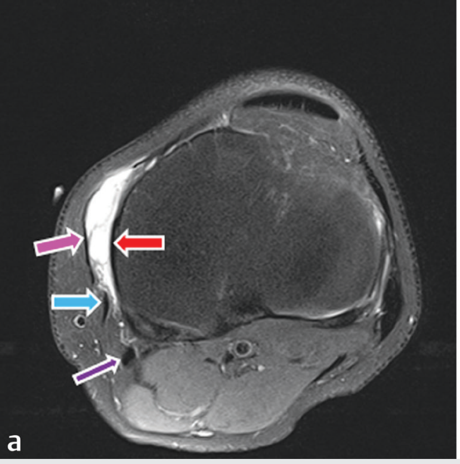
- Red arrow = Tibial collateral ligament (MCL) - the deep structure
- Purple/pink arrow = Sartorius tendon
- Blue arrow = Gracilis tendon
- Dark purple arrow = Semitendinosus tendon
- The bright white fluid between the tendons and MCL = distended pes anserine bursa
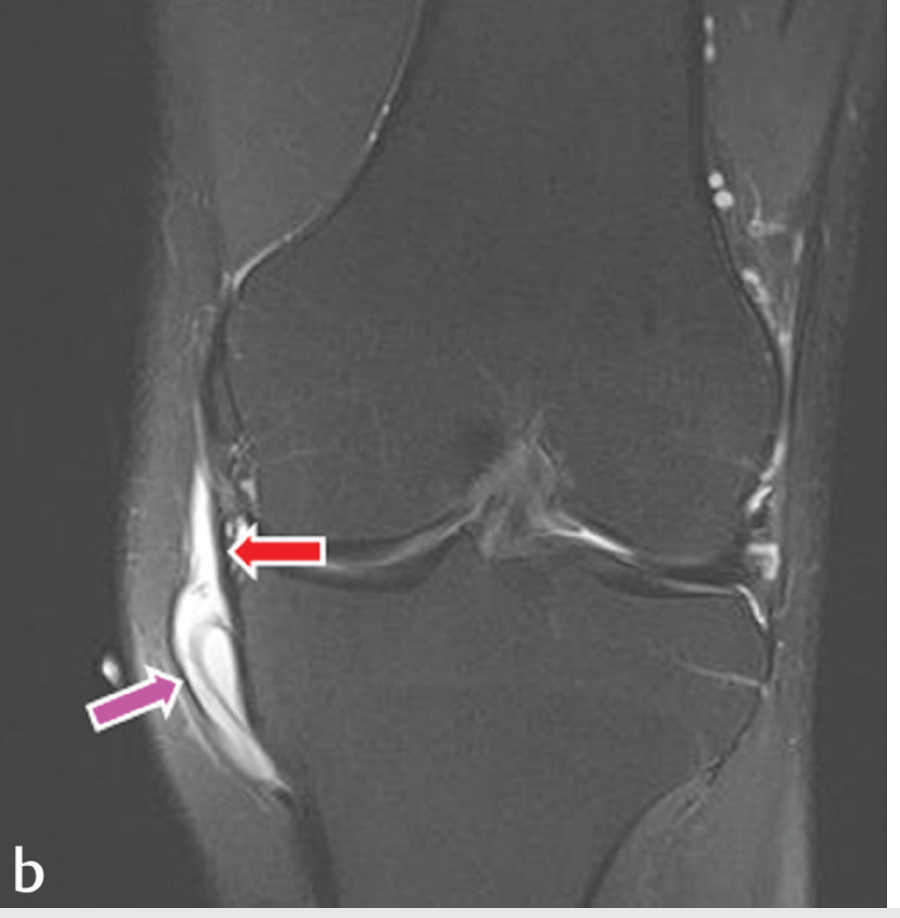
- Red arrow = Tibial collateral ligament
- Purple arrow = Pes anserine bursal fluid pool (bright white = high signal = fluid/inflammation)
- Note the fluid is below the joint line, not at it - this distinguishes bursitis from intra-articular effusion
3. Why This Region Gets Inflamed
| Risk Factor | Why It Applies to Her |
|---|---|
| Female, 48 years old | Classic demographic - pes anserine bursitis predominantly affects middle-aged women |
| Knee joint pathology (meniscus tear + effusion) | Altered joint mechanics change how the three tendons slide, increasing friction on the bursa |
| Quadriceps inhibition | Weak quads force hamstrings and sartorius to overwork as compensatory stabilizers - directly increases compressive load on the bursa |
| Possible overweight (not confirmed but common in this profile) | Body weight increases compressive force through the medial compartment |
| Abnormal gait from pain | Antalgic gait alters the mechanical loading of all three contributing tendons |
4. Clinical Diagnosis - How to Confirm It
Differentiating Pes Anserine Bursitis from MCL Pain
| Feature | MCL Sprain | Pes Anserine Bursitis |
|---|---|---|
| Tenderness location | At or above the medial joint line | 2-3 cm BELOW medial joint line |
| Swelling | Along ligament line | Focal soft swelling below joint line |
| Pain with valgus stress | Yes (MCL is stretched) | Usually NO |
| Pain with knee flexion + resisted internal rotation | No | Yes (compresses bursa) |
| Pain at night/sleep | Usually not | Common - patient rolls onto side and knee adducts, compressing bursa |
| Morning stiffness | Mild | Can be significant |
Palpation Test (Do This Now)
5. Complete Physiotherapy Treatment Protocol for Pes Anserine Region
Phase 1 - Reduce Inflammation (Week 1-2)
- Ice pack wrapped in cloth directly over the bursa (NOT the joint line - 2-3 cm below it)
- 15-20 minutes, 3-4 times/day
- Especially critical in the evening when bursal pressure peaks
- Frequency: 1 MHz (deeper tissue penetration - bursa is subfascial)
- Mode: Pulsed (non-thermal) - 20% duty cycle
- Intensity: 0.8-1.0 W/cm²
- Duration: 5-7 minutes
- Transducer placement: Directly over the pes anserine insertion, 2-3 cm below medial joint line
- Mechanism: Acoustic microstreaming reduces bursal wall inflammation; promotes resolution of fluid without heating (thermal ultrasound could worsen acute bursitis)
- Frequency: 5x/week for 2 weeks
- Couple the ultrasound head with diclofenac gel instead of standard coupling gel
- Drives topical NSAID molecules through the skin into the bursa via ultrasound pressure waves
- Directly reduces local prostaglandin-mediated inflammation at the bursal wall
- Electrode placement: one pad directly over the bursa, one pad on the medial thigh
- Parameters: 80-100 Hz, 100-150 µs pulse width, 20-30 minutes
- Reduces pain to allow comfortable rehabilitation exercises
Phase 2 - Address the Cause (Week 2-6)
| Stretch | Technique | Hold/Reps | Targets |
|---|---|---|---|
| Hamstring stretch (supine, single leg raise) | Lie on back, lift leg straight, feel pull behind knee | 30 sec x 3, twice daily | Semitendinosus |
| Gracilis / adductor stretch | Butterfly stretch - seated, soles together, press knees gently down | 30 sec x 3 | Gracilis |
| Sartorius stretch | Standing quad stretch with hip extended + externally rotated | 30 sec x 3 | Sartorius |
| Calf stretch (gastrocnemius) | Step stretch at wall | 30 sec x 3 | Reduces overall posterior chain tension |
| Exercise | Sets/Reps | Why |
|---|---|---|
| Quadriceps sets + SLR | 3x15 | Restore quad function - reduces reliance on hamstrings/sartorius as compensators |
| Hip abductor strengthening (clamshells, sidelying abduction) | 3x15 | Reduces knee valgus - reduces medial compressive forces on the bursa |
| Glute bridge | 3x12 | Posterior chain activation without direct bursa loading |
| Terminal knee extension (TKE with band) | 3x15 | VMO activation; reduces medial tibial rotation that compresses the bursa |
- Deep squats below 60° (maximum tendon compression on bursa)
- Prolonged stair climbing
- Kneeling on the medial side
- Cycling with seat too low (excessive knee flexion)
Phase 3 - Injection (If Physiotherapy Insufficient in 3-4 Weeks)
| Injection | Effect at 1 Week | Effect at 8 Weeks |
|---|---|---|
| Corticosteroid | Fastest relief (best at 1 week) | Effect wanes by 8 weeks |
| Oxygen-Ozone | Good relief | Sustained effect at 8 weeks |
| Prolotherapy (dextrose 20%) | Moderate | Sustained effect at 8 weeks |
Ultrasound-guided injection of corticosteroid (e.g., methylprednisolone 40 mg + lidocaine 1%) into the pes anserine bursa - this gives a 2-4 week pain-free window during which physiotherapy is most effective.
6. Sleep Position Advice (Very Practical)
- Side-lying causes the top knee to fall into adduction - compressing the bursa between the medial condyle and the tendons
- Supine causes the leg to externally rotate - stretching the gracilis and sartorius insertions
Summary for Your Clinical Practice
LOCATION:   2-3 cm below medial joint line (NOT at joint line = NOT MCL)
TENDONS:    Sartorius + Gracilis + Semitendinosus insert here
BURSA:      Between tendons (superficial) and MCL (deep)
DIAGNOSIS:  Point tenderness 2-3 cm below medial joint line
CAUSE:      Tendon friction on bursa from altered gait + quad inhibition + overuse
TREATMENT:  Ice to bursa → Pulsed ultrasound (1 MHz pulsed) → Hamstring/adductor stretch
            → Quad + hip abductor strengthening → Pillow between knees at night
INJECTION:  Corticosteroid (fastest) or O2-Ozone (longest lasting) under US guidance
            if unresponsive to 3-4 weeks of physiotherapy