


1000231699.jpg
+2 more
Analyze
analyse_media
analyse_media
analyse_media
Reading File
Finding Sources
Finding Sources
Reading File
Reading File
Loading Image
Reading File
I now have all the information needed for a thorough analysis. Here is the complete report:
Fluoroscopy (C-arm) Analysis - Elbow Joint
Modality: Intraoperative / Emergency C-arm fluoroscopy (extremities protocol)
Date: 14/7/2026 - captured at 08:09, 08:10, 08:10 AM
Views: Three sequential oblique/lateral projections of the elbow
Findings Across All Three Frames
Frame 1 (1699) - Oblique/Lateral view:
The distal humerus (capitellum, trochlea) is visible proximally, with the proximal radius and ulna displaced posteriorly and superiorly. The normal radiocapitellar and ulnohumeral articulations are completely disrupted. No hardware present - this is a pre-reduction image.
Frame 2 (1698) - Lateral view:
Clearest demonstration of the dislocation. The radial head (rounded structure, upper right) is completely displaced out of its articulation with the capitellum. The olecranon-trochlea relationship is lost. A suspicious bony fragment is visible near the anterior joint region, raising concern for a coronoid process fracture.
Frame 3 (1697) - Lateral/slightly oblique view:
Similar findings confirming the dislocation. The forearm bones (radius and ulna) are displaced posteriorly en masse relative to the humerus.
Primary Diagnosis
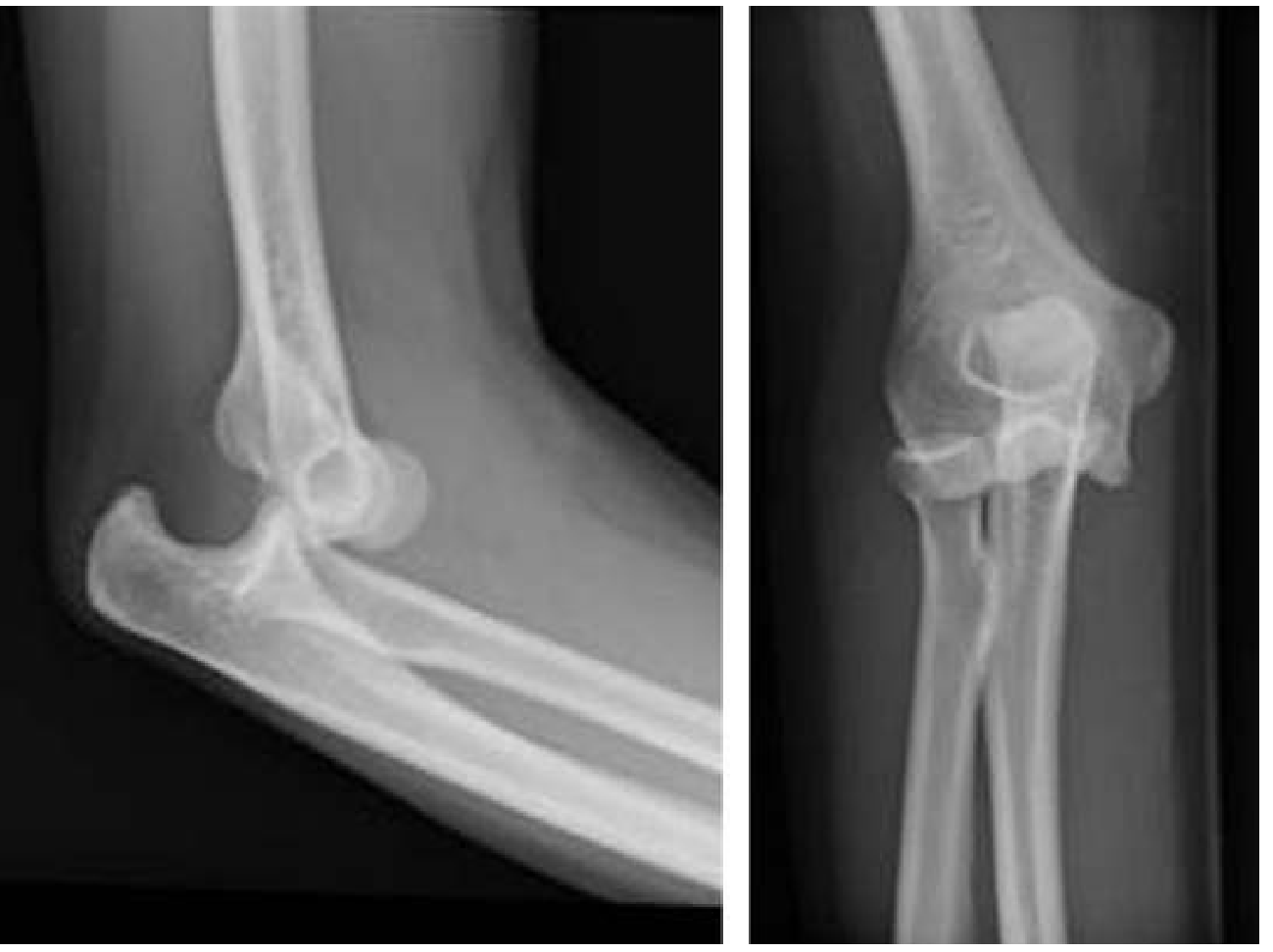
Acute Posterior Elbow Dislocation
This is the most common type of elbow dislocation, accounting for approximately 90% of all elbow dislocations. The mechanism is typically a fall on an outstretched hand (FOOSH) with the elbow in extension, generating posterolateral rotatory displacement of the forearm.
The reference X-rays from Tintinalli's Emergency Medicine below show the characteristic appearance for comparison:
Associated Injury - "Terrible Triad" Concern
The presence of a bony fragment near the coronoid process raises strong suspicion for a Terrible Triad injury, which consists of:
- Elbow dislocation ✓ (confirmed)
- Coronoid process fracture - suspected (bony fragment anteriorly visible on all 3 frames)
- Radial head fracture - cannot be excluded on fluoroscopy alone
Per Rockwood and Green's Fractures in Adults (10th ed., 2025): "Terrible triad injuries are thought to occur by posterolateral rotatory displacement of the ulna... The proposed mechanism is a fall onto an outstretched arm, with supination, valgus, and axial-directed force. The trochlea causes a shear fracture of the coronoid and is accompanied by an LCL injury and/or radial head fracture."
Clinical Priorities
Immediate neurovascular assessment (before and after reduction):
- Brachial artery - arterial injuries occur in 5-13% of elbow dislocations; absent radial pulse is a warning sign
- Ulnar nerve - most commonly injured nerve, affected in up to 20% of cases
- Median nerve
Imaging:
- Post-reduction AP and lateral plain radiographs are mandatory
- CT scan is strongly recommended to fully characterize coronoid fracture pattern (Regan-Morrey classification) and radial head morphology; 3D CT improves interobserver agreement on coronoid fracture classification
Reduction Technique
Per Tintinalli's Emergency Medicine: IV analgesia or procedural sedation is usually required.
Two-person technique (standard):
- Position forearm supine; assistant applies countertraction on upper arm
- Apply longitudinal traction on the wrist/forearm to disengage the coronoid from the olecranon fossa
- Correct any medial/lateral displacement
- Apply slow downward pressure to proximal forearm and flex the elbow until reduction is achieved
Post-reduction: Immobilize in a light splint at 90° flexion, forearm pronated (pronation typically provides the most stable position). Weekly radiographic follow-up.
Management Algorithm (Post-Reduction)
| Finding | Management |
|---|---|
| Stable arc >30° flexion, no displaced fractures | Conservative: splint 7-10 days, early motion |
| Redislocation at >30° flexion | Operative treatment |
| Displaced radial head fracture | ORIF or radial head replacement |
| Coronoid fracture (Regan-Morrey II/III) | ORIF coronoid + LCL repair |
| Full terrible triad | ORIF radial head + coronoid fixation + LCL repair ± MCL |
Per Rockwood and Green's: "Most patients with terrible triad injuries require surgical management to achieve a stable congruous reduction of the elbow allowing early motion... Nonoperative treatment has been associated with less desirable outcomes including stiffness, late instability, and arthrosis."
Summary
These three C-arm images document an acute, unreduced posterior elbow dislocation captured intraoperatively/in the emergency setting on 14 July 2026. Associated coronoid fracture is highly suspected on fluoroscopy. Radial head fracture cannot be excluded. This constellation meets the definition of a potential Terrible Triad injury, which carries significant surgical implications and a guarded prognosis for full range of motion recovery. Post-reduction CT is strongly recommended before finalizing the management plan.
Disclaimer: This analysis is for educational and clinical decision-support purposes only. Final clinical management must be determined by the treating orthopaedic surgeon based on the full clinical picture.
- Tintinalli's Emergency Medicine: A Comprehensive Study
- Rockwood and Green's Fractures in Adults, 10th ed. 2025
This is a shared conversation. Sign in to Orris to start your own chat.