


A 45 yrs old male pt presents with such lesions which are itchy what is Diagnosis and treatment in detail
lichen simplex chronicus lichenification itchy skin plaque
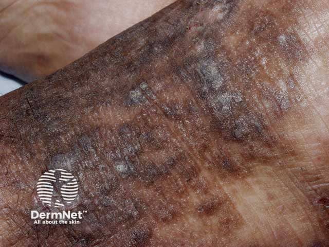
Clinical photography of a cutaneous plaque consistent with lichen simplex chronicus (neurodermatitis) affecting the dorsal hand/forearm. The image demonstrates hyperkeratotic, lichenified plaques with pronounced thickening of the epidermis and accentuated linear skin creases. The plaques are pigmented with variably brown to slate-gray hyperpigmentation and a scaly, rough surface. Dermal inflammation is suggested by peripherally hyperemic borders and faint erythema within the lesion. The distribution appears localized with well-defined margins, and there may be minor fissuring at pressure points. The appearance reflects chronic itch-scratch cycle leading to chronic scratching, which perpetuates the lichenification. The photograph provides a high-resolution, close-up view suitable for dermatology education and differential diagnosis. Notable differentials include atopic dermatitis, discoid (plaque) psoriasis, chronic contact dermatitis, and seborrheic dermatitis; however, the thickened, hyperpigmented plaques with accentuated creases and absence of classic psoriasis scale or orange staining favors lichen simplex chronicus. Clinically, this lesion would correlate with pruritus, focal scratching, and relief after rubbing; management considerations include topical corticosteroids, occlusion, antihistamines, and behavioral modification. This image is relevant for digital dermatology repositories, medical education, and phenotype documentation of neurodermatitis. Documentation should note lesion size, exact location, patient-reported itch intensity, nocturnal scratching, and response to therapy for longitudinal tracking and education.
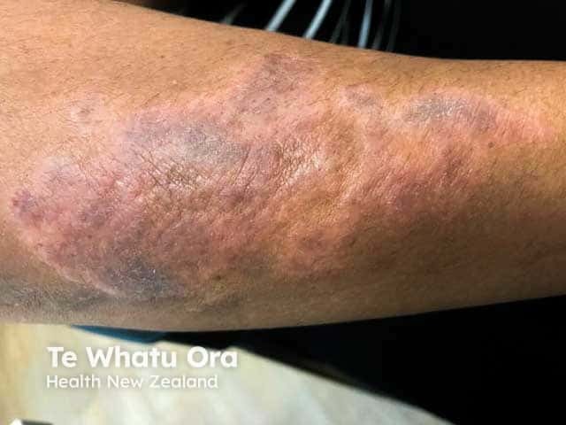
This is a high-resolution clinical photograph of a single, chronically irritated plaque on the forearm, captured with standard color dermatology photography under daylight illumination. The image depicts lichen simplex chronicus (neurodermatitis) manifesting as a well-demarcated, thickened, hyperpigmented plaque with prominent lichenification and fissuring at the surface. The lesion shows intensified skin markings, wrinkling, and a slightly violaceous hue around the periphery, reflecting chronic rubbing and scratching. The surrounding skin is mildly erythematous with subtle edema, consistent with ongoing pruritus and inflammatory response. From a dermatoscopic perspective, surface scales may be scant; however, tactile palpation would reveal firm, leathery consistency. Clinically, lichen simplex chronicus results from an itch-scratch cycle, producing epidermal hyperplasia (acanthosis), hyperkeratosis, and dermal papillary fibrosis. The image highlights typical features used to distinguish chronic eczematous dermatitis from acute flares, such as the thickened plaque, accentuated lines, and lack of acute exudation. Diagnostic significance lies in recognizing chronicity, guiding therapy with potent topical corticosteroids or calcineurin inhibitors, and addressing itch management, psychological triggers, and behavioral modification. Clinically relevant use cases include medical education, dermatology training, differential diagnosis exercises, and image-based documentation for patient records and tele-dermatology. This description supports accurate coding and reproducible educational content for teaching and research in dermatology training.
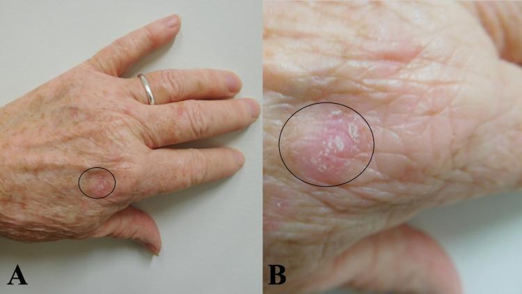
This clinical photograph consists of two panels (A and B) showing a biopsy-confirmed case of lichen simplex chronicus on the left dorsal hand of an elderly patient. Panel A provides a distant view, situating the lesion proximal to the second metacarpophalangeal joint. Panel B offers a close-up (macro) view for detailed morphological assessment. The lesion is a solitary, erythematous, circumscribed plaque approximately 14 x 10 mm in size, highlighted by a black oval. Key clinical features include prominent lichenification (thickening of the skin with exaggerated skin lines), surface scaling (whitish, fine scales), and ill-defined to moderately well-defined borders. The surrounding skin exhibits signs of chronological aging and actinic damage, including solar lentigines and fine rhytids. This image serves as an educational example of the classic presentation of a chronic eczematous process resulting from repeated scratching or rubbing, demonstrating the characteristic texture and color changes associated with lichen simplex chronicus in a common anatomical location.
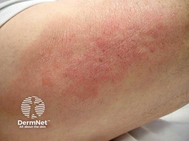
Clinical photograph of lichen simplex chronicus (neurodermatitis) on the upper arm, displaying red-brown, thickened plaques with conspicuous lichenification and accentuated skin markings. The lesion shows a rough, hyperkeratotic surface with underlying erythema, mild scaling, and scratching-induced excoriations. Chronic rubbing perpetuates the itch-scratch cycle, resulting in epidermal hyperplasia (acanthosis) and dermal inflammation, which may produce pigmentary alteration over time. Distribution is localized to a discrete upper-arm region, a common site for neurodermatitis due to habitual rubbing. The boundary is often ill-defined with variable borders, and the surrounding skin may be moderately dry. The image emphasizes hallmark features: durable plaque formation, pronounced skin folding, and a pattern consistent with localized chronic dermatitis rather than an acute eruption. Clinically, this appearance guides diagnosis toward lichen simplex chronicus and helps differentiate from psoriasis, tinea, or acute contact dermatitis. Management implications include potent topical corticosteroids, calcineurin inhibitors in sensitive areas, regular emollients, antihistamines for itch, and behavioral strategies to break the scratching cycle. The photo is valuable for dermatology education, teledermatology triage, and visual recognition of chronic itch-scratch dermatoses on the upper limb. This imaging scenario also supports patient education on chronic dermatitis, triggers identification, and adherence to treatment regimens.
Diagnosis: Lichen Simplex Chronicus (LSC) / Circumscribed Neurodermatitis
Clinical Findings in This Patient
- Diffuse, confluent, thickened plaques with ill-defined borders
- Severe lichenification - markedly exaggerated, deep "bark-like" or crisscross pattern of skin lines with quadrilateral facets
- Reddish-brown to violaceous hyperpigmentation (typical post-inflammatory change in Fitzpatrick skin types V-VI)
- Fine desquamation/scaling with superficial peeling in areas
- Multiple body sites involved - forearm, wrist, hip/upper thigh
- A metal and thread bracelet on the wrist - an important clue, as this may be causing mechanical friction and/or allergic contact dermatitis (nickel allergy) initiating the itch-scratch cycle
What is Lichen Simplex Chronicus?
- Arise on seemingly normal skin, triggered by a habitual scratch
- Develop secondary to another disease (atopic dermatitis, contact dermatitis, tinea/ringworm, psoriasis, stasis dermatitis)
Reference Images
Differential Diagnosis to Consider
| Condition | Distinguishing Features |
|---|---|
| Dermatophyte infection (tinea) | Ring-shaped with active scaly border; KOH positive |
| Psoriasis | Silvery-white thick scales; nail pitting; Koebner phenomenon |
| Nummular eczema | Coin-shaped, oozing lesions |
| Hypertrophic lichen planus | Violaceous papules; Wickham's striae |
| Lichen amyloidosis | Beaded papules on shins; amyloid on biopsy |
| Allergic contact dermatitis | History of allergen exposure; patch test positive |
Investigations
- KOH examination - rule out tinea/fungal infection
- Patch testing - if contact dermatitis (nickel from bracelet) suspected
- Skin biopsy - if diagnosis uncertain; shows compact hyperkeratosis, acanthosis with irregular elongation of rete ridges, hypergranulosis, vertically oriented collagen in papillary dermis
- Baseline investigations if systemic cause suspected: CBC, LFT, RFT, TFT, blood glucose, HbA1c
Treatment in Detail
1. Remove the Trigger
- Remove the bracelet (both metal and thread) immediately - eliminates friction and potential nickel allergen
- Identify and treat any underlying cause (tinea, atopic dermatitis, contact allergy, systemic itch)
2. Topical Corticosteroids (First-Line)
| Agent | Potency | Notes |
|---|---|---|
| Clobetasol propionate 0.05% ointment | Super-high potency | Initial short course (2-4 weeks); avoid prolonged use to prevent atrophy |
| Fluocinonide 0.05% ointment | High potency | Good for thick plaques |
| Triamcinolone acetonide 0.1% | Medium potency | Maintenance; under occlusion for added effect |
| Hydrocortisone butyrate 0.1% | Mild-medium | Reducing phase |
- Occlusion with medium-potency steroid (e.g., wrap overnight with plastic film) dramatically increases penetration
- Steroid-impregnated tape (fludroxycortide tape) provides both occlusion and anti-inflammatory effect
- Taper to lower-potency agents as lesions resolve
3. Calcineurin Inhibitors (Steroid-Sparing)
- Tacrolimus 0.1% ointment - effective for LSC, especially in skin folds and thinner skin areas; avoid steroid atrophy
- Pimecrolimus 1% cream - good adjunctive antipruritic therapy
4. Topical Antipruritic Agents
- Topical doxepin 5% cream - significant antipruritic effect; apply 3-4x daily
- Capsaicin cream 0.025-0.1% - depletes substance P; burning sensation initially; helps break the itch cycle
- Menthol-based preparations - soothing immediate antipruritic effect
5. Intralesional Corticosteroids
- Triamcinolone acetonide suspension 2.5-5 mg/mL intralesionally - for resistant plaques
- Not too superficial (risk of dermal/epidermal atrophy and depigmentation)
- Do not inject into infected lesions
6. Systemic Antipruritic Therapy
- Antihistamines:
- Hydroxyzine 25 mg at night (sedating - helps break nocturnal scratch cycle)
- Cetirizine 10 mg or fexofenadine 180 mg daily
- Gabapentinoids: Gabapentin 300 mg TDS or pregabalin - for neuropathic itch component
- Antidepressants: Doxepin (systemic), mirtazapine, or low-dose tricyclics - especially if associated anxiety or depression
- Short course of oral corticosteroids in severe flares (prednisolone 0.5 mg/kg/day tapering over 2-3 weeks)
7. Phototherapy
- Narrowband UVB (NBUVB) phototherapy - effective for recalcitrant widespread LSC
- 308 nm excimer laser/lamp - targeted phototherapy for localized plaques
- (Dermatology 2-Volume Set 5e)
8. Behavioral / Psychological Measures
- Patient education - explain the itch-scratch cycle; strict instruction to avoid scratching
- Nail trimming - reduces skin damage from scratching
- Wet-wrap therapy or Unna boot occlusion in severe cases - physically prevents scratching and promotes healing
- Habit-reversal therapy - behavioral modification to substitute rubbing/scratching with another action
- Informal insight-oriented psychotherapy - particularly if OCD or anxiety disorder present
- Hydrocolloid dressings - repeated application leads to improvement and protects from scratching
9. Newer/Adjunctive Options
- Botulinum toxin A injection - may be curative in severe, localized lesions
- Topical lidocaine 5% or capsaicin 8% patches (licensed for postherpetic neuralgia) - helpful in recalcitrant cases
- Transcutaneous electrical nerve stimulation (TENS)
- Focused ultrasound
Practical Treatment Algorithm for This Patient
| Step | Intervention |
|---|---|
| Immediate | Remove bracelet; patch test for nickel; KOH scraping |
| Week 1-4 | Clobetasol 0.05% ointment BD + hydroxyzine 25 mg at night |
| Week 4 onwards | Taper to triamcinolone 0.1% under occlusion; add tacrolimus 0.1% |
| If resistant | Intralesional triamcinolone 5 mg/mL; add gabapentin |
| Ongoing | Emollients (thick ointment base), behavioral counseling, identify triggers |
Clinical images reference: Andrews' Diseases of the Skin, Fig. 4.8; Dermatology 2-Volume Set 5e, Fig. 6.7
Can it be discoid eczema
discoid eczema nummular eczema coin shaped plaques skin
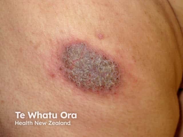
Clinical photograph of a single discoid eczema (nummular dermatitis) plaque on skin of color. Modality: Dermatology photography; technique: close-up, macroscopic view, non-dermoscopic; the lesion is coin-shaped (round to oval), well-demarcated, approximately 1.5–2 cm in diameter. Central area shows diffuse scaling with a crusted surface of grayish-brown hue, surrounded by an erythematous, slightly edematous border. The surrounding skin demonstrates post-inflammatory hyperpigmentation typical of darker phototypes. Textural change includes thickened, xerotic skin with mild fissuring at the periphery. No adjacent vesicles or pustules are evident. Lighting shows warm undertones; surface reveals adherent scale and crust, reflecting chronic inflammation. The differential diagnosis includes tinea corporis, plaque psoriasis, contact dermatitis, and chronic eczema variants; clinical correlation with pruritus, chronicity, and relapsing-remitting pattern favors discoid eczema. In skin of color, hyperpigmentation can persist after inflammation, which may complicate assessment of lesion boundaries. This image is valuable for education on morphology of coin-shaped eczematous plaques and for distinguishing discoid eczema from fungal infections in pigment-rich skin. Potential clinical use cases include dermatology training, telemedicine skin lesion assessment, comparative studies of pigmentary responses, and monitoring response to topical corticosteroids and emollients. This single-field photograph supports recognition of characteristic features: well-marginated plaque, central scaling/crusting, erythematous halo, and pigmentary change.
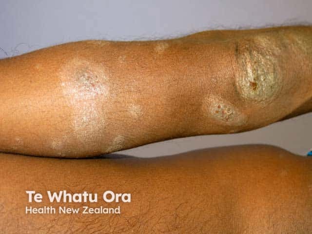
Clinical photography of skin, frontal anterolateral view of the forearm showing discoid eczema (nummular dermatitis) on darker skin. The plaques are coin-shaped, 1–3 cm in diameter, with well-demarcated, slightly scaly borders and a dry, dull sheen. Central areas show relative pallor or lighter pigment, while the outer margins exhibit erythema and hyperpigmentation consistent with postinflammatory changes common in skin of color. Surface scales are adherent, with fissuring at the edges in some plaques; minor excoriations reflect pruritus. The surrounding skin appears dry with reduced textural smoothness, characteristic of xerosis. No overt vesicles or purulent discharge are visible. The distribution is focal to the extensor aspect of the forearm, contrasting with flexural eczema patterns, and may be bilateral but is photographed unilaterally here. The appearance aligns with discoid eczema or chronic nummular dermatitis, often seen in adults with dry or compromised barrier function and irritant exposure. Secondary infection is not evident clinically. Clinically, these findings warrant management focusing on potent topical corticosteroids or calcineurin inhibitors as appropriate, stringent emollients, avoidance of irritants, and follow-up to monitor response and possible recurrence. The image provides a clear visual reference for recognition of color-variant plaque morphology and the chronicity of lesions in dermatologic practice, education, and dermatoepidemiology research.
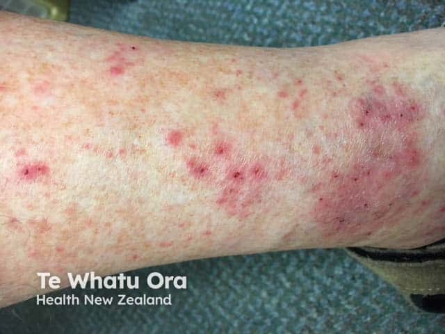
This image is a high-resolution clinical photograph of the left ankle region showing discoid eczema (nummular dermatitis). The skin exhibits multiple coin-shaped erythematous plaques, approximately 0.5–2 cm in diameter, with dry scaling, peripheral crusting, and mild surface excoriation from pruritus. Lesions are well-demarcated and arranged singly and in small clusters along the lateral malleolar area and distal lower leg. Perilesional erythema and xerosis are evident; surrounding skin appears dry without overt infection. The pattern is localized to the ankle region, a common site for discoid eczema, though plaques may appear on dorsum of hands or extensor surfaces in other contexts. Differential considerations include tinea corporis, contact dermatitis, psoriasis, and seborrheic dermatitis. Clinically, discoid eczema is associated with atopy and chronic itch, and management focuses on skin hydration, regular emollients, and anti-inflammatory therapy (topical corticosteroids and calcineurin inhibitors) with avoidance of irritants. This image serves as an educational reference for annular plaque dermatoses, differential diagnosis, and treatment response assessment in chronic eczematous skin disease.
Can It Be Discoid Eczema? - Detailed Analysis
Discoid Eczema vs Lichen Simplex Chronicus - Direct Comparison
| Feature | Discoid Eczema (NE) | Lichen Simplex Chronicus | This Patient |
|---|---|---|---|
| Shape | Coin-shaped (nummular), round/oval, well-demarcated | Irregular, confluent plaques | Large, irregular confluent - favors LSC |
| Primary lesion | Papulovesicles that coalesce into a plaque | Starts as itch → scratch → lichenification | Lichenification is dominant |
| Oozing/crusting | Yes - typically vesicular and weepy acutely | Absent (dry) | No oozing visible - favors LSC |
| Lichenification | Mild to moderate if chronic | Severe and dominant | Severe - favors LSC |
| Scaling | Fine scaling present | Fine or absent | Present but not dominant |
| Size | Typically 2-4 cm discrete plaques | Often broader, thicker patches | Broad, confluent - favors LSC |
| Distribution | Lower legs, dorsal hands, extensor arms | Neck, wrists, ankles, forearms, anogenital | Forearm + thigh - fits both |
| Age & sex | Males >40 yrs predominate | Adults, any sex | 45-yr-old male - fits both |
| S. aureus | Frequently colonized | Less common | Check for secondary infection |
| Key trigger | Trauma, alcohol, atopy, xerosis | Itch habit, anxiety, bracelet friction | Bracelet is a key trigger |
Why Discoid Eczema Is Possible Here
- The patient is a male over 40 - the predominant demographic for nummular eczema
- Extensor forearm involvement is classic for NE
- Relevant positive patch tests are found in 25-33% of NE patients - the metal bracelet (nickel) is highly suspicious
- NE frequently has nummular morphology that becomes lichenified over time - and this patient's chronic lesions show that exact progression
- "In atopy, nummular lesions tend to be more chronic and lichenified" - exactly what is seen here
What Likely Happened (Disease Progression)
Initial trigger (bracelet friction / contact allergy / xerosis)
↓
Acute nummular/discoid eczema (papulovesicular, coin-shaped, itchy)
↓
Chronic scratching and rubbing
↓
Lichenification → Lichen Simplex Chronicus (end stage)
Reference Images - Discoid Eczema

What Distinguishes the Two at This Point
- Remove the bracelet and patch test (nickel allergy)
- KOH scraping to exclude tinea
- If the lesion was coin-shaped and vesicular at onset → NE was the trigger
- Treat with the same regimen (topical corticosteroids, emollients, antihistamines)
Additional Treatment Points Specific to Discoid/NE
- Simple soaking (wet soaks) followed immediately by greasy emollient (soak-and-seal technique) - particularly effective in NE
- Check for and treat secondary S. aureus infection - swab the lesions; if positive, add fusidic acid cream topically or a short course of flucloxacillin/cefalexin orally
- Stop alcohol consumption - associated with NE in adult males; stopping can significantly improve response
- Coal tar preparations - 2% crude coal tar or 20% liquor carbonis detergens (LCD) for refractory plaques
- Patch testing - up to one-third of NE patients have a relevant contact allergen; identify and eliminate it (the metal bracelet is prime suspect here)
- Crisaborole 2% ointment - non-steroidal PDE4 inhibitor; useful alternative to calcineurin inhibitors for maintenance
Bottom Line


Additional photos of above pt
tinea corporis annular ring shaped lesion central clearing abdomen trunk
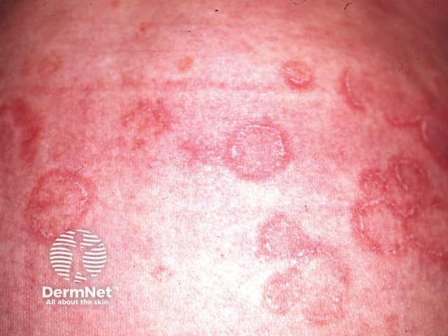
Clinical photography of multiple annular, erythematous plaques on the trunk. The lesions show well-demarcated circular borders with superficial scaling and central clearing, consistent with tinea corporis until proven otherwise. The morphological pattern is ring-shaped (annular) with a slightly raised, erythematous margin and dry scale at the edge. Colors range from pink to brick-red, with varying intensity due to lighting. The trunk is the primary region; symmetrical or scattered lesions may occur, often pruritic. The image illustrates characteristic dermatophyte infection of the cutaneous surface, invading stratum corneum with hyphal elements; differential includes granuloma annulare, nummular eczema, pityriasis rosea, pityriasis circinata, and less likely psoriasis. Clinically significant because early recognition allows topical antifungal therapy (terbinafine, itraconazole) or systemic treatment for extensive disease, and avoids unnecessary biopsy. Suggested confirmatory tests include KOH preparation showing hyaline fungal elements or fungal culture. This photo is suitable for medical education, dermatology training, and dermatopathology correlation; it supports telemedicine triage and patient counseling. Annotated features include annular lesion morphology, active border, central clearing, peripheral scale, and trunk localization. Visual cues such as peripheral scale and erythematous edge aid rapid presumptive diagnosis, while correlation with symptoms (itching) and exposure history supports management decisions and prognosis for educational purposes.
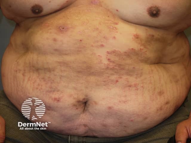
A high-resolution clinical photograph in frontal view of the abdomen and anterior trunk shows widespread, annular, erythematous, scaly plaques with well-defined, active borders and central clearing, classic for tinea corporis (dermatophytosis). The lesions vary in size and appear to coalesce across the midline and lateral abdominal surfaces, with peripheral scaling and mild erythema. Some plaques display lighter centers and darker pruritic borders, and a few areas show subtle vesiculation at advancing margins. No mucosal involvement is evident. The image demonstrates ring-shaped morphology, multiple lesions, and a diffuse trunk distribution that is educational for recognizing extensive cutaneous fungal infection. This depiction supports differential diagnoses including nummular dermatitis, eczema, psoriasis, pityriasis rosea, and granuloma annulare, though the characteristic central clearing and annular configuration strongly favor dermatophyte infection. Clinically, the finding informs management with topical antifungals (azoles or allylamines) and, in extensive disease, systemic therapy such as terbinafine or itraconazole, along with precautions to prevent transmission. The photograph serves as a valuable teaching resource for dermatology, infectious disease, and medical education, illustrating clinical presentation, morphological features, and the importance of correlating with mycological testing when needed. Imaging modality: clinical photography; technique: digital capture; no contrast.
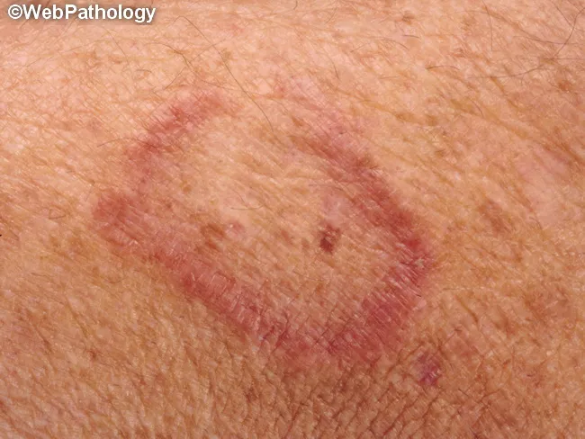
Clinical photograph of a cutaneous lesion illustrating a well‑circumscribed annular plaque with central clearing. The image shows a pink‑red, slightly raised border with fine desquamation against a moderately pigmented skin background. The central area is less inflamed and relatively pale, giving the classic ring‑like (annular) appearance. The lesion is non‑uniform in texture, with mild surface scaling along the active border. Such features are highly suggestive of superficial dermatophyte infection, most consistent with tinea corporis (ringworm) involving the trunk or proximal limbs. Differential considerations include granuloma annulare, nummular dermatitis, and less likely psoriasis with annular morphology. The color and margin support fungal etiology, but confirmation requires mycological testing (potassium hydroxide preparation, fungal culture) or dermoscopy in some settings. Clinically relevant findings include pruritus and possible exposure to infected individuals or animals; lesions typically respond to topical antifungals (terbinafine, itraconazole if extensive) and environmental hygiene measures. This image is suitable for dermatology education, medical student dermatology training, and research on cutaneous fungal infections, differential diagnosis instruction, and exam prep. The content emphasizes macroscopic morphology, border architecture, and central clearing as critical diagnostic cues. This description complements pathology reports by correlating clinical appearance with expected histologic features if biopsy were performed later.
🔴 Important Update: New Photos Change the Diagnosis
What the New Photos Show
| Image | Location | Key Findings |
|---|---|---|
| Photo 1 (abdomen/flank) | Trunk | Single, round-to-polycyclic plaque with erythematous border and central clearing - classic annular lesion |
| Photo 2 (abdomen/back) | Trunk/back | Multiple scattered faint erythematous patches with ill-defined borders, multifocal distribution |
| Photo 3 (abdomen/flank) | Trunk | Arcuate/polycyclic plaques with well-demarcated active scaling borders and distinct central clearing - multiple coalescing rings |
| Photo 4 (heel/ankle) | Lateral foot/heel | Hyperkeratotic plaque with sheet-like peeling, deep fissures at heel, post-inflammatory hyperpigmentation - moccasin-type pattern |
🔑 Revised Primary Diagnosis: Tinea Corporis + Tinea Pedis
The Annular Lesions on the Trunk Are Classic Tinea Corporis
- Well-demarcated, annular (ring-shaped) plaques
- Active, raised, scaling border
- Central clearing - the skin inside the ring is relatively spared
- Polycyclic/arcuate pattern - rings merging and coalescing
- Erythematous advancing edge with desquamation
"This form of ringworm is characterized by one or more circular, sharply circumscribed, slightly erythematous, dry, scaly patches. An advancing scaling edge is usually prominent. Progressive central clearing produces annular outlines that give them the name 'ringworm.' In some cases, concentric circles or polycyclic lesions form, making intricate patterns."
- Andrews' Diseases of the Skin, Clinical Dermatology
Reference Images
The Heel/Foot Lesion = Tinea Pedis (Moccasin-type / Hyperkeratotic type)
- Hyperkeratotic, peeling plaque on lateral heel extending to ankle
- Deep fissures at the heel
- Sheet-like desquamation with post-inflammatory hyperpigmentation
- Moccasin distribution (lateral foot, heel)
Revised Complete Diagnosis
| Site | Diagnosis |
|---|---|
| Trunk (abdomen, flank) | Tinea corporis - annular lesions with central clearing |
| Lateral thigh/hip | Tinea corporis / tinea cruris extension |
| Forearm/wrist | Tinea incognita OR chronic eczematized tinea - lichenified by chronic scratching |
| Heel/lateral foot | Tinea pedis (hyperkeratotic/moccasin type) |
Critical Concept: Tinea Incognita
- Previously treated with topical steroids (which masked the fungal infection)
- OR left untreated and become chronic and eczematized
"Tinea incognita is a term applied to lesions of tinea that have an atypical appearance due to therapy with topical steroids or calcineurin inhibitors. The lesions are often widespread and may lack an advancing, raised, scaly border. Alternatively multiple edges may be present and there may be a lack of central clearing; rather the center may be eczematous."
- Andrews' Diseases of the Skin
🚨 Critical Investigation: MUST DO NOW
- KOH microscopy - look for branching septate hyphae (diagnostic)
- Fungal culture (Sabouraud's dextrose agar) - identifies species in 1-2 weeks
- Wood's lamp - some species fluoresce (M. canis glows green)
- Blood glucose / HbA1c - widespread tinea in an adult male raises suspicion for undiagnosed diabetes mellitus
- HIV screening - widespread tinea corporis can be the presenting sign of AIDS
Revised Treatment Plan
For Tinea Corporis + Tinea Pedis (Extensive, Multi-site Disease)
First-Line Systemic Treatment
| Drug | Dose | Duration |
|---|---|---|
| Terbinafine | 250 mg once daily | 2-4 weeks (corporis); 2-6 weeks (pedis) |
| Itraconazole | 100 mg BD or 200 mg OD | 2-4 weeks |
| Fluconazole | 150-300 mg once weekly | 4-6 weeks |
| Griseofulvin | 500 mg-1g/day | 4-8 weeks (longer, less favored) |
Topical Antifungals (Adjunct)
- Terbinafine 1% cream - once daily, 1-2 weeks even for localized lesions
- Clotrimazole 1% cream or Miconazole 2% cream - BD for 2-4 weeks
- Econazole 1% cream - once daily
For Tinea Pedis (Hyperkeratotic Type)
- Systemic terbinafine 250 mg/day for 4-6 weeks (topical alone fails for hyperkeratotic type)
- Keratolytic preparation to remove thick scale: urea 40% cream or salicylic acid 6-10% applied before antifungal
- Heel emollient (e.g., petroleum jelly) after keratolytic to prevent deep fissuring and secondary bacterial infection
- Open, breathable footwear; change socks daily; dry feet thoroughly after bathing
General Measures
- Treat all sites simultaneously - otherwise re-infection from untreated sites
- Wash clothing and bedlinen at 60°C to kill fungal spores
- Avoid sharing towels, footwear
- Treat household contacts/pets if source identified
- Treat any co-existing tinea unguium (nail involvement) - check nails carefully as this is a common reservoir
Summary: What to Do Now
| Priority | Action |
|---|---|
| Immediate | KOH scrape from trunk ring border |
| Same visit | Blood glucose, HbA1c, HIV screening |
| Start today | Systemic terbinafine 250 mg OD |
| Stop immediately | Any topical steroids on affected areas |
| Topical adjunct | Terbinafine or clotrimazole cream to all sites BD |
| Foot care | Urea cream + terbinafine cream; breathable footwear |
| Follow-up | 4 weeks - assess response; check fungal culture result |