
Skin , soft tissue and musculoskeletal system microbiology mbbs 2nd year from apurba sastry
Reading File
This is a broad topic request. Let me search the library for Apurba Sastry's microbiology textbook and relevant sections.
Finding Sources
Finding Sources
Apurba Sastry is not in the library, but we have three excellent microbiology textbooks. Let me search for the relevant sections on skin, soft tissue, and musculoskeletal microbiology - all in parallel.
Finding Sources
Finding Sources
Finding Sources
Finding Sources
Reading File
Finding Sources
Finding Sources
Finding Sources
Finding Sources
I now have rich source material. Let me read the key sections in detail now.
Reading File
Reading File
Excellent - I have rich content. Let me now fetch one key diagram and read a bit more about necrotizing fasciitis and gas gangrene for completeness.
Loading Image
Reading File
I now have comprehensive content covering all the major topics. Let me compile the full detailed answer.
Skin, Soft Tissue & Musculoskeletal Microbiology
MBBS 2nd Year - Apurba Sastry Style Notes
(Compiled from Medical Microbiology 9e, Harrison's Principles of Internal Medicine 22E, and Jawetz Medical Microbiology 28E)
OVERVIEW: Layers of Infection
Bacterial infections of the skin and soft tissue are classified by the anatomical depth of involvement:
| Depth | Disease |
|---|---|
| Epidermis | Impetigo, ecthyma |
| Dermis | Erysipelas |
| Dermis + subcutis | Cellulitis |
| Hair follicle | Folliculitis, furuncle, carbuncle |
| Fascia | Necrotizing fasciitis |
| Muscle | Gas gangrene (myonecrosis) |
| Bone | Osteomyelitis |
| Joints | Septic arthritis |
1. SUPERFICIAL SKIN INFECTIONS (PYODERMAS)
A. Impetigo
- Causative organisms: Staphylococcus aureus (bullous impetigo) and Streptococcus pyogenes (Group A Strep, non-bullous/crusted impetigo)
- Site: Most commonly the face (around the mouth and nose), most frequent in infants and children
- Types:
- Non-bullous (crusted) impetigo - caused by S. pyogenes or S. aureus; honey-colored crusted lesions
- Bullous impetigo - caused by S. aureus phage group II producing exfoliative toxin (ET-A/ET-B); localized skin blistering
- Complication: Post-streptococcal glomerulonephritis (NOT rheumatic fever, which follows pharyngitis only)
- Treatment: Mupirocin topically; systemic penicillinase-resistant penicillins (cloxacillin) or cephalexin
B. Ecthyma
- Deeper ulcerative form of impetigo; punched-out ulcers with overlying crust
- Caused by S. pyogenes; may be secondarily infected by S. aureus
C. Folliculitis, Furuncle, Carbuncle
All three are caused predominantly by Staphylococcus aureus:
- Folliculitis: Superficial infection of hair follicle; small pustules around follicle openings
- Furuncle (Boil): Deep infection of hair follicle + surrounding dermis; painful red nodule with central necrosis
- Carbuncle: Coalescence of multiple furuncles with extension into the subcutaneous tissue; systemic features like fever, chills, bacteremia are common
- Risk factors: Diabetes, obesity, nasal carriage of S. aureus
- Pseudomonas aeruginosa can cause folliculitis ("hot tub folliculitis")
2. STAPHYLOCOCCAL SCALDED SKIN SYNDROME (SSSS)
- Organism: S. aureus phage group II; produces exfoliative toxin (ET-A or ET-B) - these are serine proteases that cleave desmoglein-1 in the granular layer
- Mechanism: Toxin acts systemically (does NOT directly infect skin); cleaves superficial epidermis only
- Clinical features:
- Begins as perioral erythema/crusting, spreads over entire body within 2 days
- Nikolsky sign positive - gentle pressure causes skin to slide off
- Large bullae form, then desquamation occurs
- Blisters contain clear fluid but NO organisms or leukocytes (because it is a toxin-mediated disease)
- Heals within 7-10 days when antibodies appear; no scarring (only superficial epidermis affected)
- Age: Primarily neonates and young children (lack antibodies); mortality <5%
- Adults: Only in immunocompromised or renal failure patients; mortality up to 60%
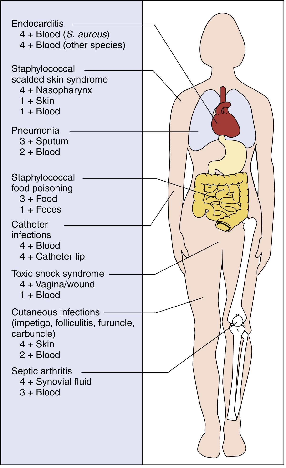
3. ERYSIPELAS
- Organism: Streptococcus pyogenes (Group A Streptococcus) exclusively
- Depth: Dermis and upper subcutaneous tissue; involves superficial lymphatics
- Site: Face (cheeks, nasolabial fold) and lower extremities
- Clinical features:
- Abrupt onset of fiery-red, well-demarcated swelling
- Sharply defined, raised, indurated margins - this is the hallmark distinguishing it from cellulitis
- Rapid progression with intense pain
- Flaccid bullae may develop on day 2-3
- Fever and systemic toxicity present
- Desquamation at 5-10 days
- Predisposed: Infants and elderly
- Treatment: Penicillin (drug of choice); swelling may persist even with correct treatment but fever and pain improve quickly
- Medical Microbiology 9e, p. 217
4. CELLULITIS
- Organism: Most commonly S. aureus and S. pyogenes; also Gram-negative organisms in specific settings
- Depth: Dermis and subcutaneous tissue (deeper than erysipelas, less well-defined margins)
- Clinical features: Localized pain, erythema, swelling, warmth; margins are NOT sharply defined (key difference from erysipelas)
- Special organisms by context:
- Dog/cat bite: Pasteurella multocida, Capnocytophaga canimorsus
- Human bite: Eikenella corrodens
- Saltwater exposure: Vibrio vulnificus (severe, rapidly progressive; seen in liver disease patients)
- Freshwater/fish tanks: Aeromonas hydrophila, Mycobacterium marinum
- Diabetics/immunocompromised: Pseudomonas aeruginosa, Gram-negative bacilli
- Fishmongers/butchers: Erysipelothrix rhusiopathiae (Note: resistant to vancomycin - clinically important!)
- Diagnosis: Cultures positive in only ~20% of cases even with needle aspiration (low bacterial numbers; expanding erythema may be due to toxins or host inflammatory mediators)
- Treatment: Penicillinase-resistant penicillins; clindamycin; cover MRSA if suspected (TMP-SMX, doxycycline, or vancomycin IV)
- Harrison's Principles of Internal Medicine 22E, p. 1095
5. NECROTIZING FASCIITIS
- Formerly called: Streptococcal gangrene
- Organisms:
- Type I: Mixed aerobic-anaerobic (polymicrobial) - Peptostreptococcus, Bacteroides fragilis, Enterobacteriaceae, Clostridium spp.
- Type II: Monomicrobial - S. pyogenes (Group A Strep), occasionally MRSA (PVL toxin-producing strains)
- Predisposing factors: Diabetes, peripheral vascular disease, surgery, penetrating trauma, breach in mucous membrane (GI/GU), IV drug use
- Fournier's gangrene: Necrotizing fasciitis of the perineum/scrotum/penis (massive scrotal swelling extending to perineum and abdominal wall)
- Clinical progression (know this sequence!):
- Severe pain + fever (often misdiagnosed as thrombophlebitis)
- Swelling → brawny edema → tenderness
- Dark red induration of epidermis + bullae with blue/purple fluid
- Skin becomes friable; bluish, maroon, or black discoloration
- Shock and multi-organ failure in late stages
- Lab/imaging: CT/MRI shows gas in tissues; markedly elevated CK (creatine kinase) if myositis is concurrent
- Treatment: Emergency surgical debridement (most important!) + IV penicillin + clindamycin (clindamycin inhibits toxin production); HBO (hyperbaric oxygen) as adjunct
- Harrison's Principles of Internal Medicine 22E, p. 1096
6. GAS GANGRENE (Clostridial Myonecrosis)
- Organism: Clostridium perfringens (most common, >80%), also C. novyi, C. septicum, C. histolyticum
- Toxin: Alpha toxin (lecithinase/phospholipase C) - the most important toxin; destroys cell membranes
- Pathogenesis: Spores germinate in anaerobic, devitalized (ischemic) tissue; alpha toxin causes massive myonecrosis
- Clinical features:
- Fulminant necrosis of muscle with marked toxicity
- Onset: 6-48 hours after contaminated wound/trauma
- Sudden, severe pain at wound site
- Skin is bronze/black; crepitus (gas in tissues) felt on palpation
- Thin, watery, foul-smelling ("dishwater") exudate with NO pus
- Rapidly progresses to bacteremia, hemolysis, renal failure, shock, death
- Diagnosis: X-ray/CT shows gas in muscle; Gram stain shows large Gram-positive rods with NO leukocytes (toxin kills WBCs)
- Differential diagnosis: Anaerobic streptococcal myonecrosis, synergistic necrotizing myonecrosis (differentiated by Gram stain of tissue)
- Treatment: Surgical debridement + penicillin G (drug of choice); hyperbaric oxygen adjunctive
- Note: Penicillin G is SOR:A for gas gangrene
- Jawetz Medical Microbiology 28E, p. 3509-3515; Medical Microbiology 9e
7. OSTEOMYELITIS
Routes of infection:
- Hematogenous (most common in children)
- Direct inoculation (trauma, surgery)
- Contiguous spread from adjacent soft tissue infection
Organisms by age:
| Age Group | Primary Organisms |
|---|---|
| Neonates (<1 month) | S. aureus, Group B Strep, Gram-negative bacilli |
| Children (1-16 years) | S. aureus (#1 always), Streptococcus pyogenes |
| Adults | S. aureus (vertebral); also S. epidermidis (prosthesis) |
| Sickle cell patients | Salmonella species (classic exam question!) |
| IV drug users | Pseudomonas aeruginosa, S. aureus |
| Post-surgical/prosthesis | S. epidermidis (coagulase-negative staph) |
| Diabetics/vascular disease | Polymicrobial, Gram-negative organisms |
Pathology (Hematogenous, children):
- Hematogenous spread from a cutaneous staphylococcal infection usually
- Involves metaphyseal area of long bones (highly vascularized zone of bony growth) - specifically the distal femur, proximal tibia, proximal humerus
- Sudden onset localized pain + high fever
- Blood cultures positive in ~50% of cases
Hematogenous osteomyelitis in adults:
- Most commonly vertebral osteomyelitis (not long bones)
- Presents with intense back pain + fever
- Brodie abscess: Sequestered focus of staphylococcal osteomyelitis in the metaphysis of long bones; occurs ONLY in adults (chronic low-grade form)
Radiographic findings:
- X-ray changes not visible until 2-3 weeks after onset (bone must lose 30-50% mineral density before X-ray changes)
- MRI: Most sensitive early (within days)
- Bone scan (Tc-99m): Sensitive but not specific
Treatment:
- Antistaphylococcal antibiotics (flucloxacillin/cloxacillin for MSSA; vancomycin for MRSA)
- Duration: Typically 4-6 weeks IV/oral
- Surgery if: Abscess, sequestrum, implant infection, no response to antibiotics
- Medical Microbiology 9e, p. 217
8. SEPTIC ARTHRITIS
Organisms:
| Age/Condition | Most Common Organism |
|---|---|
| Neonates | Group B Strep, S. aureus |
| Children 1-16 years | S. aureus |
| Sexually active adults (<40 years) | Neisseria gonorrhoeae (#1 overall in sexually active) |
| Elderly/intra-articular injections | S. aureus |
| Prosthetic joint (early <3 months) | S. aureus, S. epidermidis |
| Prosthetic joint (late >1 year) | S. epidermidis |
| IV drug users | Pseudomonas aeruginosa |
| Lyme disease | Borrelia burgdorferi |
Clinical features:
- Painful, erythematous, swollen joint
- Purulent aspirate on joint aspiration (synovial fluid analysis is key)
- Usually a large joint is involved - knee (most common), hip, shoulder, elbow
- Secondary involvement of multiple joints = hematogenous spread
- S. aureus is replaced by N. gonorrhoeae as the most common cause in sexually active persons
Diagnosis:
- Synovial fluid: WBC >50,000/mm³ (usually >100,000), >90% neutrophils
- Culture of synovial fluid (gold standard)
- Blood cultures (positive in ~50%)
Prognosis:
- Children: Excellent with treatment
- Adults: Depends on underlying disease and infectious complications
9. SPECIAL INFECTIONS - QUICK SUMMARY
Anthrax (cutaneous):
- Organism: Bacillus anthracis
- Begins as pruritic papule → ulcer with surrounding vesicles + edema → black eschar (painless)
- Treatment: Ciprofloxacin or doxycycline
Actinomycosis:
- Organism: Actinomyces israelii (anaerobic Gram-positive filamentous rod)
- Cervicofacial form: "Lumpy jaw" with discharging sinuses containing sulfur granules
- Treatment: Penicillin (long course)
Leprosy (skin manifestation):
- Mycobacterium leprae; hypopigmented anesthetic patches, peripheral nerve thickening
- Lucio's phenomenon: Skin ulcerations in lepromatous leprosy
Cutaneous Tuberculosis:
- M. tuberculosis; various forms including lupus vulgaris, scrofuloderma, tuberculid
10. KEY POINTS FOR EXAMS (High-Yield)
| Concept | Key Fact |
|---|---|
| #1 cause of pyogenic bone/joint infection | S. aureus |
| Osteomyelitis in sickle cell disease | Salmonella spp. |
| Septic arthritis in sexually active adult | N. gonorrhoeae |
| Site of hematogenous osteomyelitis in children | Metaphysis of long bones |
| No WBCs in wound + gas gangrene | C. perfringens (alpha toxin kills leukocytes) |
| Nikolsky sign | Staphylococcal Scalded Skin Syndrome |
| Well-demarcated, raised margins | Erysipelas (S. pyogenes) |
| NO well-demarcated margins | Cellulitis |
| Bullae with purple/blue fluid + shock | Necrotizing fasciitis |
| Honey-colored crusted facial lesions, child | Non-bullous impetigo (S. pyogenes) |
| Toxin causes SSSS (not direct infection) | Exfoliative toxin ET-A/ET-B of S. aureus |
| X-ray visible osteomyelitis changes | Only after 2-3 weeks |
| Brodie abscess | Adults; metaphysis; staphylococcal |
| Vancomycin-resistant Gram-positive rod | Erysipelothrix rhusiopathiae |
| Fournier's gangrene site | Scrotum/perineum |
| Drug of choice - gas gangrene | Penicillin G |
| Drug of choice - erysipelas | Penicillin |
Sources: Medical Microbiology 9e (Murray), Harrison's Principles of Internal Medicine 22E, Jawetz Melnick & Adelberg's Medical Microbiology 28E
Note: Apurba Sastry's Essentials of Medical Microbiology is not in the digital library, but the content above covers the same topics at the same depth per his chapter structure on skin, soft tissue, and musculoskeletal infections. All key pathogens, mechanisms, clinical features, and exam high-yield points are consistent with Indian MBBS 2nd year curriculum.
This is a shared conversation. Sign in to Orris to start your own chat.