
Patho physiology of chronic otitis media
Reading File
Chronic otitis media (COM) involves persistent middle ear inflammation, often driven by eustachian tube dysfunction, biofilm formation, and immune-mediated mucosal changes. The core mechanism starts with inadequate middle ear aeration leading to negative pressure, mucosal metaplasia, and a cycle of infection and inflammation.
Searching sources...
Finding Sources
Finding Sources
Searching PubMed
"chronic otitis media" AND pathophysiology
Good results. Now let me read the key sections in parallel from the most authoritative ENT textbooks.
Reading File
Reading File
Reading File
Let me now read deeper into the Scott-Brown's and Shambaugh sections for the detailed pathophysiology of COM.
Reading File
Reading File
Finding Sources
Now let me get the etiology/pathophysiology section from Shambaugh and the detailed pathophysiology from Scott-Brown's:
Reading File
Reading File
Finding Sources
Now let me get the cholesteatoma bone erosion and inflammatory mediator content:
Reading File
Reading File
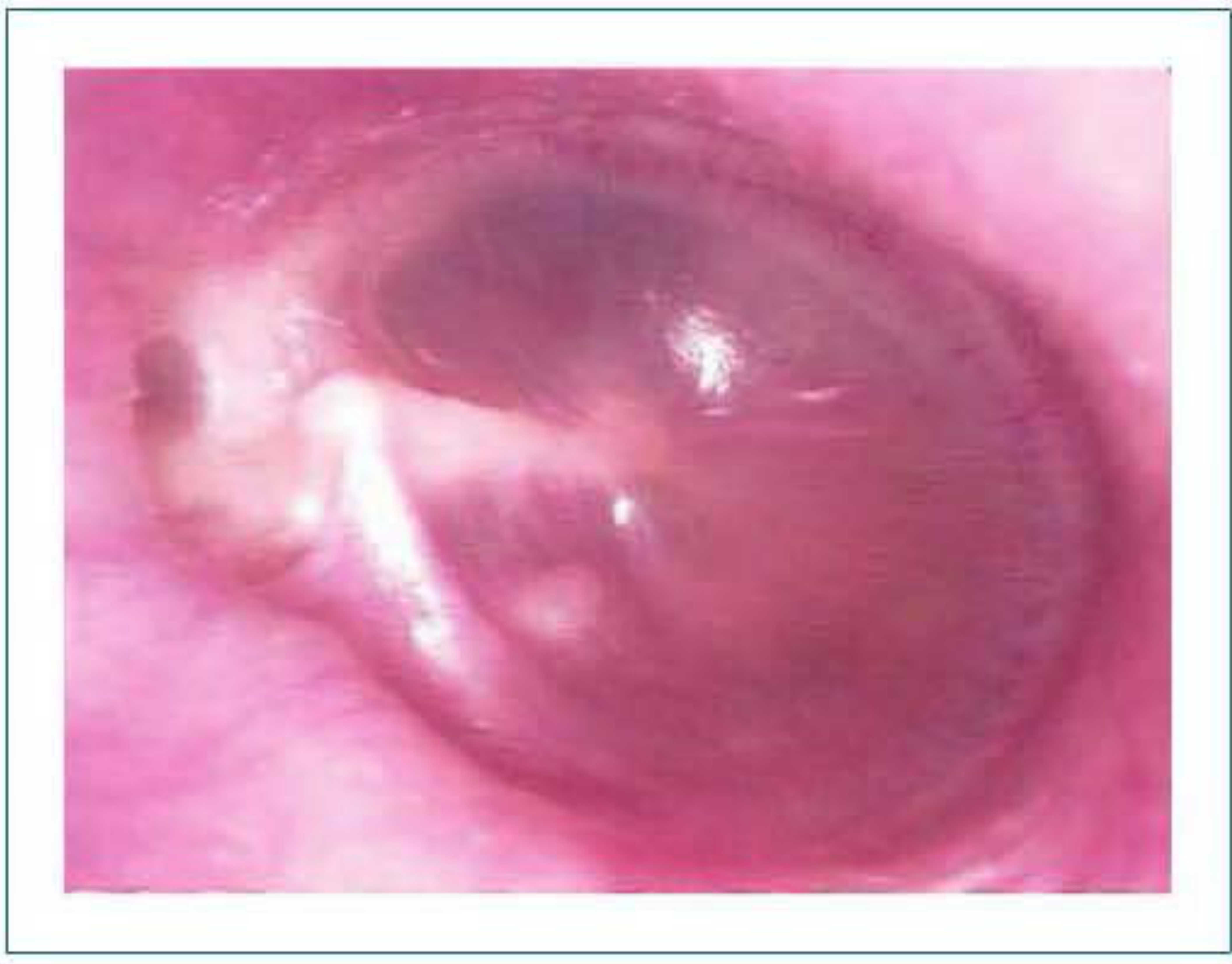
I now have rich, comprehensive content from multiple authoritative ENT textbooks. Let me also fetch the retraction pocket image and the tympanosclerosis image to include in the answer.
Loading Image
Loading Image
I now have comprehensive information from multiple authoritative ENT textbooks. Here is a thorough, well-structured answer on the pathophysiology of chronic otitis media.
Pathophysiology of Chronic Otitis Media (COM)
Definition
Chronic otitis media is an inflammatory process of the middle ear that results in long-term - often permanent - structural changes to the tympanic membrane and middle ear, including atelectasis, perforation, tympanosclerosis, retraction pockets, or cholesteatoma, with variable involvement of the ossicular chain. (Shambaugh Surgery of the Ear)
1. Central Role of Eustachian Tube Dysfunction (ETD)
The foundation of COM pathophysiology is Eustachian tube (ET) dysfunction. Normally, the ET:
- Equalizes middle ear pressure with atmospheric pressure
- Drains secretions from the middle ear into the nasopharynx
- Protects the middle ear from nasopharyngeal pathogens
When ET fails, these three functions break down:
| Failure | Consequence |
|---|---|
| Poor pressure equalization | Sustained negative middle ear pressure |
| Impaired drainage | Accumulation of secretions and effusion |
| Loss of protection | Entry of pathogens from nasopharynx |
Negative middle ear pressure is the primary mechanical driver. It draws the tympanic membrane medially, creating retraction pockets - first in the pars flaccida (attic region), then potentially extending into the posterosuperior pars tensa. (Shambaugh Surgery of the Ear, Scott-Brown's Vol 2)
2. Mucosal Changes and the Inflammatory Cascade
Once infection or persistent effusion sets in, the middle ear mucosa undergoes a cascade of changes:
A. Initial Inflammatory Response
- Bacteria and their toxins (endotoxins + exotoxins) in the middle ear - often in a biofilm state - trigger an immune response
- Neutrophils, macrophages, and lymphocytes are recruited into the submucosa
- Early mediators released include:
- Arachidonic acid metabolites (prostaglandins, leukotrienes)
- Histamine
- Platelet activating factor (PAF)
- Adhesion cell molecules
B. Key Cytokines
The primary cytokines driving chronic middle ear inflammation are (Shambaugh Surgery of the Ear):
- TNF-α - produced by macrophages; upregulates production of other cytokines
- IL-1β - central mediator; stimulates fibroblasts, endothelial cells, osteoclasts, T-cells, B-cells, monocytes, and neutrophils
- Supporting roles: IL-2, IL-4, IL-5, IL-6, IL-8, IL-10, IFN-γ
C. Mucosal Metaplasia
In otitis media with effusion (OME, a precursor to COM), basal mucosal cells differentiate into secretory/goblet cells, converting normal flat epithelium into a mucus-secreting respiratory-type epithelium. This creates a self-perpetuating cycle of effusion production. (Shambaugh Surgery of the Ear)
3. Biofilm Formation
A major reason COM becomes chronic is bacterial biofilm persistence. In biofilms:
- Bacteria aggregate on mucosal surfaces in a polysaccharide matrix
- Nutrients and fluid pass freely within the matrix, and bacteria communicate via hormonal signals (quorum sensing)
- Bacteria are shielded from host defenses and antibiotics
- Traditional middle ear fluid cultures are often negative because bacteria are not free-floating; they are sequestered onto the mucosa surface
Hall-Stoodley (2006) found bacterial biofilms in 92% of pediatric patients with COM undergoing tympanostomy tube placement (vs. none in controls), detected by confocal laser-scanning microscopy. (Shambaugh Surgery of the Ear)
Common organisms: H. influenzae, S. pneumoniae, Pseudomonas aeruginosa, S. aureus, and anaerobes (particularly in cholesteatoma).
4. Granulation Tissue Formation and Tympanic Membrane Breakdown
As chronic inflammation persists:
- Bacterial toxins and inflammatory mediators interact with the edematous mucosa
- The basement membrane of the epithelium ruptures - inflammatory cells from the lamina propria enter the lumen
- Angiogenic growth factors (e.g., VEGF) and epithelial growth factors drive:
- Fibroblast recruitment
- Neovascularization
- Polyp and granulation tissue formation
- Granulation tissue enzymes (collagenases, proteases) break down the collagen skeleton of the tympanic membrane
- The weakened TM, combined with negative middle ear pressure, retracts medially - forming retraction pockets
5. Pathways Leading to the Major Complications of COM
A. Tympanic Membrane Perforation
- Deepening retraction pockets contact underlying mucosa/granulation tissue
- Fibrous adhesions tether the TM medially (adhesive otitis media)
- Eventually, perforation occurs, allowing ongoing contamination from external canal water and bacteria
B. Ossicular Chain Damage
- Ossicular erosion (without cholesteatoma): chronic surrounding hyperaemia and enzymatic action erode the ossicles, especially the long process of the incus (most vulnerable due to its blood supply)
- Tympanosclerosis: calcification of the fibrous layer of the TM and/or fixation of ossicles; results from repeated cycles of inflammation, hyalinization of collagen, and dystrophic calcification - particularly affecting the tympanic membrane and epitympanum
- Fibrous ossicular fixation: from fibrous adhesion bands in the middle ear
C. Cholesteatoma Formation
A cholesteatoma is a keratinizing squamous epithelium within the middle ear - it does not belong there and is the most destructive complication of COM.
Pathogenesis (Acquired):
- Retraction pocket theory: The most accepted mechanism. A deep retraction pocket in the pars flaccida progressively accumulates desquamated keratin debris. As the pocket deepens and migrates, it behaves like a self-expanding cholesteatoma.
- Epithelial migration theory: Squamous epithelium migrates through a perforation into the middle ear.
- Squamous metaplasia theory: Chronic inflammation causes metaplasia of the middle ear mucosa from cuboidal/columnar to squamous epithelium.
- Basal cell hyperplasia: Proliferation of basal keratinocytes through the TM into the middle ear.
Bone Erosion by Cholesteatoma (RANK/RANKL axis):
The old "pressure necrosis" theory has been abandoned - direct pressure from a cholesteatoma only reaches 1.3-11.9 mmHg, far below capillary perfusion pressure (25 mmHg). Bone erosion is now understood to be osteoclast-mediated:
- Cholesteatoma-induced inflammation increases expression of RANKL on stromal cells and activated T-cells
- RANKL binds RANK on osteoclast precursor cells in the perimatrix - triggering osteoclastogenesis
- Macrophage Colony Stimulating Factor (M-CSF) and osteoprotegerin (OPG) also regulate this process
- Activated multinucleated osteoclasts directly resorb bone via acid phosphatase, collagenase, and cathepsin-like proteases in the resorption lacunae
- Intramembranous bone (mastoid, middle ear) is more susceptible than endochondral bone (otic capsule)
(Cummings Otolaryngology, Shambaugh Surgery of the Ear)
6. Classification of COM (Nadol)
| Type | Features |
|---|---|
| Chronic active - with cholesteatoma | Destructive, bone-eroding, requires surgery |
| Chronic active - without cholesteatoma | Granulation tissue, perforation with discharge |
| Chronic inactive - with perforation | Dry, no active inflammation |
| Chronic inactive - with retraction pocket | ETD-driven TM retraction |
| Chronic inactive - adhesive otitis media | TM adherent to medial wall |
| Chronic inactive - ossicular fixation/resorption | Conductive hearing loss |
| Chronic inactive with frequent reactivation | Recurrent wet episodes on background COM |
7. Why Some Patients Progress to COM While Others Resolve
This remains largely unknown. Factors thought to contribute include:
- Genetic susceptibility to impaired mucociliary clearance
- ET anatomy and function (inherently poor ET function in cleft palate, Down syndrome)
- Biofilm persistence
- Host immune response variability
- Recurrent acute OM episodes (multiple bouts perpetuate mucosal changes)
- Mastoid pneumatization: poorly pneumatized mastoids have less volume reserve and worse buffering of negative pressure
Summary Flowchart
ET Dysfunction
    ↓
Negative Middle Ear Pressure
    ↓
Mucosal Edema + Effusion accumulation
    ↓
Bacterial colonization → Biofilm formation
    ↓
Cytokine cascade (TNF-α, IL-1β) → Persistent inflammation
    ↓
Mucosal metaplasia (secretory cells) → More effusion
Granulation tissue → TM collagen breakdown
    ↓
TM retraction / Perforation
    ↓
├─ Tympanosclerosis (calcification)
├─ Ossicular erosion / fixation
└─ Cholesteatoma formation
        ↓
   RANKL/RANK-driven osteoclast activation
        ↓
   Bone erosion → Complications
   (labyrinthine fistula, facial nerve, intracranial extension)
Key Sources:
- Shambaugh Surgery of the Ear - detailed cytokine cascade and biofilm pathophysiology
- Cummings Otolaryngology Head and Neck Surgery - cholesteatoma bone erosion and RANKL/RANK pathway
- Scott-Brown's Otorhinolaryngology Head & Neck Surgery Vol 2 - ETD, tympanosclerosis, ossicular damage
- Bailey and Love's Short Practice of Surgery - COM overview and imaging findings
This is a shared conversation. Sign in to Orris to start your own chat.