
Embryology of female genital tract, write about who classification of Mullerian duct anomaly, clinical features and management 10 marks obgy pg exam
WHO AFS classification Mullerian duct anomalies diagram uterus types
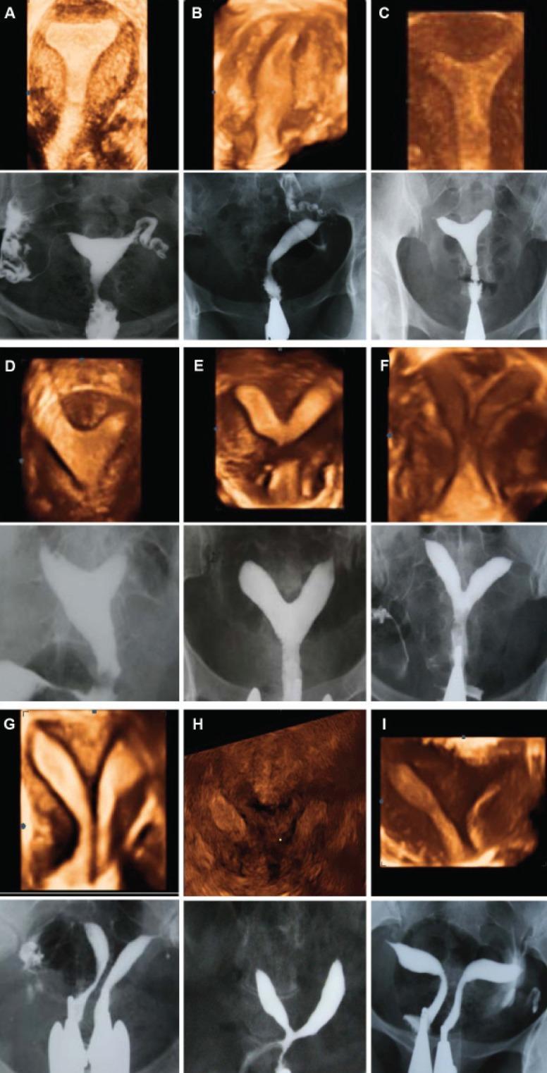
This composite educational graphic provides a side-by-side comparison of 3D ultrasound (coronal view) and Hysterosalpingography (HSG) imaging for the classification of Mullerian Duct Anomalies (MDAs) according to American Fertility Society (AFS) criteria. The panel features nine clinical cases (A-I), each illustrating a specific uterine morphology. (A) Normal uterus with a convex fundal contour and triangular cavity. (B) Unicornuate uterus demonstrating a single uterine horn and fallopian tube. (C) Arcuate uterus characterized by a mild fundal indentation. (D-G) Subtypes of septate uterus, ranging from partial to complete septation, where the 3D ultrasound clarifies the external fundal contour (straight or convex) versus the internal division. (H) Bicornuate uterus showing a prominent external fundal cleft and two divergent horns. (I) Uterus didelphys displaying two completely separate uterine bodies. The dual-modality approach highlights how 3D ultrasound assesses both the external serosal contour and internal cavity, while HSG provides high-contrast visualization of the internal lumen and tubal patency.
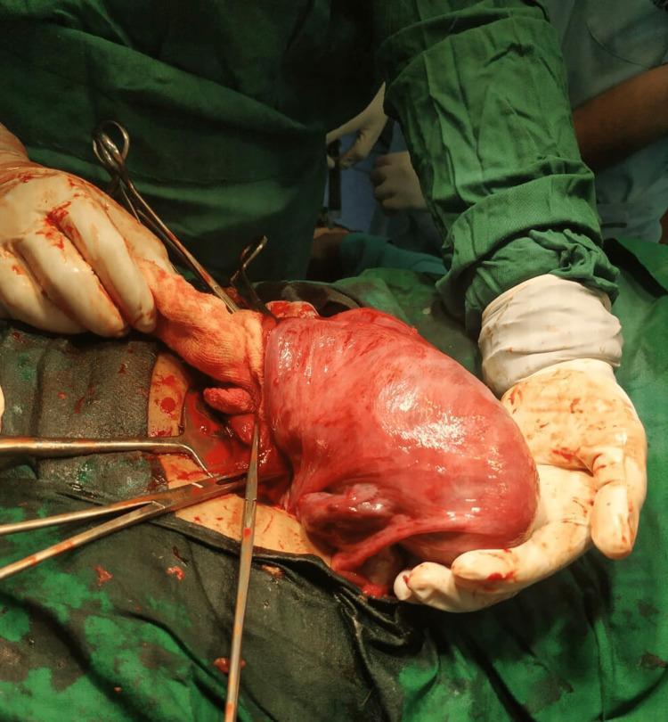
This clinical photograph captures an intraoperative view during an emergency Lower Segment Cesarean Section (LSCS), demonstrating a unicornuate uterus. The uterus is notably tubular and elongated, exhibiting a characteristic 'banana-shaped' morphology associated with Müllerian duct anomalies. The uterine surface appears erythematous and highly vascularized, typical of gestational changes. The surgical field is established with green sterile drapes, and the uterus is being exteriorized and supported by a surgeon wearing blood-stained sterile gloves. Surgical instruments, including forceps and retractors, are visible within the abdominal incision. This image serves as a significant educational example of a Class II Müllerian duct anomaly (AFS classification), highlighting the anatomical variations encountered during obstetric surgery in patients with congenital uterine malformations.
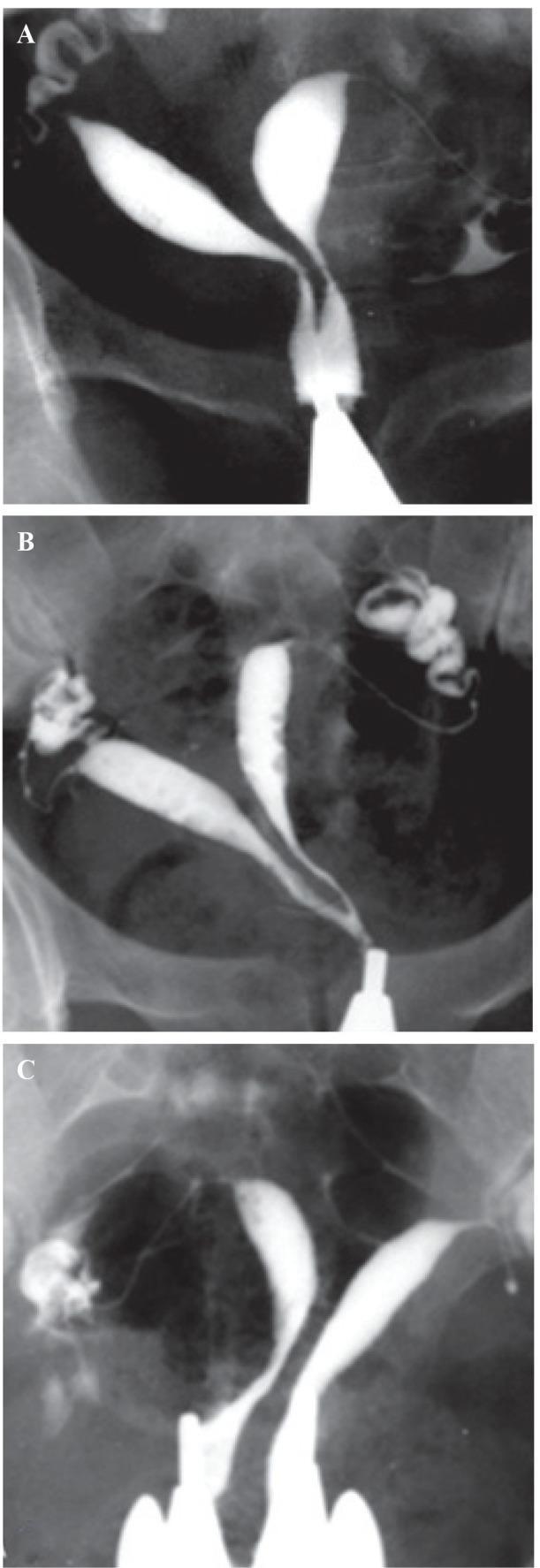
This composite of three fluoroscopic hysterosalpingography (HSG) images (A, B, and C) demonstrates varying degrees of a septate uterus, classified as American Fertility Society (AFS) Class V Müllerian duct anomalies. Each image shows radiopaque contrast material outlining the uterine cavities and cervical canals. Image A illustrates a partial septum extending from the fundus to the lower internal os, resulting in two separate uterine horns that converge into a single cervical canal. Image B displays a more extensive septum reaching the external os, creating two distinct cervical canals that terminate at a single cervical opening. Image C demonstrates a complete septate uterus, often termed 'pseudodidelphys,' where the septum divides the entire length of the uterus and cervix, resulting in two completely separate uterine cavities and two distinct cervical canals. These images highlight the diagnostic role of HSG in characterizing the length and clinical significance of uterine septa, which is essential for differentiating them from bicornuate uteri and planning potential hysteroscopic resection.
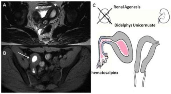
This composite educational material demonstrates Mullerian duct anomalies (MDA). Figure A is an axial T2-weighted MRI scan showing an asymmetrical uterine morphology with heterogeneous signal intensity, indicating a deviated endometrial cavity. Figure B is an axial fat-saturated T1-weighted MRI scan revealing a localized area of high signal intensity (hyperintense) within a rudimentary uterine horn, consistent with methemoglobin from blood degradation products (hematometra). Figure C is a clinical illustration summarizing the findings: a didelphys/unicornuate uterus with an obstructed, non-communicating right horn leading to hematosalpinx. The diagram also correlates this genital anomaly with ipsilateral renal agenesis, a common associated finding in Herlyn-Werner-Wunderlich syndrome or related MDA classifications. This content is designed for medical education regarding diagnostic imaging of congenital female reproductive tract malformations and the importance of screening for associated renal variations.
WHO ESHRE ESGE classification Mullerian duct anomaly 2023 female genital tract embryology management
Embryology of Female Genital Tract, WHO/AFS Classification of Mullerian Duct Anomalies, Clinical Features, and Management
EMBRYOLOGY OF FEMALE GENITAL TRACT
Early Indifferent Stage (Up to 6th Week)
Development of Mullerian Ducts (Weeks 6-9)
- At approximately the 6th week of gestation, the Mullerian (paramesonephric) duct develops as an evagination (invagination) of the coelomic epithelium, just lateral to the Wolffian duct on each side.
- The Mullerian duct grows caudally, crossing the Wolffian duct anteriorly, and the two Mullerian ducts then approach each other in the midline.
Three Phases of Mullerian Development
Fate of Mullerian Structures
| Structure | Formed From |
|---|---|
| Fallopian tubes | Unfused, cranial portions of each Mullerian duct |
| Uterine body and fundus | Fused caudal portions after septal resorption |
| Uterine cervix | Fused caudal portions |
| Upper 2/3 of vagina | Mullerian duct contribution via sino-vaginal bulbs |
| Lower 1/3 of vagina | Urogenital sinus |
Role of Anti-Mullerian Hormone (AMH)
WHO / AFS CLASSIFICATION OF MULLERIAN DUCT ANOMALIES
The ESHRE/ESGE CONUTA classification (2013) is a newer system used in Europe, but AFS remains the most widely cited in South Asian PG exams.
CLASS I - Uterine Agenesis / Hypoplasia
- (a) Vaginal - vaginal agenesis alone
- (b) Cervical - cervical aplasia/hypoplasia
- (c) Fundal - fundal hypoplasia
- (d) Tubal - tubal hypoplasia
- (e) Combined - most severe
- Uterine and vaginal agenesis or hypoplasia
- Ovaries and fallopian tubes are intact
- Karyotype 46XX
- Associated with renal (30-40%), skeletal (12%), and auditory abnormalities
CLASS II - Unicornuate Uterus
- (a) Rudimentary horn with endometrial cavity - communicating
- (b) Rudimentary horn with endometrial cavity - non-communicating
- (c) Rudimentary horn without endometrial cavity
- (d) No rudimentary horn
- Often asymptomatic
- Cyclic pelvic pain if non-communicating functioning horn (hematometra)
- Recurrent pregnancy loss (45%), preterm delivery (20%)
- Banana-shaped or elongated uterus on imaging
- Increased risk of endometriosis
- Associated with ipsilateral renal agenesis (40%)
CLASS III - Uterus Didelphys
- Two normal-sized uterine horns, two cervices
- Longitudinal vaginal septum present in 75% of cases
- Best obstetric outcome among fusion anomalies
- Widely divergent horns on HSG
CLASS IV - Bicornuate Uterus
- (a) Bicornuate unicollis - one cervix, separated fundus (fundal cleft > 1 cm)
- (b) Bicornuate bicollis - two cervices (partial non-fusion extends to cervix)
CLASS V - Septate Uterus
- (a) Complete septate - septum extends from fundus to cervical os or beyond
- (b) Partial/incomplete septate - septum does not reach internal os
CLASS VI - Arcuate Uterus
CLASS VII - DES-Related Uterine Anomaly
SUMMARY TABLE: AFS Classification at a Glance
| Class | Anomaly | Embryological Defect | Key Clinical Feature | Management |
|---|---|---|---|---|
| I | Agenesis/Hypoplasia | Failure of both Mullerian ducts | Primary amenorrhea (MRKH) | Vaginal dilation/neovaginoplasty |
| II | Unicornuate | Failure of one Mullerian duct | Cyclic pain (non-communicating horn), IUGR | Excise non-communicating horn |
| III | Didelphys | Complete non-fusion | Longitudinal vaginal septum (75%) | Resect vaginal septum if needed |
| IV | Bicornuate | Incomplete fusion | Heart-shaped uterus, fundal cleft >1cm | Strassman's metroplasty |
| V | Septate | Failure of resorption | Highest miscarriage rate | Hysteroscopic metroplasty |
| VI | Arcuate | Minor resorption defect | Normal variant | Usually none |
| VII | DES-related | In utero DES exposure | T-shaped uterus | Hysteroscopic correction |
INVESTIGATIONS FOR MULLERIAN ANOMALIES
- 3D Transvaginal Ultrasound - best first-line modality; differentiates septate vs bicornuate by showing both external contour and internal cavity
- Hysterosalpingogram (HSG) - shows internal cavity, tubal patency; cannot assess external contour
- Sonohysterography (SIS) - sensitive for cavity lesions; 3D-SIS is excellent for anomaly typing
- MRI Pelvis - gold standard; best for complex anomalies, rudimentary horns, MRKH; shows zonal anatomy and uterine morphology precisely
- Diagnostic hysteroscopy - direct visualization of cavity; combined with laparoscopy when needed
- Laparoscopy - to assess external uterine contour and associated endometriosis
- Renal ultrasound - mandatory (30-40% associated renal anomalies, especially in Class I and II)
- Karyotype - if primary amenorrhea or ambiguous genitalia
KEY CLINICAL POINTS FOR PG EXAM
- Most common anomaly: Septate uterus (35%)
- Worst pregnancy outcome / highest miscarriage rate: Septate uterus
- Best pregnancy outcome: Didelphys uterus (paradoxically)
- Most common cause of primary amenorrhea from Mullerian anomaly: MRKH syndrome
- Gold standard imaging: MRI pelvis
- Treatment of septate uterus: Hysteroscopic metroplasty (NOT Strassman)
- Treatment of bicornuate with pregnancy loss: Strassman's metroplasty (abdominal)
- Most important association to check: Renal anomalies (USG kidneys mandatory)
- DES daughters: T-shaped uterus + risk of clear cell carcinoma of vagina