
Is fish mouth vertebrae and hand feet syndrome found in SCA ?
sickle cell dactylitis hand foot syndrome X-ray fish mouth vertebrae
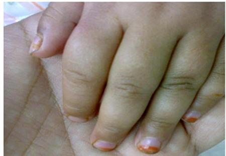
This clinical photograph displays a close-up view of a pediatric patient's hand demonstrating dactylitis, a hallmark clinical sign of sickle cell disease (SCD) often referred to as 'hand-foot syndrome.' The image shows marked, diffuse swelling of the second and third digits, characterized by a 'sausage-like' appearance. The fingers appear edematous and bulbous, particularly around the proximal interphalangeal (PIP) joints, with taut, shiny overlying skin indicating acute soft tissue inflammation. The nail beds exhibit slight pallor, consistent with the patient's associated severe anemia. Clinically, this presentation represents a vaso-occlusive crisis where sickle-shaped red blood cells obstruct small capillaries in the phalanges, leading to ischemia and painful infarction of the bone marrow and periosteum. This visual is a classic educational example of early-onset skeletal complications in toddlers with sickle cell anemia, illustrating the inflammatory response to microvascular occlusion in the extremities.
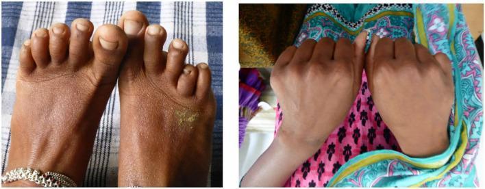
Two clinical photographs demonstrate structural hand and foot abnormalities in patients with sickle cell disease. The left image shows a dorsal view of the feet, highlighting a shortened fourth metatarsal on the right foot, which causes the fourth toe to appear significantly recessed compared to the third and fifth digits. The right image displays a dorsal view of both hands, where the right fifth metacarpal is noticeably shortened, resulting in a proximally displaced fifth digit (pinky finger). These findings illustrate the clinical sequelae of dactylitis (hand-foot syndrome), where vaso-occlusive crises or secondary infection in childhood lead to premature epiphyseal fusion and arrested longitudinal bone growth. This visual evidence serves as a diagnostic indicator of past severe vaso-occlusive events in patients with homozygous sickle cell (HbSS) disease or other sickle cell variants common in South Asian populations.
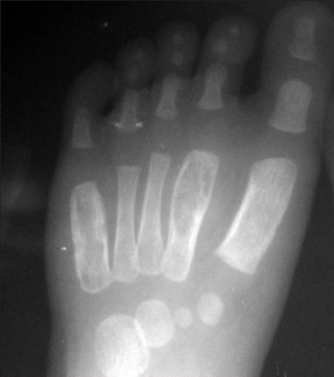
**Imaging Modality:** Anteroposterior (AP) projection radiograph (X-ray). **Anatomical Region:** Left pediatric foot, including the metatarsals, phalanges, and tarsal bones. **Observed Pathology:** The image demonstrates significant fusiform, expansile remodeling of the diaphyses of the second and fifth metatarsals. These lesions exhibit cortical thinning and a "wind-filled" appearance, characteristic of tuberculous dactylitis, also known as spina ventosa. **Characteristic Visual Features:** * **Expansile Bone Destruction:** Marked expansion of the medullary cavity in the affected metatarsals. * **Cortical Changes:** Prominent thinning of the overlying cortex without an aggressive periosteal reaction. * **Skeletal Maturity:** Presence of open growth plates and primary ossification centers consistent with a pediatric patient. * **Distribution:** Multiple, non-contiguous involvement of the small tubular bones of the foot. **Key Diagnostic Features:** The combination of cystic-like expansion of short tubular bones and cortical thinning in a pediatric patient is highly suggestive of osseous tuberculosis (spina ventosa), distinguishing it from other lytic bone lesions or dactylitis caused by sickle cell disease or syphilis.
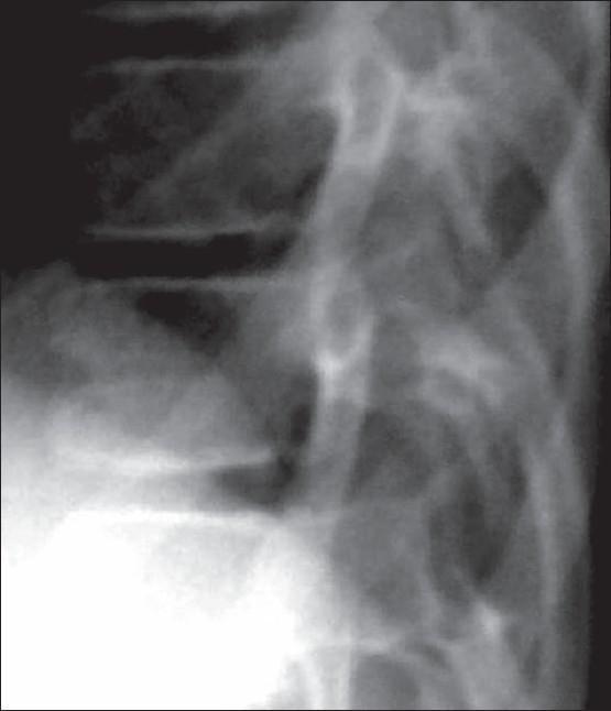
A diagnostic x-ray in lateral projection focusing on the dorsal (thoracic) spine, illustrating a characteristic skeletal abnormality known as 'fishmouth' or biconcave vertebrae. The radiograph reveals significant concavity of both the superior and inferior endplates of multiple vertebral bodies, resulting in a narrowed central height and an expanded appearance of the intervertebral disc spaces. This biconcave morphology is a hallmark finding in several systemic conditions, including progeria (Hutchinson-Gilford Progeria Syndrome), sickle cell disease, and severe osteoporosis. The image also depicts surrounding anatomical structures, including the posterior elements of the vertebrae, pedicles, and portions of the adjacent ribs. Clinically, this imaging modality is used to identify vertebral remodeling and bone density changes in pediatric and adult metabolic bone disorders.
Fish Mouth (Biconcave) Vertebrae in SCA
- Repeated vaso-occlusive crises cause infarction of the central portion of the vertebral end plates (the weakest zone, supplied by terminal capillaries)
- This leads to concave depressions of both the superior and inferior end plates of multiple vertebral bodies
- The intervertebral disc spaces appear widened (H-shaped or fish mouth appearance on lateral spine X-ray)
- The peripheral end plates are relatively spared (unlike osteoporosis, where the entire end plate caves in)
Hand-Foot Syndrome (Sickle Cell Dactylitis)
| Feature | Detail |
|---|---|
| Age group | Children < 5 years (rarely seen after age 5) |
| Also called | Sickle cell dactylitis |
| Mechanism | Infarction of bone marrow and cortical bone of metacarpals, metatarsals, and proximal phalanges, leading to periostitis |
| Clinical findings | Diffuse swelling, tenderness, warmth of hands and feet lasting 1-3 weeks |
| X-ray findings | Periosteal elevation, subperiosteal new bone formation, areas of radiolucency and increased density in metacarpals/metatarsals/proximal phalanges |
| Outcome | Changes disappear after several months; little or no residual damage in most cases |
| Also seen in | Sickle cell thalassemia (HbS/thal) |
Key Teaching Points
- Fish mouth vertebrae = virtually pathognomonic for SCA; caused by central end-plate infarction from vaso-occlusion
- Hand-foot syndrome = often the first manifestation of SCA in infants; due to dactylitis from bone marrow infarction in small bones of hands and feet
- Both are skeletal complications resulting from the same underlying mechanism: vaso-occlusion by sickled RBCs causing ischemia and infarction of bone
Peripheral blood picture in Sca and thalssemia
sickle cell anemia peripheral blood smear sickle cells target cells
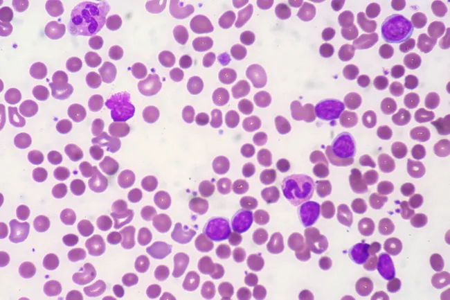
Peripheral blood smear from a patient with known chronic lymphocytic leukemia (CLL) showing autoimmune hemolytic anemia (AIHA). Brightfield microscopy of Wright-Giemsa stained smear at 1000x (oil immersion) reveals two dominant cell populations: numerous small mature lymphocytes typical of CLL and red blood cells showing spherocytic deformation. A smudge cell is evident just left of center, a classical clue for CLL. Red cells display reduced central pallor consistent with spherocytes, indicating intravascular or extravascular hemolysis driven by autoantibodies. The background shows mild polychromasia and rare nucleated RBCs, consistent with a robust reticulocyte response; the corrected reticulocyte count in this case is markedly elevated (>7%). Platelets are not significantly abnormal. These features collectively support autoimmune destruction of erythrocytes in the setting of CLL. Diagnostic significance lies in recognizing AIHA as a common autoimmune complication in CLL, with a clinical picture of sudden Hb decline and compensatory erythropoiesis. Differential considerations include drug-induced hemolysis, other causes of hemolytic anemia, or marrow infiltration by CLL. Clinical correlation includes therapy implications (steroids, rituximab) and close monitoring of hemolysis markers. This image is a valuable teaching resource for hematology morphology, AIHA in CLL, and differential diagnosis in anemia.
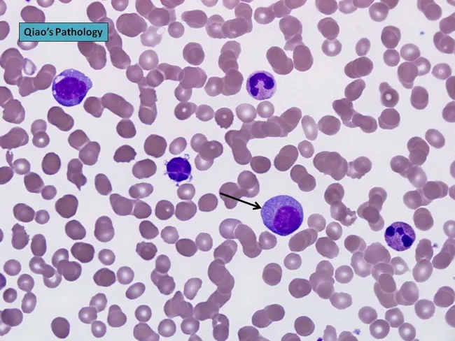
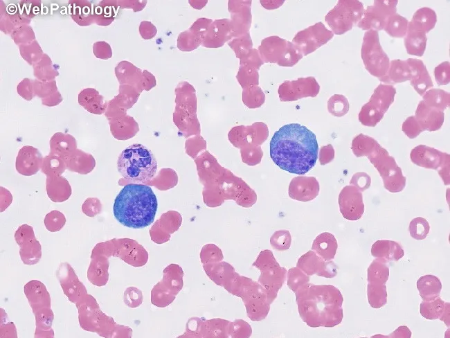
Peripheral blood smear prepared with Wright-Giemsa stain and examined under brightfield illumination using a 100× oil immersion objective reveals a single large plasma cell among a field of mature erythrocytes. The plasma cell displays features typical of clonal plasma cells: basophilic cytoplasm, an eccentrically placed round nucleus, and a conspicuous perinuclear hof; background shows rouleaux formation of red cells. This image exemplifies hematologic involvement by a plasma cell dyscrasia. In plasma cell leukemia, circulating plasma cells constitute more than 20% of leukocytes, a hallmark distinguishing it from conventional multiple myeloma, although both conditions share monoclonal plasma cell proliferation. The presence of circulating plasma cells is associated with younger patient age, higher incidence of anemia, thrombocytopenia, organomegaly, and possible lymphadenopathy; however lytic bone lesions may be less common. Immunophenotypic patterns often include lack of CD56 expression relative to myeloma. Clinically, this morphological finding warrants confirmation with complete blood count, peripheral smear review, quantitative plasma cell percentage, serum protein electrophoresis, and immunofixation to identify monoclonal immunoglobulin isotype (IgD/IgE or light-chain predominance). Early detection of circulating plasma cells supports prognosis assessment and guides therapy decisions in plasma cell dyscrasia management and monitoring, including risk stratification and treatment response evaluation for practice in clinics.
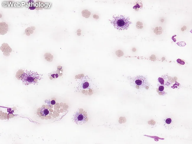
This peripheral blood smear was prepared and stained with Wright-Giemsa and examined by bright-field light microscopy to assess circulating hematopoietic cells in suspected systemic mastocytosis (SM). The smear shows rare, but conspicuous, mast cells with round to polygonal shapes and abundant cytoplasmic granules that impart a deep purple, metachromatic appearance. Nuclei are generally round to ovoid and may be slightly irregular or bilobed in some cells; granules occupy most of the cytoplasm and may obscure nuclear detail. The surrounding leukocytes include eosinophils and neutrophils, and the erythrocyte background is normocytic with occasional anisocytosis. In SM, circulating mast cells are typically infrequent; when they constitute a substantial fraction of leukocytes (≥10%), the finding is highly suggestive of mast cell leukemia (MCL). The image exemplifies an increased mast cell burden consistent with aggressive SM or MCL, often accompanied by cytopenias such as anemia or thrombocytopenia and potential associated hematologic neoplasms (e.g., CMML, MDS/MPN). This morphologic snapshot supports integration with phenotypic assays (CD117/c-KIT), flow cytometry, and molecular testing for KIT mutations, to establish diagnosis, prognostication, and therapeutic planning. Relevant keywords: systemic mastocytosis, mast cell leukemia, circulating mast cells, metachromatic granules, Wright-Giemsa, toluidine blue, KIT D816V, CD117, SM-AHN. This description emphasizes morphology guiding diagnosis appropriately.
thalassemia major peripheral blood smear target cells nucleated RBC hypochromic microcytic
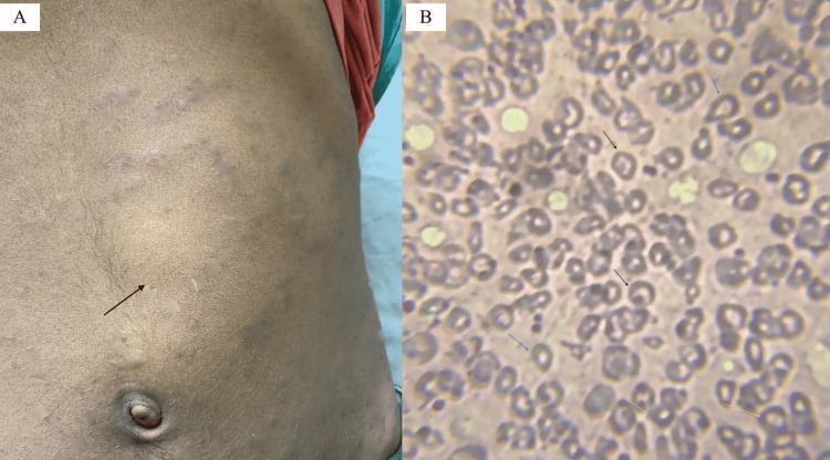
Educational medical image panel consisting of a clinical photograph and a diagnostic pathology image illustrating physical and hematological findings of a hemoglobinopathy such as Hb E/beta-thalassemia. Panel A is a clinical photograph of a patient's abdomen showing a prominent bulge in the left upper quadrant and mid-abdomen, with a black arrow indicating the anterior notch of a massively enlarged spleen (splenomegaly). The overlying skin shows subtle striae or discoloration. Panel B is a peripheral blood smear (Leishman stain, 200x) showing significant red blood cell (RBC) dysmorphology. Key findings include target cells (codocytes) marked by black arrows, teardrop cells (dacrocytes) marked by blue arrows, and basophilic stippling within microcytic, hypochromic RBCs marked by yellow arrows. These visual markers are classic indicators of disordered erythropoiesis and hemoglobin synthesis abnormalities, providing a diagnostic bridge between clinical examination (splenomegaly) and laboratory hematopathology.
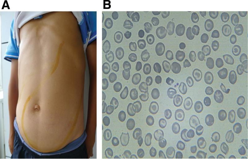
This composite educational image illustrates the clinical signs and laboratory findings of thalassemia intermedia. Panel A is a clinical photograph of a patient's torso showing massive abdominal distention. Yellow skin markings delineate the margins of significant hepatomegaly and massive splenomegaly, with the spleen extending from the left upper quadrant across the midline and deep into the pelvic region, occupying nearly the entire abdominal cavity. Panel B is a peripheral blood smear (light microscopy) demonstrating classic hematological abnormalities associated with thalassemia. The red blood cells exhibit marked microcytosis (small size) and hypochromia (central pallor), along with numerous target cells (codocytes) and irregularly contracted cells (schistocytes). These findings collectively highlight the physical manifestations of extramedullary hematopoiesis and chronic hemolytic anemia characteristic of advanced hemoglobinopathies.
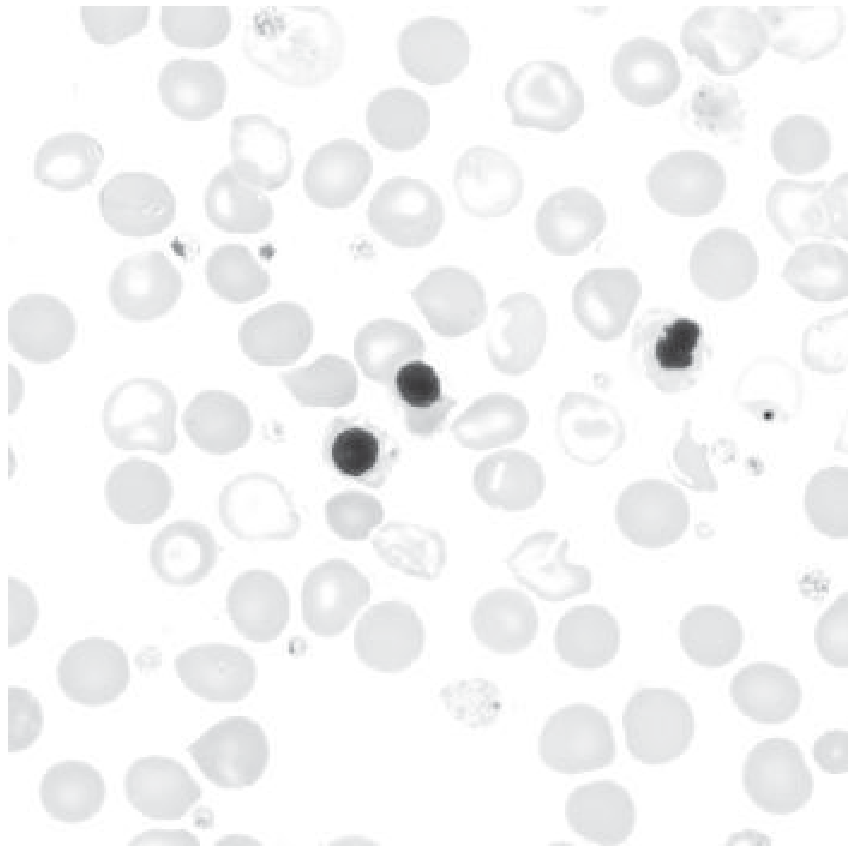
This is a peripheral blood smear prepared with Wright-Giemsa stain and examined under brightfield illumination at high magnification (approximately 1000x total, 100x objective with oil immersion). The smear displays predominantly erythrocytes with uniform pink cytoplasm and characteristic biconcave morphology, arranged singly with occasional rouleaux. Interspersed among the red cells are leukocytes with visible nuclei. Notably, two large lymphocyte-like cells or mononuclear leukocytes appear conspicuously larger than surrounding erythrocytes; these cells have round to oval, deeply basophilic nuclei with dense chromatin and scant, lightly basophilic cytoplasm. No visible granulocytic cytoplasm granules or multiple lobes are clearly resolved in this field, and there are no obvious nucleoli or cytoplasmic inclusions evident in these cells. The background lacks abnormal pigment or reticulocytosis. Overall cell morphology is consistent with a normal or mildly reactive hematologic smear; in a clinical context, additional data such as complete blood count, differential, and patient history would be needed to determine whether there is leukocytosis or blasts. This image serves as an educational reference for recognizing basic peripheral blood components, evaluating leukocyte size variation, and assessing RBC morphology for hematology training and diagnostic differential diagnoses. Additional cases and image contexts can reinforce recognition of hematologic normal variants and patterns.
Peripheral Blood Picture: SCA vs Thalassemia
Sickle Cell Anemia (HbSS) - Peripheral Blood Smear
RBC Morphology
| Finding | Description |
|---|---|
| Sickle cells (drepanocytes) | Elongated, crescent/sickle-shaped RBCs - the hallmark finding; seen only in homozygous HbSS |
| Target cells (codocytes) | Cells with bull's-eye appearance; central dense area + pale ring + dense rim |
| Howell-Jolly bodies | Nuclear remnants in RBCs - due to functional asplenia from autoinfarction |
| Nucleated RBCs (normoblasts) | Reflect erythropoietic stress in bone marrow |
| Polychromasia | Blue-tinged RBCs = reticulocytes; reflects compensatory erythropoiesis |
| Anisocytosis + poikilocytosis | Variable size and shape |
| Boat-shaped cells | Reversibly sickled cells |
Key point: In sickle cell trait (HbAS), the peripheral smear is normal - sickling only occurs in vitro under hypoxic conditions.
Other CBC findings in SCA
- Hemoglobin: 6-9 g/dL (normocytic or mildly macrocytic due to reticulocytosis)
- Elevated reticulocyte count (5-15%)
- Leukocytosis and thrombocytosis (especially during crises)
- Elevated bilirubin (indirect) - hemolysis
Thalassemia - Peripheral Blood Smear
Beta-Thalassemia Major (Cooley's Anemia)
| Finding | Description |
|---|---|
| Severe microcytosis | MCV 48-72 fL - markedly small RBCs |
| Severe hypochromia | Pale RBCs - reduced Hb content |
| Target cells (prominent) | Numerous codocytes - classic feature |
| Nucleated RBCs | Poorly hemoglobinized normoblasts; characteristic of beta-thal major/intermedia |
| Poikilocytosis | Tear-drop cells (dacrocytes), fragmented cells (schistocytes) |
| Anisocytosis | Mix of microcytes and macrocytes |
| Basophilic stippling | Dots in RBCs - reflect RNA remnants from ineffective erythropoiesis |
| Polychromasia | Occasional |
| Occasional spherocytes | Due to membrane damage |
Beta-Thalassemia Minor (Trait)
- Mild microcytosis + hypochromia
- Target cells (few)
- RBCs are small and pale but regular in shape
- Mild or no anemia; often mistaken for iron deficiency
Alpha-Thalassemia Trait
- Same as beta-thal minor: mild microcytosis, hypochromia, target cells
- Often the smear is nearly normal (silent carrier: completely normal)
HbH Disease (3 alpha-gene deletion)
- Moderate microcytosis, hypochromia, poikilocytosis
- HbH inclusions visible with brilliant cresyl blue stain (golf-ball or "multiple inclusion" pattern)
Side-by-Side Comparison
| Feature | SCA (HbSS) | Beta-Thal Major | Beta-Thal Minor |
|---|---|---|---|
| Sickle cells | ✅ Hallmark | ❌ | ❌ |
| Target cells | ✅ Present | ✅✅ Prominent | ✅ Few |
| Microcytosis | ❌ (normocytic) | ✅✅ Severe | ✅ Mild |
| Hypochromia | Mild | ✅✅ Severe | ✅ Mild |
| Nucleated RBCs | ✅ Occasional | ✅✅ Characteristic | ❌ |
| Basophilic stippling | Occasional | ✅✅ Prominent | Mild |
| Howell-Jolly bodies | ✅✅ (autosplenectomy) | Occasional | ❌ |
| Poikilocytosis | Moderate | ✅✅ Marked | Mild |
| Polychromasia | ✅ (reticulocytosis) | ✅ | ❌/Mild |
| Anemia severity | Moderate-severe | Severe (Hb 30-65 g/L) | Mild/none |
Microscopic, blood smear , lab findings of IDA
iron deficiency anemia peripheral blood smear hypochromic microcytic pencil cells
Peripheral blood smear from a patient with known chronic lymphocytic leukemia (CLL) showing autoimmune hemolytic anemia (AIHA). Brightfield microscopy of Wright-Giemsa stained smear at 1000x (oil immersion) reveals two dominant cell populations: numerous small mature lymphocytes typical of CLL and red blood cells showing spherocytic deformation. A smudge cell is evident just left of center, a classical clue for CLL. Red cells display reduced central pallor consistent with spherocytes, indicating intravascular or extravascular hemolysis driven by autoantibodies. The background shows mild polychromasia and rare nucleated RBCs, consistent with a robust reticulocyte response; the corrected reticulocyte count in this case is markedly elevated (>7%). Platelets are not significantly abnormal. These features collectively support autoimmune destruction of erythrocytes in the setting of CLL. Diagnostic significance lies in recognizing AIHA as a common autoimmune complication in CLL, with a clinical picture of sudden Hb decline and compensatory erythropoiesis. Differential considerations include drug-induced hemolysis, other causes of hemolytic anemia, or marrow infiltration by CLL. Clinical correlation includes therapy implications (steroids, rituximab) and close monitoring of hemolysis markers. This image is a valuable teaching resource for hematology morphology, AIHA in CLL, and differential diagnosis in anemia.
Peripheral blood smear prepared with Wright-Giemsa stain and examined under brightfield illumination using a 100× oil immersion objective reveals a single large plasma cell among a field of mature erythrocytes. The plasma cell displays features typical of clonal plasma cells: basophilic cytoplasm, an eccentrically placed round nucleus, and a conspicuous perinuclear hof; background shows rouleaux formation of red cells. This image exemplifies hematologic involvement by a plasma cell dyscrasia. In plasma cell leukemia, circulating plasma cells constitute more than 20% of leukocytes, a hallmark distinguishing it from conventional multiple myeloma, although both conditions share monoclonal plasma cell proliferation. The presence of circulating plasma cells is associated with younger patient age, higher incidence of anemia, thrombocytopenia, organomegaly, and possible lymphadenopathy; however lytic bone lesions may be less common. Immunophenotypic patterns often include lack of CD56 expression relative to myeloma. Clinically, this morphological finding warrants confirmation with complete blood count, peripheral smear review, quantitative plasma cell percentage, serum protein electrophoresis, and immunofixation to identify monoclonal immunoglobulin isotype (IgD/IgE or light-chain predominance). Early detection of circulating plasma cells supports prognosis assessment and guides therapy decisions in plasma cell dyscrasia management and monitoring, including risk stratification and treatment response evaluation for practice in clinics.
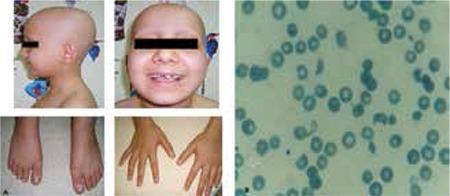
This composite image consists of clinical photographs and a diagnostic peripheral blood smear. Figure 1a (left) shows a pediatric patient with clinical signs including total alopecia (complete absence of scalp hair) and nail pitting affecting both hands and feet, characterized by small punctate depressions on the nail plates. Figure 1b (right) is a microscopic view of a peripheral blood smear demonstrating microangiopathic hemolytic anemia. Visible are numerous schistocytes—fragmented, irregular red blood cells including helmet cells and triangular forms—alongside some normal erythrocytes showing central pallor. The educational focus is on the multi-systemic manifestations of a medical condition, likely a microangiopathic process such as Hemolytic Uremic Syndrome (HUS) or similar systemic pathology, illustrating the correlation between dermatological signs (hair and nail changes) and hematological abnormalities (hemolysis and schistocytosis).
This peripheral blood smear was prepared and stained with Wright-Giemsa and examined by bright-field light microscopy to assess circulating hematopoietic cells in suspected systemic mastocytosis (SM). The smear shows rare, but conspicuous, mast cells with round to polygonal shapes and abundant cytoplasmic granules that impart a deep purple, metachromatic appearance. Nuclei are generally round to ovoid and may be slightly irregular or bilobed in some cells; granules occupy most of the cytoplasm and may obscure nuclear detail. The surrounding leukocytes include eosinophils and neutrophils, and the erythrocyte background is normocytic with occasional anisocytosis. In SM, circulating mast cells are typically infrequent; when they constitute a substantial fraction of leukocytes (≥10%), the finding is highly suggestive of mast cell leukemia (MCL). The image exemplifies an increased mast cell burden consistent with aggressive SM or MCL, often accompanied by cytopenias such as anemia or thrombocytopenia and potential associated hematologic neoplasms (e.g., CMML, MDS/MPN). This morphologic snapshot supports integration with phenotypic assays (CD117/c-KIT), flow cytometry, and molecular testing for KIT mutations, to establish diagnosis, prognostication, and therapeutic planning. Relevant keywords: systemic mastocytosis, mast cell leukemia, circulating mast cells, metachromatic granules, Wright-Giemsa, toluidine blue, KIT D816V, CD117, SM-AHN. This description emphasizes morphology guiding diagnosis appropriately.
Iron Deficiency Anemia (IDA) - Complete Lab & Microscopic Findings
1. Peripheral Blood Smear (PBS)
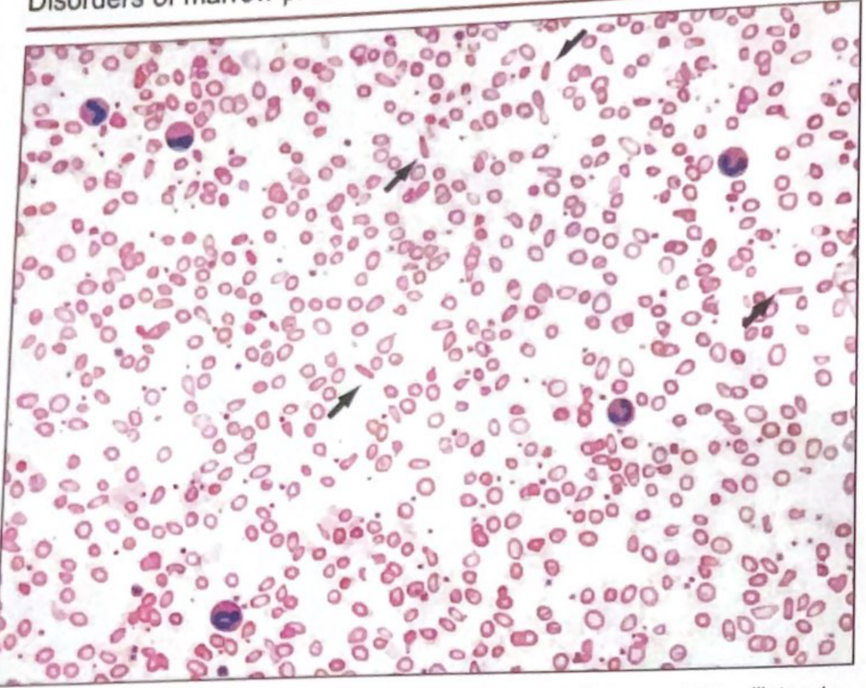

RBC Morphology on Smear
| Finding | Description |
|---|---|
| Microcytosis | Small RBCs (MCV < 80 fL) |
| Hypochromia | Enlarged zone of central pallor; normally central pallor = 1/3 of cell diameter. In IDA it expands - hemoglobin seen only as a narrow peripheral rim |
| Pencil cells (elliptocytes) | Thin, elongated elliptocytes - characteristic of IDA, not typically seen in thalassemia or anemia of chronic disease |
| Anisocytosis | Variation in RBC size |
| Poikilocytosis | Variation in RBC shape |
| Target cells | May be seen but less prominent than in thalassemia |
| Thrombocytosis | Platelet count often elevated (~450,000/mm³) - reactive |
Pearl: Pencil cells are typical of IDA but NOT common in thalassemia or anemia of inflammation - this is an important distinguishing feature on smear.
2. Bone Marrow Microscopy (Gold Standard)
| Finding | Description |
|---|---|
| Absent stainable iron | No Prussian blue-staining hemosiderin in macrophages - the gold standard for diagnosing IDA |
| Absent sideroblasts | No iron granules in erythroid precursors (normally ~30-40% of erythroblasts are sideroblasts) |
| Erythroid hyperplasia | Mild to moderate increase in erythroid progenitors (compensatory) |
Prussian blue (Perls') stain on bone marrow aspirate - absence of stainable iron = definitive diagnosis of IDA
3. Stages of Iron Deficiency & Lab Changes
| Stage | What's Depleted | Lab Abnormality |
|---|---|---|
| Stage 1 - Iron depletion | Storage iron (RES, marrow) | ↓ Serum ferritin, ↓ Bone marrow hemosiderin, ↓ Hepcidin |
| Stage 2 - Iron-deficient erythropoiesis | Circulating + erythroid iron | ↓ Transferrin saturation, ↑ sTfR, ↑ ZnPP, ↑ FEP, ↑ % hypochromic cells |
| Stage 3 - IDA | Hb synthesis affected | ↓ Hb, ↓ MCV, ↓ MCH → frank microcytic hypochromic anemia |
4. Complete Lab Profile in Established IDA
Iron Studies
| Test | IDA Value | Normal |
|---|---|---|
| Serum ferritin | ↓↓ < 12 µg/L (most sensitive & earliest marker) | 12-150 µg/L |
| Serum iron | ↓ Low | 60-170 µg/dL |
| TIBC (Total Iron Binding Capacity) | ↑↑ Increased | 250-370 µg/dL |
| Transferrin saturation | ↓ < 15% | 20-50% |
| Soluble transferrin receptor (sTfR) | ↑ Increased | |
| Zinc protoporphyrin (ZPP) | ↑ Increased | |
| Free erythrocyte protoporphyrin (FEP) | ↑ Increased | |
| Hepcidin | ↓ Low (maximizes iron uptake) |
Ferritin caveat: Ferritin is an acute phase reactant. It can be falsely normal/elevated in hepatic disease, infection, or inflammation - even when IDA is present.
CBC Findings
| Parameter | Finding |
|---|---|
| Hemoglobin | ↓ Low |
| MCV | ↓ < 80 fL (microcytic) |
| MCH | ↓ Low (hypochromic) |
| MCHC | ↓ Low |
| RDW | ↑↑ > 17 (high - early rise, distinguishes IDA from thalassemia trait where RDW is normal/mildly raised) |
| Reticulocyte count | Low (hypoproliferative) |
| Platelet count | ↑ Reactive thrombocytosis (~450,000/mm³) |
| WBC | Normal |
5. Key Differentiating Points: IDA vs Thalassemia vs Anemia of Inflammation
| Feature | IDA | Thalassemia Trait | Anemia of Inflammation |
|---|---|---|---|
| Ferritin | ↓↓ | Normal | ↑ (acute phase) |
| TIBC | ↑↑ | Normal | ↓ |
| Serum iron | ↓ | Normal | ↓ |
| Transferrin sat | ↓ (<15%) | Normal | ↓ |
| RDW | ↑↑ (>17) | Normal/mildly ↑ | Normal |
| Pencil cells on smear | ✅ Typical | ❌ Absent | ❌ Absent |
| Target cells | Present | ✅✅ Prominent | Absent |
| Marrow iron (Prussian blue) | ❌ Absent | Normal | ↑ (trapped in RES) |
| ZPP/FEP | ↑ | Normal | ↑ |
Lab diagnosis of megaloblastic anemia
megaloblastic anemia peripheral blood smear hypersegmented neutrophil macro-ovalocyte
Peripheral blood smear from a patient with known chronic lymphocytic leukemia (CLL) showing autoimmune hemolytic anemia (AIHA). Brightfield microscopy of Wright-Giemsa stained smear at 1000x (oil immersion) reveals two dominant cell populations: numerous small mature lymphocytes typical of CLL and red blood cells showing spherocytic deformation. A smudge cell is evident just left of center, a classical clue for CLL. Red cells display reduced central pallor consistent with spherocytes, indicating intravascular or extravascular hemolysis driven by autoantibodies. The background shows mild polychromasia and rare nucleated RBCs, consistent with a robust reticulocyte response; the corrected reticulocyte count in this case is markedly elevated (>7%). Platelets are not significantly abnormal. These features collectively support autoimmune destruction of erythrocytes in the setting of CLL. Diagnostic significance lies in recognizing AIHA as a common autoimmune complication in CLL, with a clinical picture of sudden Hb decline and compensatory erythropoiesis. Differential considerations include drug-induced hemolysis, other causes of hemolytic anemia, or marrow infiltration by CLL. Clinical correlation includes therapy implications (steroids, rituximab) and close monitoring of hemolysis markers. This image is a valuable teaching resource for hematology morphology, AIHA in CLL, and differential diagnosis in anemia.
This composite image consists of clinical photographs and a diagnostic peripheral blood smear. Figure 1a (left) shows a pediatric patient with clinical signs including total alopecia (complete absence of scalp hair) and nail pitting affecting both hands and feet, characterized by small punctate depressions on the nail plates. Figure 1b (right) is a microscopic view of a peripheral blood smear demonstrating microangiopathic hemolytic anemia. Visible are numerous schistocytes—fragmented, irregular red blood cells including helmet cells and triangular forms—alongside some normal erythrocytes showing central pallor. The educational focus is on the multi-systemic manifestations of a medical condition, likely a microangiopathic process such as Hemolytic Uremic Syndrome (HUS) or similar systemic pathology, illustrating the correlation between dermatological signs (hair and nail changes) and hematological abnormalities (hemolysis and schistocytosis).
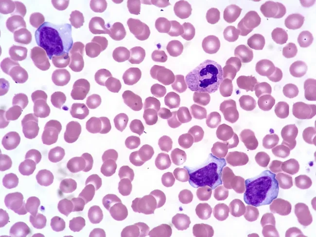
Comprehensive description: This is a brightfield, Wright-Giemsa stained peripheral blood smear examined by light microscopy at high magnification (oil immersion). The specimen shows several activated (atypical) lymphocytes in the peripheral blood, a hallmark of infectious mononucleosis. The lymphocytes have abundant basophilic cytoplasm and irregular, often indented nuclei with clumped chromatin and inconspicuous nucleoli. A normal segmented neutrophil is present for contrast. Erythrocytes exhibit occasional cytoplasmic membrane scalloping around red cells, a feature sometimes observed in EBV-related lymphocytosis. The background cytology is otherwise typical, with uniform erythrocytes and occasional platelets. The activated lymphocytes vary in size and cytoplasmic texture, some appearing plasmacytoid with vesicular basophilia, while others are more indented with coarse chromatin. These morphologic changes reflect an immune response to Epstein-Barr virus infection and are described as reactive lymphocytosis. Clinically, the combination of acute lymphadenitis, fever, a positive Monospot test, and EBV serology supports infectious mononucleosis. The image helps differentiate viral-induced lymphocytosis from bacterial lymphadenitis and from hematologic malignancies when integrated with clinical data. In summary, this smear illustrates the classical hematologic manifestation of EBV-associated mononucleosis. These atypical lymphocytes, sometimes named Downey cells, accompany fever and pharyngitis in EBV infection and aid differentiation from leukemia when combined with serology and clinical data.
Peripheral blood smear prepared with Wright-Giemsa stain and examined under brightfield illumination using a 100× oil immersion objective reveals a single large plasma cell among a field of mature erythrocytes. The plasma cell displays features typical of clonal plasma cells: basophilic cytoplasm, an eccentrically placed round nucleus, and a conspicuous perinuclear hof; background shows rouleaux formation of red cells. This image exemplifies hematologic involvement by a plasma cell dyscrasia. In plasma cell leukemia, circulating plasma cells constitute more than 20% of leukocytes, a hallmark distinguishing it from conventional multiple myeloma, although both conditions share monoclonal plasma cell proliferation. The presence of circulating plasma cells is associated with younger patient age, higher incidence of anemia, thrombocytopenia, organomegaly, and possible lymphadenopathy; however lytic bone lesions may be less common. Immunophenotypic patterns often include lack of CD56 expression relative to myeloma. Clinically, this morphological finding warrants confirmation with complete blood count, peripheral smear review, quantitative plasma cell percentage, serum protein electrophoresis, and immunofixation to identify monoclonal immunoglobulin isotype (IgD/IgE or light-chain predominance). Early detection of circulating plasma cells supports prognosis assessment and guides therapy decisions in plasma cell dyscrasia management and monitoring, including risk stratification and treatment response evaluation for practice in clinics.
Lab Diagnosis of Megaloblastic Anemia
Core Concept
1. Peripheral Blood Smear
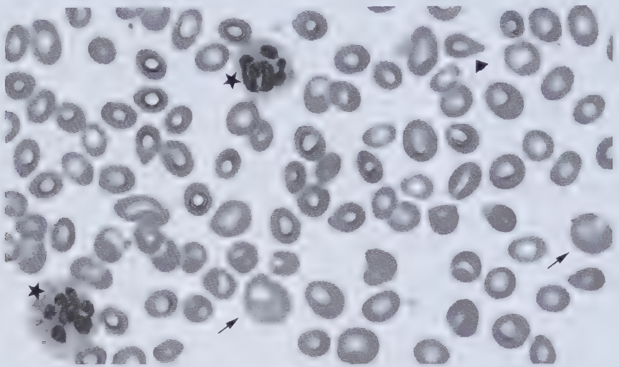
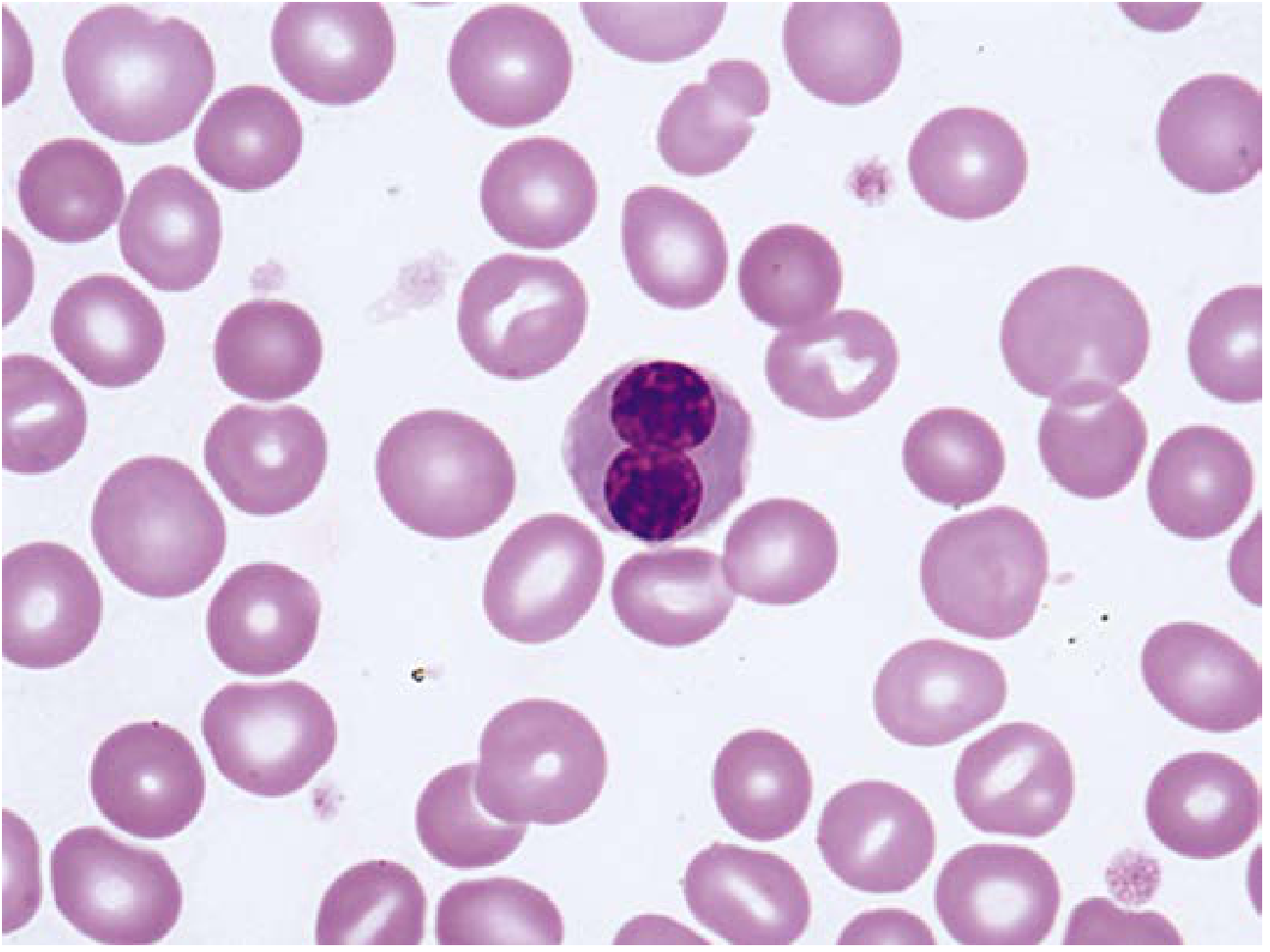
RBC Findings
| Finding | Description |
|---|---|
| Macro-ovalocytes | Large, oval-shaped RBCs (not round) - highly characteristic; appear "hyperchromic" but MCHC is actually normal |
| MCV > 115 fL | Markedly elevated in fully developed megaloblastic anemia |
| Anisocytosis | Marked variation in RBC size |
| Poikilocytosis | Teardrop cells (dacrocytes), RBC fragments, microcytes |
| Basophilic stippling | Present |
| Howell-Jolly bodies | Multiple per cell (karyorrhexis of megaloblasts) |
| Nucleated RBCs / Megaloblasts | Appear in circulation when anemia is severe; show "open", lacy chromatin |
| Low reticulocyte count | Hypoproliferative (ineffective erythropoiesis) |
WBC Findings - KEY DIAGNOSTIC SIGN
| Finding | Description |
|---|---|
| Hypersegmented neutrophils | ≥ 5 lobes in >5% of neutrophils, OR any neutrophil with ≥ 6 lobes - pathognomonic |
| Giant metamyelocytes | Large granulocyte precursors with abnormal nuclear shapes - seen in bone marrow and occasionally blood |
| Leukopenia | In severe/chronic disease |
Platelet Findings
| Finding | Description |
|---|---|
| Thrombocytopenia | In severe disease; large, abnormal platelets |
2. Bone Marrow Aspirate
| Finding | Description |
|---|---|
| Hypercellularity | Fat replaced; red marrow extends into long bones |
| Erythroid hyperplasia | Markedly increased erythroid precursors; M:E ratio decreased |
| Megaloblasts | Abnormally large erythroid precursors; finely stippled, "lacy/open" nuclear chromatin surrounded by normal-appearing (well-hemoglobinized) cytoplasm - nuclear-cytoplasmic dyssynchrony |
| Promegaloblasts > Basophilic megaloblasts | More early-stage cells than normal - "maturation arrest" pattern |
| Karyorrhexis | Fragmented, pyknotic nuclei; multiple Howell-Jolly bodies in cells |
| Giant metamyelocytes | Most characteristic granulocytic abnormality; large, abnormally contorted nuclei |
| Abnormal megakaryocytes | Large, polylobated, separated nuclear lobes |
Intramedullary hemolysis: Many megaloblasts die within the marrow before release, accounting for elevated LDH and bilirubin despite low reticulocytes.
3. Serum Biochemistry
Common to Both B12 and Folate Deficiency
| Test | Finding | Reason |
|---|---|---|
| LDH (Lactate Dehydrogenase) | ↑↑ Markedly elevated | Intramedullary destruction of RBC precursors |
| Indirect bilirubin | ↑ Elevated | Hemolysis of megaloblasts in marrow |
| Serum homocysteine | ↑ Elevated | Both B12 and folate needed to convert homocysteine → methionine |
| Serum haptoglobin | ↓ Decreased | Hemolysis |
Specific to B12 Deficiency
| Test | Finding |
|---|---|
| Serum vitamin B12 | ↓ Low (<200 pg/mL; borderline 200-400 pg/mL) |
| Serum methylmalonic acid (MMA) | ↑↑ Markedly elevated - most specific for B12 deficiency |
| Urine methylmalonic acid | ↑ Elevated |
| RBC folate | ↓ Decreased in 2/3 of B12-deficient patients (secondary folate deficiency) |
| Anti-intrinsic factor antibodies | ✅ Highly specific for pernicious anemia |
Specific to Folate Deficiency
| Test | Finding |
|---|---|
| Serum folate | ↓ Low (<3 µg/L) |
| RBC folate | ↓ Low (<165 ng/mL) - more stable than serum folate |
| Urine FIGLU (formiminoglutamic acid) | ↑ Elevated |
Important: Homocysteine is elevated in both deficiencies. Methylmalonic acid is elevated only in B12 deficiency - this is the key differentiating metabolite.
4. CBC Summary
| Parameter | Finding |
|---|---|
| Hb | ↓ (moderate to severe anemia) |
| MCV | ↑↑ > 115 fL |
| MCH | ↑ Elevated |
| MCHC | Normal |
| RDW | ↑ Elevated (anisocytosis) |
| WBC | ↓ Leukopenia (severe cases) |
| Platelets | ↓ Thrombocytopenia (severe cases) |
| Reticulocytes | ↓ Low (ineffective erythropoiesis) |
5. Key Distinguishing Points: B12 vs Folate Deficiency
| Feature | B12 Deficiency | Folate Deficiency |
|---|---|---|
| Neurological symptoms | ✅ Subacute combined degeneration of spinal cord | ❌ No neurological effects |
| Serum MMA | ↑↑ | Normal |
| Serum homocysteine | ↑ | ↑ |
| Serum B12 | ↓ | Normal |
| Serum folate | Normal or ↑ | ↓ |
| RBC folate | ↓ (2/3 cases) | ↓ |
| Anti-IF antibodies | ✅ (pernicious anemia) | ❌ |
| Response to B12 therapy | Reticulocytosis in 5 days | No response |
Diagnostic Criteria for Pernicious Anemia (B12 deficiency subtype)
- Moderate-to-severe megaloblastic anemia
- Leukopenia with hypersegmented neutrophils
- Low serum vitamin B12
- Elevated serum homocysteine + methylmalonic acid
- Serum anti-intrinsic factor antibodies (highly specific)
- Brisk reticulocyte response within 5 days of parenteral B12
Is ephedrine beta blocker and alpha agonist? How does it cause vasoconstriction
Ephedrine - Receptor Classification & Mechanism of Vasoconstriction
Is Ephedrine a Beta Blocker? Is It an Alpha Agonist?
- Alpha-1 agonist activity → vasoconstriction ✅
- Beta-1 agonist activity → increased HR + increased myocardial contractility ✅
- Beta-2 agonist activity → bronchodilation ✅
Mechanism of Action - Direct vs Indirect
Indirect Action (Primary) - NE Release
Ephedrine enters presynaptic nerve terminal
↓
Displaces norepinephrine (NE) from synaptic vesicles
↓
NE is released into the synapse
↓
NE activates postsynaptic α1 and β receptors
Direct Action (Secondary)
How Ephedrine Causes Vasoconstriction
| Step | Detail |
|---|---|
| 1. NE is released (indirect) | Ephedrine enters postganglionic adrenergic nerve terminals → displaces NE from synaptic vesicles → NE floods the synapse |
| 2. α1 receptors activated | Released NE (and ephedrine directly) activates postsynaptic α1 receptors on vascular smooth muscle |
| 3. IP3/DAG pathway | α1 activation → Gq protein → phospholipase C → IP3 + DAG → ↑ intracellular Ca²⁺ |
| 4. Smooth muscle contraction | Elevated Ca²⁺ → calmodulin-myosin light chain kinase (MLCK) activation → vascular smooth muscle contracts |
| 5. Vasoconstriction | Arterial + venous vasoconstriction → ↑ systemic vascular resistance → ↑ blood pressure |
The direct β2 stimulation (bronchodilation/vasodilation in some beds) partially counteracts the α1-mediated vasopressor effect, making ephedrine a less potent vasopressor than pure α agonists like phenylephrine.
Cardiovascular Effects Summary
| Effect | Receptor | Outcome |
|---|---|---|
| Vasoconstriction (arteries + veins) | α1 | ↑ SVR, ↑ BP |
| ↑ Heart rate | β1 | Tachycardia |
| ↑ Myocardial contractility | β1 | ↑ Cardiac output |
| Bronchodilation | β2 | Used in asthma (now largely replaced) |
| ↑ BP (systolic & diastolic) | α1 + β1 combined | Net pressor effect |
The overall cardiovascular picture resembles epinephrine - dose-related increases in HR, cardiac output, and SVR - but ephedrine is less potent and longer acting than epinephrine.
Important Caveat: Tachyphylaxis
- Presynaptic NE stores become depleted
- Ephedrine itself is then released as a "false neurotransmitter" from vesicles (weak activity)
- This does NOT happen with epinephrine, which acts directly on receptors regardless of NE stores
- Drugs that deplete NE (e.g., reserpine) or block nerve uptake (e.g., cocaine) attenuate ephedrine's effects
Quick Comparison: Ephedrine vs Similar Drugs
| Drug | α1 | β1 | β2 | Mechanism |
|---|---|---|---|---|
| Ephedrine | ✅ | ✅ | ✅ | Mixed (predominantly indirect) |
| Phenylephrine | ✅✅ | ❌ | ❌ | Direct (pure α1 agonist) |
| Epinephrine | ✅✅ | ✅✅ | ✅✅ | Direct (α + β) |
| Norepinephrine | ✅✅ | ✅ | ❌ | Direct (α1 >> β1) |
| Beta blockers (e.g., propranolol) | ❌ | BLOCKS β | BLOCKS β | Antagonist at β receptors |