
You are writing model exam answers for an MS (General Surgery) Preliminary Examination, Paper IV "Modern Trends & Recent Advances" for Indian postgraduate surgery students. You have access to query_library_db (medical textbook library - use headings_fts and paragraphs_fts to find grounding facts on hernia surgery, varicose veins, bile duct injury, hemorrhoid surgery, NOTES, robotic surgery, MRCP, GI staplers, 3D printing, energy sources in surgery, bariatric OT setup, E-FAST, tissue engineering scaffolds, transplant compatibility, ventral hernia repair) and web_search for anything not well covered in the library (e.g. very recent devices). Use these tools to ground your answers in accurate current surgical knowledge, but you do NOT need to cite every sentence - write in a natural exam-answer style with citations to textbook page/section only where a direct fact is drawn (format: - Book Title, p. XXX). Write full, comprehensive, exam-quality model answers for the following questions from TWO exam papers. Match the depth/length to the marks allocated (roughly: 20 marks = very comprehensive answer with classification, technique details, comparison table of merits/demerits, recent advances, ~500-700 words; 10 marks = moderately detailed, ~300-400 words; 5-7 marks = focused short note, ~150-250 words). Use headings, subheadings, bullet points, and short tables where useful (describe tables in markdown). Include classification systems, named techniques/eponyms, and mention specific evidence-based recent advances (e.g. named techniques, devices, trial evidence) since examiners reward this. Draw diagrams verbally where diagrams would be expected (describe what to draw) since this is a written exam answer. PAPER 1 - B. J. Medical College (write full answers for all): Q1 (20 marks): Discuss the recent advances in Surgery for all types of Hernia with its merits and demerits. Cover: inguinal hernia (Lichtenstein open mesh repair, laparoscopic TEP, TAPP, robotic-assisted inguinal hernia repair), ventral/incisional hernia (open, laparoscopic IPOM/IPOM-plus, eTEP, TAR - transversus abdominis release, robotic TAR/IPOM), umbilical hernia, hiatus hernia (laparoscopic Nissen fundoplication and mesh cruroplasty), and give a merits/demerits comparison for each approach (open vs lap vs robotic). Q2 (20 marks): Discuss the recent advances in management of varicose veins of lower limb. Cover pathophysiology briefly, CEAP classification, conservative management, and endovenous techniques: Endovenous Laser Ablation (EVLA), Radiofrequency Ablation (RFA), ultrasound-guided foam sclerotherapy (UGFS), Mechanochemical Ablation (MOCA/ClariVein), cyanoacrylate glue closure (VenaSeal), and conventional surgery (high ligation and stripping, ambulatory phlebectomy). Discuss merits/demerits, recurrence, evidence (e.g. NICE/ESVS guideline recommendations). Q3 (20 marks): Discuss the recent advances in management of Common Bile Duct (CBD) injury. Cover classification (Strasberg classification, Bismuth classification), timing of recognition (intraoperative vs delayed), management options: intraoperative primary repair, T-tube drainage, end-to-end repair, Roux-en-Y hepaticojejunostomy, endoscopic management (ERCP with stenting/sphincterotomy), percutaneous transhepatic cholangiography (PTC) and drainage, role of MRCP in diagnosis, and outcomes/prevention strategies (critical view of safety, ICG cholangiography, indocyanine green fluorescence). Q4(a) (10 marks): Write in detail about MIPH (Minimally Invasive Procedure for Hemorrhoids) - also known as stapled hemorrhoidopexy / Procedure for Prolapse and Hemorrhoids (PPH, Longo's technique). Cover indications, technique, instrument (circular stapler), mechanism (mucosal pexy, interruption of blood supply), merits over conventional (Milligan-Morgan) hemorrhoidectomy, demerits/complications, and comparison with other minimally invasive options (Doppler-guided hemorrhoidal artery ligation - THD, LigaSure/energy hemorrhoidectomy). Q4(b) (10 marks): Write about NOTES (Natural Orifice Transluminal Endoscopic Surgery). Cover concept, access routes (transgastric, transvaginal, transrectal, transvesical), procedures performed, hybrid NOTES, advantages (scarless surgery), limitations/challenges (closure of viscerotomy, triangulation, infection, training), and current status/future. Q5(a) (5 marks): Use of Robot in Surgery - brief focused note (components of robotic system - console, patient cart, vision system; advantages, disadvantages/limitations, current applications in general surgery). Q5(b) (5 marks): MRCP (Magnetic Resonance Cholangiopancreatography) - principle, indications, advantages over ERCP, limitations. Q5(c) (5 marks): Staplers in Gastrointestinal Surgery - types (linear, circular, linear cutter/GIA, curved), uses, advantages, complications. Q5(d) (5 marks): Use of 3D printing in Surgery - applications (patient-specific anatomical models for preop planning, surgical guides, patient-specific implants/prosthetics, bioprinting), advantages, limitations. PAPER 3 - Smt. N.H.L. Municipal Medical College (write full answers for all): Q1 (20 marks): Write down recent advances in Varicose Veins management. (Same core content as Paper 1 Q2 above - you may write a similarly comprehensive but independently-worded answer, or note briefly that it mirrors Paper 1 Q2 and give the full answer again in this section for completeness.) Q2 (20 marks): Enumerate the different energy sources used in surgery, their principles, advantages and disadvantages. Cover: Monopolar electrosurgery, Bipolar electrosurgery, Ultrasonic energy devices (Harmonic scalpel), Advanced bipolar/vessel sealing devices (LigaSure), Argon beam coagulation, Laser energy, Radiofrequency ablation, Microwave ablation, Cryoablation. For each: principle of action, common uses, advantages, disadvantages/complications (e.g. thermal spread, smoke, stray current injury, insulation failure). Q3: - OT set up for Bariatric Surgery (10 marks): patient positioning, table requirements (weight capacity, reverse Trendelenburg), instrumentation (bariatric-length instruments, extra-long trocars), anesthesia considerations, staff/equipment ergonomics, energy source and stapler needs, fire safety, imaging (fluoroscopy for leak test). - Magnet therapy in Endoscopy (10 marks): magnetic compression anastomosis / "magnamosis" technique - principle (paired magnets creating compression necrosis and fistula/anastomosis formation), applications (biliary-enteric anastomosis, esophageal atresia repair, gastrointestinal anastomosis, recanalization of strictures), advantages (avoids surgery/sutures, natural orifice), limitations. Q4: - Fallacies (pitfalls/limitations) in E-FAST (Extended Focused Assessment with Sonography for Trauma) (10 marks): false negatives (retroperitoneal injury, hollow viscus/bowel injury, diaphragmatic injury, small hemoperitoneum <200ml, early scan before blood accumulates), false positives (ascites, pre-existing fluid, ovarian cyst), operator dependency, body habitus limitations, pneumothorax/pneumoperitoneum confusion, limited sensitivity for solid organ grading, need for CT correlation in stable patients. - Tissue engineering scaffold (10 marks): definition, ideal scaffold properties (biocompatibility, biodegradability, porosity, mechanical strength), types of scaffold materials (natural - collagen, chitosan, alginate; synthetic - PLA, PGA, PLGA; decellularized ECM scaffolds), fabrication techniques (electrospinning, 3D printing, freeze-drying, decellularization), applications in surgery. Q5: - Compatibility for Transplant Surgery (10 marks): ABO compatibility, HLA typing/matching, crossmatch (complement-dependent cytotoxicity, flow cytometry crossmatch), panel reactive antibody (PRA), donor specific antibodies (DSA), ABO-incompatible transplant protocols (desensitization), role in kidney/liver transplant. - Principles of Ventral Hernia Repair (10 marks): classification (European Hernia Society classification by location/size), principles - tension-free repair with mesh, myofascial release techniques (component separation, transversus abdominis release - TAR), mesh position (onlay, sublay/retrorectus, IPOM, underlay), open vs laparoscopic vs robotic approaches, management of loss of domain (botulinum toxin, progressive pneumoperitoneum). OUTPUT FORMAT: Return ONE single markdown document with clear headers "## PAPER 1 - Q1", "## PAPER 1 - Q2" etc. and "## PAPER 3 - Q1" etc., with the full answer text under each header. This is the ONLY output I need - do not include meta-commentary about your research process, just the final polished answers ready to be inserted into a Word document.
You are writing model exam answers for an MS (General Surgery) Preliminary Examination, Paper IV "Modern Trends & Recent Advances" for Indian postgraduate surgery students. You have access to query_library_db (medical textbook library - use headings_fts and paragraphs_fts to find grounding facts on surgical pathology/biopsy techniques, immunohistochemistry, organ transplantation, robotic surgery, bariatric surgery nutrition, tissue engineering, sialendoscopy, prehabilitation/ERAS, TAMIS, indocyanine green, vacuum-assisted breast biopsy, cancer registries) and web_search for anything not well covered in the library (e.g. very recent guidelines/devices). Use these tools to ground your answers in accurate current surgical knowledge, but you do NOT need to cite every sentence - write in a natural exam-answer style with citations to textbook page/section only where a direct fact is drawn (format: - Book Title, p. XXX). Write full, comprehensive, exam-quality model answers for the following questions from ONE exam paper - Narendra Modi Medical College, MS Preliminary Exam, General Surgery Branch 1, Paper IV. Match the depth/length to the marks allocated (roughly: 20 marks = very comprehensive answer with classification, mechanisms, detailed discussion of techniques/ethics, ~600-800 words; 14 marks = detailed, ~450-550 words; 6-7 marks = focused note, ~200-250 words; 5 marks = short note, ~150-200 words). Use headings, subheadings, bullet points, and short tables where useful (describe tables in markdown). Include named classification systems, mechanisms, and current evidence/guidelines since examiners reward this. Q1 (20 marks): Discuss tissue and molecular diagnosis in surgery. Explain biopsy techniques (FNAC, core needle biopsy, incisional/excisional biopsy, image-guided biopsy, vacuum-assisted biopsy, sentinel lymph node biopsy, liquid biopsy/circulating tumor DNA), immunohistochemistry (principle, common markers used in surgical oncology e.g. ER/PR/HER2 in breast cancer, CD markers in lymphoma, cytokeratins), molecular pathology (PCR, FISH, next-generation sequencing, gene expression profiling), and their role in precision/personalized medicine (targeted therapy selection, prognostication, minimal residual disease detection). Q2 (20 marks): Discuss the principles of organ transplantation including: donor selection (living donor vs deceased/brain-dead donor criteria, expanded criteria donors, donation after circulatory death), recipient selection (indications, contraindications, recipient work-up), organ allocation (allocation policies/principles - e.g. MELD score for liver, waiting list systems, geographic/priority allocation), immunology of transplantation (HLA matching, crossmatching, types of rejection - hyperacute, acute, chronic), immunosuppression (induction agents, maintenance - calcineurin inhibitors, antimetabolites, steroids, mTOR inhibitors; complications of immunosuppression), and ethical issues (organ trade/commercialization, informed consent, allocation equity, brain death determination, THOA Act in India). Q3(a) (14 marks): Robotic surgery: Principles (telemanipulation, master-slave system, 3D vision, EndoWrist instruments, tremor filtration, motion scaling), applications in general surgery (cholecystectomy, hernia repair, colorectal, bariatric, hepatobiliary, thyroid), advantages (precision, ergonomics, reduced tremor, 3D visualization, dexterity in confined spaces), limitations (cost, lack of haptic feedback, longer setup/docking time, training curve, no tactile sensation), and future directions (single-port robotic platforms, AI integration, haptic feedback development, tele-surgery, newer platforms beyond da Vinci). Q3(b) (6 marks): Nutritional deficiency after bariatric surgery - types of deficiencies by procedure (restrictive vs malabsorptive - sleeve gastrectomy vs Roux-en-Y gastric bypass vs biliopancreatic diversion), specific deficiencies (iron, vitamin B12, folate, calcium, vitamin D, fat-soluble vitamins A/D/E/K, thiamine - Wernicke's risk), clinical manifestations, monitoring and supplementation protocols. Q4 - write notes on: (a) (7 marks) Principle of tissue engineering and regenerative medicine - triad of scaffold, cells (stem cells/autologous cells), and signaling molecules/growth factors; applications in surgery (skin substitutes, cartilage, vascular grafts, bladder). (b) (7 marks) Endoscopic management of salivary gland disorders (sialendoscopy) - indications (sialolithiasis, ductal strictures, recurrent sialadenitis), technique, instruments, advantages over open sialadenectomy, limitations. (c) (6 marks) Recent advances in perioperative optimization, including prehabilitation - concept of ERAS (Enhanced Recovery After Surgery), prehabilitation (exercise, nutritional optimization, psychological preparation, smoking/alcohol cessation before surgery), evidence for reduced complications/LOS. Q5 - write short notes on: (a) (5 marks) Transanal Minimally Invasive Surgery (TAMIS) - indications (rectal polyps/early rectal cancer local excision), technique, platform, advantages over TEM (Transanal Endoscopic Microsurgery), limitations. (b) (5 marks) Indocyanine Green (ICG) applications in surgery - fluorescence imaging principle, uses (perfusion assessment in anastomosis/flaps, sentinel lymph node mapping, biliary anatomy delineation/cholangiography, tumor localization, lymphatic mapping). (c) (5 marks) Vacuum assisted biopsy - device/technique (e.g. Mammotome), indications (breast lesions - both diagnostic and therapeutic excision of benign lesions), advantages over core needle biopsy, limitations. (d) (5 marks) Cancer Registry - definition, types (hospital-based vs population-based), purpose (incidence/prevalence data, research, planning cancer control programs), National Cancer Registry Programme in India (ICMR-NCRP), uses of data. OUTPUT FORMAT: Return ONE single markdown document with clear headers "## PAPER 2 - Q1", "## PAPER 2 - Q2", "## PAPER 2 - Q3(a)", "## PAPER 2 - Q3(b)", "## PAPER 2 - Q4(a)" etc. with the full answer text under each header. This is the ONLY output I need - do not include meta-commentary about your research process, just the final polished answers ready to be inserted into a Word document.
Transplantation of Human Organs Act India THOA 1994 amendment brain death certification
National Cancer Registry Programme India ICMR NCDIR history population based registries number
mkdir -p /tmp/workspace/surgery-exam/output && ls -la /tmp/workspace/
Where is answer
PAPER 1 — B. J. Medical College
Q1 (20 marks): Recent Advances in Surgery for All Types of Hernia — Merits & Demerits
- Lichtenstein open mesh repair: tension-free onlay polypropylene mesh. Merits: recurrence <2%, can use LA, cheap, good for strangulated hernia. Demerits: groin scar, higher chronic groin pain (10-12%) than laparoscopic repair.
- Laparoscopic TEP: preperitoneal space developed without entering peritoneum. Merits: no visceral injury risk, less pain, ideal for bilateral/recurrent hernia. Demerits: steep learning curve, limited working space.
- Laparoscopic TAPP: peritoneum entered, flap raised, mesh placed, peritoneum closed. Merits: wider space, better visualization, easier for occult hernia diagnosis. Demerits: risk of visceral/vascular injury, adhesions, port-site hernia.
- Robotic TAPP: Merits: better ergonomics for complex/recurrent/obese cases, precise suturing. Demerits: high cost, longer docking time, no haptic feedback.
| Parameter | Open | Lap (TEP/TAPP) | Robotic |
|---|---|---|---|
| Pain/return to work | Moderate | Least | Least |
| Recurrence | <2% | <2% | Comparable |
| Cost | Lowest | Moderate | Highest |
| Bilateral/recurrent | Less ideal | Ideal | Ideal |
- EHS classification (midline M1-M5, lateral L1-L4; width W1<4cm, W2 4-10cm, W3>10cm).
- Sublay (Rives-Stoppa retrorectus) repair preferred over onlay/underlay/bridging — lowest SSI and recurrence (7-11%).
- Transversus Abdominis Release (TAR, Novitsky 2012) — posterior component separation, up to 8-10cm bilateral medialization, recurrence <5%; superseded anterior component separation (Ramirez) which has high wound morbidity.
- Laparoscopic IPOM/IPOM-plus (defect closure before mesh), eTEP (extraperitoneal retrorectus access, can combine with TAR).
- Robotic TAR/IPOM: combines MIS access with ability to do complex intracorporeal suturing.
Q2 (20 marks): Recent Advances in Management of Varicose Veins of Lower Limb
- EVLA (810-1470nm laser under tumescent LA): >90-95% occlusion at 5yr; demerits — bruising, paresthesia, burns.
- RFA (ClosureFast): 120°C via catheter; less pain/bruising than EVLA.
- UGFS (foam sclerotherapy): cheap, no thermal risk; higher recurrence, skin staining, rare stroke via PFO.
- MOCA/ClariVein: mechanical + chemical injury, no tumescent LA needed, less data.
- Cyanoacrylate glue (VenaSeal): no LA/compression needed; cost, glue hypersensitivity.
- Conventional surgery (high ligation + stripping, ambulatory phlebectomy): effective for tortuous veins unsuitable for endovenous access; more morbidity, higher recurrence (neovascularization) than endovenous.
Q3 (20 marks): Recent Advances in Management of CBD Injury
- Bismuth (level of stricture relative to confluence, I-V).
- Strasberg (A-E5; includes minor leaks A/D plus major injuries E1-E5 = Bismuth I-V).
- Stewart-Way (mechanism-based; Class III — CBD mistaken for cystic duct, unrecognized — is commonest, ~60%).
- Minor injury (Strasberg A/D): primary repair over T-tube, or ligation of small aberrant ducts.
- Clean transection <1cm, not near hilum: end-to-end repair + T-tube.
- Major injury/near confluence/thermal injury: Roux-en-Y hepaticojejunostomy — gold standard, best done by experienced hepatobiliary surgeon at first attempt; if unavailable, drain and refer.
- ERCP (sphincterotomy + stent): for leaks with ductal continuity (Strasberg A/D).
- PTC + drainage: for complete obstruction/discontinuity, decompresses and defines proximal anatomy.
- Definitive: RYHJ remains standard for major/delayed strictures.
- Critical View of Safety (Strasberg) — 3 criteria before clipping.
- Routine/selective intraoperative cholangiography.
- ICG fluorescence cholangiography — real-time NIR visualization of biliary anatomy without radiation/contrast, complements CVS, useful in laparoscopic/robotic cholecystectomy.
- Fundus-first/subtotal cholecystectomy in difficult gallbladder, low threshold for conversion to open.
Q4(a) (10 marks): MIPH (Minimally Invasive Procedure for Hemorrhoids / Stapled Hemorrhoidopexy / PPH — Longo's Technique)
- Indications: Grade II-III, selected reducible Grade IV.
- Technique: Circular stapler (33mm) via dedicated anoscope; purse-string suture placed 2-4cm above dentate line; stapler fires — excises ring of mucosa/submucosa + staples edges (doughnut sent for histology).
- Mechanism: mucopexy + interruption of terminal branches of superior rectal artery.
- Merits over Milligan-Morgan: far less pain (staple line in insensate mucosa), shorter OT time, quicker recovery, day-care.
- Demerits: blind procedure, risk of rectal/rectovaginal perforation, bleeding, urgency/tenesmus (5-10%), doesn't treat external component, recurrence possible.
- Comparison: THD (Doppler-guided artery ligation) — no excision, minimal pain, repeatable, higher recurrence for large prolapse. LigaSure hemorrhoidectomy — still an open wound but less bleeding/pain than conventional.
Q4(b) (10 marks): NOTES (Natural Orifice Transluminal Endoscopic Surgery)
- Routes: transgastric, transvaginal (most successful clinically — cholecystectomy), transrectal/colonic, transvesical (experimental).
- Hybrid NOTES: combines 1-2 small abdominal ports with endoscope for safety/triangulation — current mainstream practice.
- Advantages: no scar, less parietal trauma/pain, avoids port-site complications.
- Limitations: closure of viscerotomy (biggest challenge), loss of triangulation, infection risk, limited dedicated instrumentation, steep training curve.
- Current status: transvaginal cholecystectomy most common; concept lives on via POEM (per-oral endoscopic myotomy) and third-space endoscopy.
Q5 — Short Notes (5 marks each)
PAPER 2 — Narendra Modi Medical College
Q1 (20 marks): Tissue and Molecular Diagnosis in Surgery
Q2 (20 marks): Principles of Organ Transplantation
Q3(a) (14 marks): Robotic Surgery — Principles, Applications, Advantages, Limitations, Future
Q3(b) (6 marks): Nutritional Deficiency After Bariatric Surgery
- Sleeve: B12 (fundus/intrinsic factor loss).
- RYGB: Iron deficiency anemia (also B12, folate, Ca, vit D — duodenum/jejunum bypassed).
- BPD/BPD-DS: protein malnutrition, fat-soluble vitamins A/D/E/K.
- Thiamine — risk of Wernicke's encephalopathy with rapid weight loss/vomiting — emergency, treat with IV thiamine before glucose.
- Calcium/Vit D → secondary hyperparathyroidism/bone disease.
- Monitoring: lifelong multivitamin + iron/B12, 3-6 monthly labs year 1, then annual.
Q4 — Notes
Q5 — Short Notes (5 marks each)
PAPER 3 — Smt. N.H.L. Municipal Medical College
Q1 (20 marks): Recent Advances in Varicose Veins Management
Q2 (20 marks): Different Energy Sources in Surgery
| Device | Principle | Uses | Key advantage | Key risk |
|---|---|---|---|---|
| Monopolar electrosurgery | RF current through patient to return pad | Cutting, dissection | Versatile, cheap | Stray current, insulation failure, capacitive coupling |
| Bipolar electrosurgery | Current only between forceps tips | Hemostasis of small vessels | No grounding pad needed, safe near nerves | Can't cut, slow |
| Harmonic scalpel (ultrasonic) | Piezoelectric vibration (55.5kHz), no electrical current through patient | Dissection + hemostasis to ~5mm vessels | Minimal smoke, low lateral spread | "Cold-tip" injury misconception |
| LigaSure (advanced bipolar) | Pressure + impedance-feedback RF sealing | Seals vessels up to 7mm | Reliable large-vessel seal, cut+seal in one | Cost, seal failure on wet/calcified tissue |
| Argon beam coagulation | Ionized argon gas conducts monopolar current, non-contact | Diffuse surface bleeding (liver/spleen) | Rapid coverage of large area | Gas embolism, bowel distension |
| Laser | Coherent light absorbed by chromophores | EVLA, lithotripsy, ablation | Very precise | Eye/fire injury, expensive |
| RFA | Ionic agitation via needle electrode | Liver tumor ablation, varicose vein truncal ablation | Minimally invasive, repeatable | Heat-sink effect near vessels |
| Microwave ablation | Water dipole oscillation | Liver/lung/renal tumor ablation | Larger, more uniform zone; less heat-sink | Less precise margins |
| Cryoablation | Freezing (Joule-Thomson) causes necrosis | Renal/prostate tumor ablation | Visible ice-ball on imaging | Delayed bleeding on thaw, cryoshock |
Q3
Q4
- False negatives: small hemoperitoneum (<200-250mL), early scan before blood accumulates, retroperitoneal injury (poorly seen), hollow viscus/bowel injury, diaphragmatic injury, doesn't grade solid organ injury (sensitivity only 29-35% for organ injury without hemoperitoneum).
- False positives: ascites, ovarian cyst, physiological pelvic fluid.
- Highly operator-dependent; degraded by obesity/subcutaneous emphysema/bowel gas; pneumothorax vs pneumoperitoneum confusion; unreliable in penetrating trauma.
- Clinical implication: negative FAST in unstable patient does NOT exclude injury; stable patients need CT correlation regardless of FAST result.
Q5


Paper 1
You are writing model exam answers for an MS (General Surgery) Preliminary Examination, Paper I "Applied Basic Science" for Indian postgraduate surgery students. You have access to query_library_db (medical textbook library) and web_search to ground answers in accurate current surgical knowledge. Cite textbook page/section only where a direct fact is drawn (format: - Book Title, p. XXX). Write in natural exam-answer style, do not over-cite every sentence. Write full, comprehensive, exam-quality model answers for the following questions from TWO exam papers. Match depth/length to marks (20 marks = very comprehensive, classification/tables/mechanisms, ~600-800 words; 10 marks = ~300-400 words; 5-7 marks = focused note ~150-250 words). Use headings, subheadings, bullet points, and short tables where useful. Include classification systems, scoring systems, named eponyms/techniques, and current evidence/guidelines since examiners reward this. Describe diagrams verbally where diagrams would be expected. === PAPER A - B. J. Medical College, MS Preliminary Exam, Paper I (Applied Basic Science) === Q1 (20 marks): Write down the different types of shock, pathophysiology of shock, and management of septic shock. Cover classification (hypovolemic, cardiogenic, obstructive, distributive - septic/anaphylactic/neurogenic), pathophysiology (stages of shock - compensated/decompensated/irreversible, cellular effects, SIRS), and detailed management of septic shock per Surviving Sepsis Campaign guidelines (early recognition, qSOFA/SOFA score, fluid resuscitation, vasopressors - norepinephrine first line, source control, antibiotics within 1 hour, lactate clearance). Q2 (20 marks): Write down modalities to stop bleeding. Cover: mechanical/surgical (direct pressure, packing, ligation, suturing), endoscopic (clips, banding, injection, thermal), interventional radiology (angioembolization), laser coagulation, topical hemostatic agents (fibrin sealants, oxidized cellulose, gelatin sponge), energy devices (electrocautery, ultrasonic, argon beam), pharmacological/medical (tranexamic acid, vitamin K, FFP, cryoprecipitate, desmopressin, recombinant factors), and damage control resuscitation principles. Q3 (20 marks): Write the mechanism of wound healing and factors affecting wound healing. Cover phases (hemostasis, inflammation, proliferation, remodeling), cellular/molecular mediators (platelets, growth factors - PDGF, TGF-beta, VEGF, FGF; role of macrophages, fibroblasts, collagen types I/III), and local/systemic factors affecting healing (infection, ischemia, foreign body, diabetes, malnutrition, steroids, smoking, age, radiation, vitamin deficiency). Q4(a) (10 marks): Principles of intestinal anastomosis - types (end-to-end, side-to-side, end-to-side), techniques (hand-sewn single/double layer vs stapled), key principles (adequate blood supply, tension-free, mucosa-to-mucosa apposition, no tension, adequate lumen, avoid contamination), factors affecting anastomotic healing/leak risk. Q4(b) (10 marks): Abdominal faecal fistula management - definition/classification (high vs low output), etiology (post-surgical, Crohn's, malignancy, radiation), the "SNAP" or FRIEND mnemonic for factors preventing spontaneous closure, management principles (sepsis control, nutrition/skin protection, anatomy definition via fistulogram/CT, definitive surgical repair timing). Q5(a) (10 marks): Thyroglossal cyst - embryology (thyroglossal duct remnant), clinical features, investigations, and Sistrunk's operation (technique and rationale - removal of central hyoid bone segment). Q5(b) (5 marks): Pathological pharyngeal pouch (Zenker's diverticulum) - anatomy (Killian's dehiscence), pathogenesis, clinical features, investigations (barium swallow), management (endoscopic stapling/diverticulotomy vs open diverticulectomy with cricopharyngeal myotomy). Q5(c) (5 marks): Principles of hernia repair - tension-free repair, mesh reinforcement, identification and treatment of the hernia sac, layered anatomical repair, avoiding recurrence, prevention of complications. Q5(d) (5 marks): Total gut irrigation (whole bowel irrigation for bowel preparation) - indications (elective colorectal surgery bowel prep, ingested toxin/foreign body removal), technique (polyethylene glycol solution via NG tube or oral), complications. === PAPER B - Dr. M.K. Shah Medical College & Research Centre, Ahmedabad, MS Branch-I Preliminary Exam, Paper I (Applied Basic Sciences) === Q1 (20 marks): Describe Epidemiology, Pathogenesis, Clinical features, Spread, Diagnosis, Differential Diagnosis, and management of Mycetoma. Cover eumycetoma vs actinomycetoma, geographic distribution (India/Sudan/Mexico - "mycetoma belt"), triad (painless swelling, sinuses, grains), diagnosis (grain microscopy/culture, imaging - "dot-in-circle" sign on MRI), differential diagnosis (chronic osteomyelitis, actinomycosis, TB, soft tissue tumor), management (medical - antifungals for eumycetoma, antibiotics for actinomycetoma; surgical - wide excision/amputation for advanced eumycetoma). Q2 (20 marks): Define Sepsis. Describe the pathophysiology and systemic inflammatory response associated with sepsis. Discuss clinical features, diagnosis, scoring systems and management of Septic Shock. Cover Sepsis-3 definition, SIRS criteria, qSOFA/SOFA scoring, pathophysiology (cytokine cascade, endothelial dysfunction, coagulopathy, mitochondrial dysfunction), and Surviving Sepsis Campaign management bundle (1-hour bundle: cultures, lactate, broad-spectrum antibiotics, 30ml/kg crystalloid, vasopressors for MAP≥65). Q3 (20 marks): Describe the process of normal wound healing. Discuss the phases of healing, factors affecting wound healing. Discuss in detail management of Leg Ulcers. Cover wound healing phases as above, then leg ulcer classification (venous, arterial, neuropathic/diabetic, pressure, mixed), differentiating clinical features, investigations (ABPI, duplex), and management principles (compression therapy for venous, revascularization for arterial, offloading for neuropathic, debridement, dressings, skin grafting). Q4(a) (10 marks): Discuss in detail the Magnitude of Problem of TRAUMA, Primary Survey, Secondary Survey, Damage Control Surgery versus Early Total Care, Significance of time in the outcome, Venous lactate in resuscitation of TRAUMA. Cover global trauma burden/epidemiology, ATLS primary survey (ABCDE), secondary survey, damage control surgery concept (abbreviated laparotomy, packing, temporary closure, staged definitive repair) vs early total care, "golden hour" concept, and lactate clearance as a resuscitation endpoint/prognostic marker. Q4(b) (10 marks): Describe applied surgical anatomy of Inguinal region with diagrams. Discuss importance of Myopectineal orifice of Fruchaud. Discuss the guiding principles of Open Lichtenstein and Laparoscopic inguinal hernia repair. Cover inguinal canal boundaries, Hesselbach's triangle, myopectineal orifice of Fruchaud (single musculofascial defect through which both direct/indirect/femoral hernias occur - rationale for mesh covering entire orifice), Lichtenstein technique principles, and TEP/TAPP principles. Q5 - Short notes (5 marks each): (A) Enhanced Recovery After Surgery (ERAS) protocols - pre/intra/postoperative elements. (B) Total Parenteral Nutrition: indications, methods (central line access, formulation), and complications (catheter-related sepsis, metabolic complications, refeeding syndrome, liver dysfunction). (C) Fournier's Gangrene - necrotizing fasciitis of perineum/genitalia, etiology, clinical features, Fournier's Gangrene Severity Index, management (aggressive debridement, broad-spectrum antibiotics, hyperbaric oxygen). (D) Ultrasonic Coagulator Dissector Scalpel (Harmonic scalpel): Principle and Applications - piezoelectric mechanism, coagulation without electrical current, applications in laparoscopic/open surgery. OUTPUT FORMAT: Return ONE single markdown document with clear headers "## PAPER A - Q1", "## PAPER A - Q2" ... "## PAPER B - Q1" etc., with the full answer text under each header. This is the ONLY output needed - no meta-commentary, just the final polished answers.
You are writing model exam answers for an MS (General Surgery) Preliminary Examination, Paper I "Applied Basic Science" for Indian postgraduate surgery students. You have access to query_library_db (medical textbook library) and web_search to ground answers in accurate current surgical knowledge. Cite textbook page/section only where a direct fact is drawn (format: - Book Title, p. XXX). Write in natural exam-answer style, do not over-cite every sentence. Write full, comprehensive, exam-quality model answers for the following questions from TWO exam papers. Match depth/length to marks (20 marks = very comprehensive, classification/tables/mechanisms, ~600-800 words; 10 marks = ~300-400 words; 6-7 marks = ~200-250 words; 5 marks = focused note ~150-200 words). Use headings, subheadings, bullet points, and short tables where useful. Include classification systems, scoring systems, named eponyms/techniques, and current evidence/guidelines since examiners reward this. Describe diagrams verbally where diagrams would be expected. === PAPER C - Smt. N.H.L. Municipal Medical College, Preliminary Examination, M.S. General Surgery, Paper 1 (Basic medical science as applied to subject) === Q1(a) (10 marks): Write in detail about anatomy, blood supply, and innervation of rectus abdominis muscle and its sheaths. Cover origin/insertion, rectus sheath composition above and below the arcuate line (Douglas's line), blood supply (superior/inferior epigastric arteries, anastomosis), nerve supply (T7-T12 thoracoabdominal nerves), and surgical relevance (incisions, TAR plane, retrorectus space for hernia repair). Q1(b) (10 marks): Describe indications and technique of posterior component separation in ventral hernia repair. Cover Transversus Abdominis Release (TAR) technique step-by-step (Novitsky), indications (large/complex/recurrent ventral hernia, loss of domain), and outcomes. Q2(a) (10 marks): Describe in detail about the modified Kuppuswamy scale - socioeconomic classification scale used in India, components (education, occupation, income of head of family), scoring, and classification into upper/upper-middle/lower-middle/upper-lower/lower classes, and periodic updates for inflation. Q2(b) (10 marks): Describe the clinical importance of socio-economic status in surgical patients - impact on disease presentation (delayed presentation of malignancy/hernia complications), nutritional status, comorbidity burden, access to healthcare/compliance, post-operative outcomes, relevance to research/audit and health policy planning, relevance to informed consent and shared decision making across literacy levels. Q3(a) (10 marks): Describe the surgical anatomy, blood supply, innervation and lymphatic drainage of Pancreas. Cover head/neck/body/tail relations, blood supply (superior and inferior pancreaticoduodenal arteries, splenic artery branches), innervation (autonomic - splanchnic nerves, vagus), and lymphatic drainage (peripancreatic nodes to celiac/superior mesenteric nodes) - relevant to surgical resection and lymphadenectomy extent in pancreatic cancer surgery. Q3(b) (10 marks): Classify cystic lesions of Pancreas and write in detail about management of Mucinous Cystadenoma of Pancreas. Cover classification (pseudocyst, serous cystadenoma, mucinous cystic neoplasm, IPMN, solid pseudopapillary neoplasm), features distinguishing mucinous cystadenoma (female predominance, body/tail location, malignant potential), diagnostic work-up (CT/MRI, cyst fluid CEA/amylase, EUS-FNA), and management (surgical resection - distal pancreatectomy given malignant potential, versus surveillance criteria). Q4 (20 marks): Describe in detail about different clinical presentations of Head injury and write in detail about management of Extra Dural Hematoma. Cover GCS, types of head injury (concussion, contusion, diffuse axonal injury, EDH, SDH, SAH), clinical presentation patterns (lucid interval in EDH), and detailed EDH management (indications for surgery - volume >30cc, midline shift, GCS deterioration; craniotomy technique, conservative management criteria, outcomes). Q5 - Short notes (5 marks each): (a) Informed Consent - elements, legal requirements in India, special situations (emergency, minors, mentally incompetent). (b) Methods to do Surgical AUDIT - types of audit (mortality/morbidity meetings, clinical audit cycle, criteria-based audit), scoring systems used (POSSUM, NSQIP), purpose. (c) Total Parenteral Nutrition - indications, methods, complications (same core content as needed; write independently and fully here). (d) Management of Biofilm formation in general surgical patients - definition, formation on prosthetic mesh/implants/catheters, clinical significance (chronic infection, antibiotic resistance), prevention (antimicrobial-coated devices, aseptic technique) and management (biofilm disruption, device removal, prolonged antibiotics, negative pressure wound therapy). === PAPER D - Narendra Modi Medical College, MS Preliminary Exam, General Surgery Branch 1, Paper I (Basic Medical Science) === Q1 (20 marks): Discuss Surgical infections including their pathogenesis, host immune response, principles of antimicrobial stewardship, and recent advances in their prevention and management. Cover classification of surgical site infections (superficial/deep/organ-space per CDC), pathogenesis (bacterial virulence factors, biofilm, contamination sources), host immune response (innate/adaptive, role of neutrophils/complement), antimicrobial stewardship principles (appropriate spectrum/dose/duration, de-escalation, antibiogram-guided therapy), and recent advances (antimicrobial-coated sutures/mesh, negative pressure wound therapy, rapid molecular diagnostics, phage therapy). Q2 (20 marks): Discuss the management of complicated appendicitis including Appendicular abscess and appendicular mass. Cover definitions, clinical features, imaging (USG/CT), management options - conservative (Ochsner-Sherren regimen) vs early appendectomy, percutaneous drainage of abscess, interval appendectomy controversy/current evidence, and management of appendicular perforation with generalized peritonitis. Q3 (20 marks): Discuss the principles of disaster surgery, mass casualty triage, resource allocation, and hospital disaster preparedness. Cover types of disasters (natural/man-made/mass casualty incidents), triage systems (START, Simple Triage And Rapid Treatment; military/civilian triage categories - immediate/delayed/minimal/expectant), resource allocation principles (surge capacity, damage control surgery in mass casualty), and hospital disaster preparedness (Hospital Incident Command System, disaster drills, stockpiling, communication protocols). Q4 - Notes: (a) (7 marks) Pheochromocytoma: Preoperative preparation and surgical management. Cover alpha-blockade (phenoxybenzamine) prior to beta-blockade, volume expansion, intraoperative hemodynamic monitoring, surgical approach (laparoscopic adrenalectomy), and perioperative crisis management. (b) (7 marks) Skin Bank and Skin Substitutes - cadaveric skin banking (procurement, storage, cryopreservation), indications (burns), and synthetic/biologic skin substitutes (Integra, Biobrane, AlloDerm, cultured epithelial autografts). (c) (6 marks) OPSI (Overwhelming Post-Splenectomy Infection) - pathophysiology (loss of splenic filtration/IgM/opsonization), causative organisms (encapsulated bacteria - Pneumococcus, Meningococcus, H. influenzae), prevention (vaccination schedule, prophylactic antibiotics, patient education, medical alert card). Q5 - Short notes (5 marks each): (a) Enhanced Recovery after Surgery (ERAS) - write independently and fully. (b) Hyperbaric Oxygen therapy - mechanism, indications in surgery (necrotizing infections, compromised flaps, chronic wounds, gas gangrene), complications. (c) Functional Constipation - Rome IV criteria, evaluation, management (dietary/lifestyle, laxatives, biofeedback, surgical options in refractory cases). (d) Visual Analogue Scale (VAS) for pain assessment - description, scoring, clinical use, advantages/limitations. OUTPUT FORMAT: Return ONE single markdown document with clear headers "## PAPER C - Q1(a)", "## PAPER C - Q1(b)" ... "## PAPER D - Q1" etc., with the full answer text under each header. This is the ONLY output needed - no meta-commentary, just the final polished answers.
modified Kuppuswamy scale 2024 updated socioeconomic classification scoring India
Transversus Abdominis Release TAR technique Novitsky steps posterior component separation
https://www.ijcmph.com/index.php/ijcmph/article/download/125…
Surviving Sepsis Campaign 2021 guidelines hour-1 bundle vasopressor norepinephrine lactate
Sepsis-3 definition qSOFA SOFA score criteria septic shock
interval appendectomy after conservative management appendiceal mass current evidence NICE 2023
OPSI post splenectomy vaccination schedule pneumococcal meningococcal Hib timing guideline
Fournier's Gangrene Severity Index scoring management hyperbaric oxygen
ERAS Enhanced Recovery After Surgery protocol elements preoperative intraoperative postoperative
PAPER A — B. J. Medical College
Q1 (20 marks): Types of Shock, Pathophysiology, Management of Septic Shock
| Type | Mechanism | Examples | CO | SVR |
|---|---|---|---|---|
| Hypovolaemic | ↓circulating volume | Haemorrhage, burns, third-spacing | Low | High |
| Cardiogenic | Pump failure | MI, arrhythmia, contusion | Low | High |
| Obstructive | Mechanical impediment to filling | Tension pneumothorax, tamponade, massive PE | Low | High |
| Distributive | Loss of vasomotor tone | Septic, anaphylactic, neurogenic | High | Low |
- Sepsis-3: Sepsis = infection + organ dysfunction (SOFA ↑≥2). Septic shock = persistent hypotension needing vasopressors for MAP≥65 + lactate >2mmol/L despite fluids.
- qSOFA screening (SBP≤100, RR≥22, altered mentation).
- Hour-1 bundle: lactate measurement, blood cultures before antibiotics, broad-spectrum antibiotics within 1hr, 30ml/kg crystalloid bolus, vasopressors to MAP≥65.
- Norepinephrine first-line; add vasopressin before escalating norepinephrine further; hydrocortisone 200mg/day if vasopressor-refractory.
- Source control (drain/debride) as early as feasible; lactate clearance as resuscitation endpoint.
Q2 (20 marks): Modalities to Stop Bleeding
- Mechanical/surgical: direct pressure, packing, ligation, suturing/underrunning, tourniquets, vascular clamps, organ resection.
- Endoscopic: injection (adrenaline/sclerosant), clips, band ligation (varices), thermal probes, argon plasma coagulation.
- Interventional radiology: selective angioembolization, REBOA, covered stent-grafts.
- Laser coagulation: Nd:YAG/argon for GI bleeding, HHT, vascular lesions.
- Energy devices: monopolar/bipolar cautery, harmonic scalpel, argon beam, LigaSure.
- Topical agents: oxidized cellulose (Surgicel), gelatin sponge, fibrin glue, thrombin agents, bone wax.
- Pharmacological: tranexamic acid (CRASH-2/3, WOMAN trials), vitamin K, FFP, cryoprecipitate, desmopressin, recombinant factor VIIa/PCC, platelets, protamine.
- Damage control resuscitation: permissive hypotension, 1:1:1 blood product ratio, abbreviated laparotomy, prevent lethal triad (hypothermia-acidosis-coagulopathy).
Q3 (20 marks): Wound Healing Mechanism and Factors
Q4(a) (10 marks): Principles of Intestinal Anastomosis
Q4(b) (10 marks): Abdominal Faecal Fistula Management
Q5(a) (10 marks): Thyroglossal Cyst
Q5(b) (5 marks): Pathological Pharyngeal Pouch (Zenker's Diverticulum)
Q5(c) (5 marks): Principles of Hernia Repair
Q5(d) (5 marks): Total Gut Irrigation
PAPER B — Dr. M.K. Shah Medical College & Research Centre
Q1 (20 marks): Mycetoma
Q2 (20 marks): Sepsis, Pathophysiology, Scoring, Septic Shock Management
Q3 (20 marks): Wound Healing + Leg Ulcer Management
Q4(a) (10 marks): Trauma Magnitude, Primary/Secondary Survey, DCS vs ETC
Q4(b) (10 marks): Inguinal Anatomy, Myopectineal Orifice of Fruchaud, Lichtenstein/Lap Repair
Q5 Short Notes (5 marks each)
PAPER C — Smt. N.H.L. Municipal Medical College
Q1(a) (10 marks): Rectus Abdominis Anatomy
Q1(b) (10 marks): Posterior Component Separation (TAR)
Q2(a) (10 marks): Modified Kuppuswamy Scale
Q2(b) (10 marks): SES Importance in Surgical Patients
Q3(a) (10 marks): Pancreas Surgical Anatomy
Q3(b) (10 marks): Pancreatic Cystic Lesions & Mucinous Cystadenoma Management
Q4 (20 marks): Head Injury Presentations & EDH Management
Q5 Short Notes (5 marks each)
PAPER D — Narendra Modi Medical College
Q1 (20 marks): Surgical Infections, Stewardship, Recent Advances
Q2 (20 marks): Complicated Appendicitis Management
Q3 (20 marks): Disaster Surgery, Triage, Preparedness
Q4 Notes
Q5 Short Notes (5 marks each)
You are writing model exam answers for postgraduate MS General Surgery preliminary examination papers (Indian medical college format). Write full, detailed, textbook-quality model answers for the following TWO exam papers. Use query_library_db (medical textbook library) to ground answers in surgery textbooks (Sabiston, Bailey & Love, SRB's Manual of Surgery, etc.) and web_search to supplement with current guidelines/recent advances where useful. Cite textbook sources as "- Book Title, p. XXX" where you found specific content. Depth/length guidance based on marks allocated: 20 marks ≈ 600-800 words with classification tables/diagrams described; 10 marks ≈ 300-400 words; 7 marks ≈ 220-280 words; 6 marks ≈ 200-250 words; 5 marks ≈ 150-250 words focused note. === PAPER 1: B.J. Medical College, MS Preliminary Examination, Paper II (General Surgery & Pathology), Max Marks 100 === Q1 (20 marks): Write down the life cycle of the causative organism of liver Hydatid disease (Echinococcus granulosus). How will you manage a case of Liver Hydatid cyst? Q2 (20 marks): Differential diagnosis of posterior triangle of neck swelling. Q3 (20 marks): How will you manage a case of blunt abdominal injury with suspected splenic injury? Q4(a) (10 marks): Diagnosis and management of GIST (Gastrointestinal Stromal Tumor). Q4(b) (10 marks): Classification of CBD (Common Bile Duct) injury (e.g. Strasberg/Bismuth classification). Q5 short notes (5 marks each): (a) Lucid interval (b) Propylthiouracil (c) Radiological scoring system for breast lump (e.g. BI-RADS) (d) Urinary diversion === PAPER 2: Smt. N.H.L. Municipal Medical College, Preliminary Examination, M.S. General Surgery, Paper 2 (Principles & Practice of Surgery), Max Marks 100 === Q1(a) (10 marks): Write in detail about anatomy, blood supply, lymphatic drainage of Stomach. Q1(b) (10 marks): Describe in detail management of Stage 2 Gastric Malignancy. Q2 (20 marks): A 70 year old male presents with abdominal distention and inability to pass stool and flatus since 5 days in emergency ward. Investigations reveal large bowel obstruction. Kindly describe different possible diagnoses and their management. Q3(a) (10 marks): Describe the surgical anatomy, blood supply and lymphatic drainage of Liver. Q3(b) (10 marks): Classify grades of Liver trauma (AAST grading). Write down management of Grade 4 Liver trauma in detail. Q4(a) (5 marks): Describe in detail about different clinical presentations of Gastrointestinal NeuroEndocrine tumors. Q4(b) (5 marks): Write in detail about DOTA scan. Q4(c) (10 marks): Write in detail about management of NeuroEndocrine tumor including recent management. Q5 short notes (5 marks each): (a) Pyle phlebitis (b) Hinchey Classification for Colonic Diverticulosis (c) Intestinal Duplication Cyst (d) Glasgow-Blatchford Bleeding Score (GBS) for Upper GI Haemorrhage === OUTPUT FORMAT === Produce a single markdown document. Use clear headers: "## PAPER 1 - B.J. MEDICAL COLLEGE (Paper II)" then "### Q1", "### Q2" etc, then "## PAPER 2 - SMT. N.H.L. MUNICIPAL MEDICAL COLLEGE (Paper 2)" then "### Q1(a)" etc. Use tables/bullet points for classifications where appropriate. Do NOT include any meta-commentary, preamble, or notes about your process - only the exam answer content itself. Return the full markdown document as your final message.
You are writing model exam answers for a postgraduate MS General Surgery preliminary examination paper (Indian medical college format). Write full, detailed, textbook-quality model answers for the following exam paper. Use query_library_db (medical textbook library) to ground answers in surgery textbooks (Sabiston, Bailey & Love, SRB's Manual of Surgery, Sriram Bhat, etc.) and web_search to supplement with current guidelines/recent advances where useful. Cite textbook sources as "- Book Title, p. XXX" where you found specific content. Depth/length guidance based on marks allocated: 20 marks ≈ 600-800 words with classification tables/diagrams described; 14 marks ≈ 450-550 words; 10 marks ≈ 300-400 words; 7 marks ≈ 220-280 words; 6 marks ≈ 200-250 words; 5 marks ≈ 150-250 words focused note. === NARENDRA MODI MEDICAL COLLEGE, MS Preliminary Exam, General Surgery: Branch 1, Paper-II (Principles and Practice of Surgery), Max Marks 100 === Q1 (20 marks): Discuss the various techniques and indications for the establishment of enteral tube feeding. Q2 (20 marks): Discuss the legal aspects of surgical practice, including medical negligence, documentation, informed consent, medicolegal issues and consumer protection. (Focus on Indian medicolegal context - Consumer Protection Act applicability to doctors, landmark cases like Samira Kohli, Indian Medical Association v V.P. Shantha, documentation standards, informed consent requirements.) Q3(a) (14 marks): Describe Deep Vein Thrombosis (DVT) including its etiopathogenesis, management and prevention. Q3(b) (6 marks): Surgical anatomy of Inguinal canal and its clinical importance. Q4 write notes on: (A) (7 marks): Advanced vessel sealing devices (e.g. LigaSure, Harmonic scalpel, thermal fusion technology). (b) (7 marks): Principle and treatment of perianal fistula surgery (fistula-in-ano - Parks classification, Goodsall's rule, fistulotomy, seton, LIFT, advancement flap, fibrin glue/plug, VAAFT). (c) (6 marks): Preoperative management of patients receiving anticoagulants and antiplatelet drugs (bridging therapy, timing of stoppage, reversal agents). Q5 write short notes on: (a) (5 marks): Hungry Bone Syndrome (b) (5 marks): ABCD approach to nutritional assessment (c) (5 marks): Interventional radiology (d) (5 marks): Child-Pugh and MELD scoring system === OUTPUT FORMAT === Produce a single markdown document. Use clear headers: "## NARENDRA MODI MEDICAL COLLEGE - Paper II" then "### Q1", "### Q2", "### Q3(a)", "### Q3(b)", "### Q4(A)", "### Q4(b)", "### Q4(c)", "### Q5(a)" through "### Q5(d)". Use tables/bullet points for classifications and scoring systems where appropriate. Do NOT include any meta-commentary, preamble, or notes about your process - only the exam answer content itself. Return the full markdown document as your final message.
Samira Kohli v Dr Prabha Manchanda Supreme Court informed consent judgment summary
Indian Medical Association v V.P. Shantha 1995 Supreme Court consumer protection act medical services summary
B.J. MEDICAL COLLEGE — Paper II (General Surgery & Pathology)
Q1 (20 marks): Life Cycle of Echinococcus granulosus & Management of Liver Hydatid Cyst
- Imaging: USG (WHO-IWGE staging CE1-CE5), CT/MRI for anatomy and vascular/biliary relations. Avoid unprotected FNAC (anaphylaxis/seeding risk).
- Medical: Albendazole 10-15mg/kg/day, pre-op (4wks) and post-op (4-12wks); sole therapy for small CE1/CE3a cysts or poor surgical candidates.
- PAIR (Puncture-Aspiration-Injection-Reaspiration): for CE1/CE3a cysts without biliary communication, using hypertonic saline/cetrimide scolicidal instillation.
- Surgery: Conservative (de-roofing, evacuation, omentoplasty) vs radical (pericystectomy/hepatectomy). Peritoneum packed with scolicidal gauze to prevent spillage. Biliary communication needs suture repair ± T-tube/ERCP.
- Post-op: Albendazole continued; recurrence 1-20% (<5% in expert centres).
Q2 (20 marks): Differential Diagnosis of Posterior Triangle Neck Swelling
| Category | Causes |
|---|---|
| Lymph nodes (commonest) | Reactive lymphadenitis, TB cold abscess, lymphoma, metastatic nodes, Virchow's/Troisier's node (GI malignancy) |
| Congenital/cystic | Cystic hygroma (brilliantly transilluminant), branchial cyst extension |
| Vascular | Subclavian aneurysm (cervical rib), phlebectasia |
| Neurogenic | Brachial plexus schwannoma/neurofibroma |
| Skeletal | Cervical rib, clavicular tumour |
| Soft tissue | Lipoma, sebaceous/dermoid cyst, sarcoma |
| Miscellaneous | Pancoast tumour + Horner's syndrome |
Q3 (20 marks): Management of Blunt Abdominal Injury with Suspected Splenic Injury
Q4(a) (10 marks): GIST Diagnosis & Management
Q4(b) (10 marks): Classification of CBD Injury
You are writing model exam answers for postgraduate MS General Surgery preliminary examination papers (Indian medical college format). Write full, detailed, textbook-quality model answers for the following TWO exam papers. Use query_library_db (medical textbook library) to ground answers in surgery textbooks (Sabiston, Bailey & Love, SRB's Manual of Surgery, etc.) and web_search to supplement with current guidelines/recent advances where useful. Cite textbook sources as "- Book Title, p. XXX" where you found specific content. Depth/length guidance based on marks allocated: 20 marks ≈ 600-800 words with classification tables; 15 marks ≈ 500-600 words; 12-13 marks ≈ 400-500 words; 10 marks ≈ 300-400 words; 8 marks ≈ 250-300 words; 7 marks ≈ 220-280 words; 5 marks ≈ 150-250 words; 4 marks ≈ 130-180 words focused note. === PAPER 1: B.J. Medical College, MS Preliminary Examination, Paper III (General Surgery & Pathology), Max Marks 100 === Q1 (20 marks): Write down the life cycle of the causative organism of amoebic liver abscess (Entamoeba histolytica). How will you manage a case of amoebic liver abscess? Q2 (20 marks): How will you manage a case of lower limb arterial embolism? Q3 (20 marks): How will you manage a case of acid poisoning? Q4(a) (10 marks): Diagnosis and management of Urethral injury. Q4(b) (10 marks): Management of Carcinoma Breast Stage 2A. Q5 short notes (5 marks each): (a) Pneumothorax (b) Glasgow Coma Scale (c) Scoring system for pancreatitis (e.g. Ranson's/APACHE II/BISAP/Glasgow-Imrie) (d) Classification of ano-rectal fistula (Parks classification) === PAPER 2: Smt. N.H.L. Municipal Medical College, Preliminary Examination, M.S. General Surgery, Paper 3 (Principles & Practice of Operative Surgery), Max Marks 100 === Q1(a) (8 marks): Classify Oral ulcers and write about their characteristics. Q1(b) (12 marks): Write about management of malignant ulcers over anterior 2/3rd of Tongue (i.e. carcinoma tongue). Q2(a) (15 marks): Write about management of Locally Advanced Breast Cancer. Q2(b) (5 marks): Narrate the operative steps of Modified Radical Mastectomy. Q3 (20 marks): Classify Testicular Tumors and write about their management. Describe operative steps of Retroperitoneal Lymph node dissection (RPLND) in brief. Q4(a) (7 marks): Write about aetiopathogenesis of Urolithiasis. Q4(b) (13 marks): Discuss various treatment modalities of Renal Pelvic Stones (ESWL, PCNL, RIRS, open surgery). Q5 short notes (4 marks each): (A) Ectopia Vesicae (bladder exstrophy) (b) Operative Steps of Hemithyroidectomy (c) Sialadenitis (d) Hypersplenism (e) Perianal Fistula === OUTPUT FORMAT === Produce a single markdown document. Use clear headers: "## PAPER 1 - B.J. MEDICAL COLLEGE (Paper III)" then "### Q1", "### Q2" etc, then "## PAPER 2 - SMT. N.H.L. MUNICIPAL MEDICAL COLLEGE (Paper 3)" then "### Q1(a)" etc. Use tables/bullet points for classifications where appropriate. Do NOT include any meta-commentary, preamble, or notes about your process - only the exam answer content itself. Return the full markdown document as your final message.
You are writing model exam answers for a postgraduate MS General Surgery preliminary examination paper (Indian medical college format). Write full, detailed, textbook-quality model answers for the following exam paper. Use query_library_db (medical textbook library) to ground answers in surgery textbooks (Sabiston, Bailey & Love, SRB's Manual of Surgery, Sriram Bhat, CSDT operative surgery texts, etc.) and web_search to supplement with current guidelines/recent advances where useful. Cite textbook sources as "- Book Title, p. XXX" where you found specific content. Depth/length guidance based on marks allocated: 20 marks ≈ 600-800 words with classification/step-by-step detail; 14 marks ≈ 450-550 words; 7 marks ≈ 220-280 words; 6 marks ≈ 200-250 words; 5 marks ≈ 150-250 words focused note. === NARENDRA MODI MEDICAL COLLEGE, MS Preliminary Exam, General Surgery: Branch 1, Paper-III (Principles and Practices of Operative Surgery), Max Marks 100 === Q1 (20 marks): Discuss the basic surgical skills required for a postgraduate resident, including operating room safety and ergonomics. Q2 (20 marks): Describe Laparoscopic Cholecystectomy. Discuss its complications and the management of bile duct injury (include Strasberg/Bismuth classification and management approach per injury type). Q3(a) (14 marks): Discuss surgical anatomy of the thyroid gland. Briefly describe the steps of left hemithyroidectomy and discuss complications of thyroid surgery (thyroidectomy). Q3(b) (6 marks): PIRS - Percutaneous Internal Ring Suturing (for paediatric inguinal hernia repair - technique, advantages, outcomes). Q4 write notes on: (a) (7 marks): Diagnosis and management of obstructive uropathy. (b) (7 marks): Oncoplastic breast surgery. (c) (6 marks): Diagnosis and certification of Brain Death (Indian legal criteria - Transplantation of Human Organs Act, apnea test, ancillary tests). Q5 write short notes on: (a) (5 marks): Saphenous vein graft. (b) (5 marks): Counselling of patients undergoing prostatectomy. (c) (5 marks): Balloon assisted enteroscopy. (d) (5 marks): Blood transfusion in the perioperative period. === OUTPUT FORMAT === Produce a single markdown document. Use clear headers: "## NARENDRA MODI MEDICAL COLLEGE - Paper III" then "### Q1", "### Q2", "### Q3(a)", "### Q3(b)", "### Q4(a)", "### Q4(b)", "### Q4(c)", "### Q5(a)" through "### Q5(d)". Use tables/bullet points for classifications and step-by-step operative technique where appropriate. Do NOT include any meta-commentary, preamble, or notes about your process - only the exam answer content itself. Return the full markdown document as your final message.
acid poisoning management first aid Zargar classification esophageal stricture surgery India
Transplantation of Human Organs Act India brain death certification criteria apnea test ancillary tests
Percutaneous Internal Ring Suturing PIRS pediatric inguinal hernia technique advantages outcomes
https://www.mdpi.com/1648-9144/60/7/1137
PIRS percutaneous internal ring suturing technique steps epidural needle spinal needle hydrodissection
B.J. MEDICAL COLLEGE — Paper III (General Surgery & Pathology)
Q1 (20 marks): Life Cycle of Entamoeba histolytica & Management of Amoebic Liver Abscess
Q2 (20 marks): Management of Lower Limb Arterial Embolism
Q3 (20 marks): Management of Acid Poisoning
Q4(a) (10 marks): Diagnosis & Management of Urethral Injury
Q4(b) (10 marks): Management of Carcinoma Breast Stage 2A
Q5 Short Notes (5 marks each)
SMT. N.H.L. MUNICIPAL MEDICAL COLLEGE — Paper 3 (Operative Surgery)
Q1(a) (8 marks): Classification of Oral Ulcers
Q1(b) (12 marks): Management of Malignant Ulcer, Anterior 2/3rd Tongue
Q2(a) (15 marks): Management of Locally Advanced Breast Cancer
Q2(b) (5 marks): Operative Steps of MRM
Q3 (20 marks): Testicular Tumours Classification, Management, RPLND Steps
Q4(a) (7 marks): Aetiopathogenesis of Urolithiasis
Q4(b) (13 marks): Treatment of Renal Pelvic Stones
Q5 Short Notes (4 marks each)
NARENDRA MODI MEDICAL COLLEGE — Paper III (Operative Surgery)
Q1 (20 marks): Basic Surgical Skills, OR Safety, Ergonomics
Q2 (20 marks): Laparoscopic Cholecystectomy, Complications, Bile Duct Injury Management
Q3(a) (14 marks): Thyroid Anatomy, Left Hemithyroidectomy Steps, Complications
Q3(b) (6 marks): PIRS
Q4 Notes
Q5 Short Notes (5 marks each)
Do you need answers in a different format or with additional visual diagrams/tables?
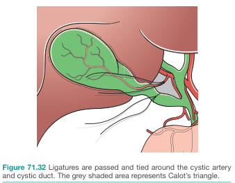
Calot's triangle critical view of safety laparoscopic cholecystectomy anatomy
inguinal canal anatomy myopectineal orifice hernia diagram
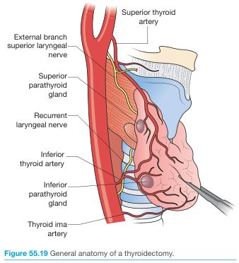
thyroid gland anatomy recurrent laryngeal nerve parathyroid glands relations
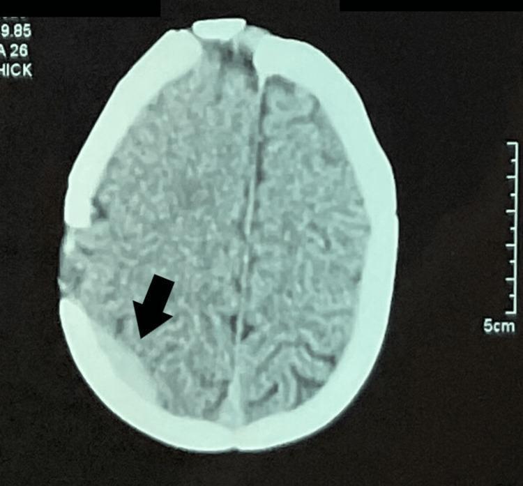
extradural haematoma epidural hematoma CT scan biconvex lens shaped
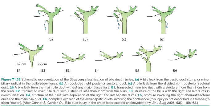
bile duct injury Bismuth classification biliary stricture diagram
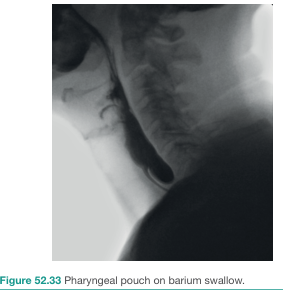
Zenker's diverticulum pharyngeal pouch Killian's dehiscence
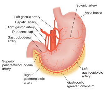
stomach blood supply arterial anatomy left gastric right gastric
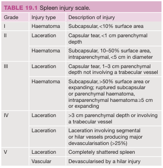
spleen injury AAST grading laceration diagram
1. Calot's Triangle / Critical View of Safety
2. Thyroid Gland Anatomy — RLN & Parathyroid Relations
3. Strasberg Classification of Bile Duct Injury
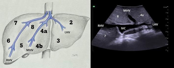
4. Liver — Couinaud Segmental Anatomy
5. Stomach — Arterial Blood Supply
6. AAST Splenic Injury Grading Table
7. Extradural (Epidural) Haematoma — CT appearance
8. Zenker's Diverticulum (Barium Swallow)
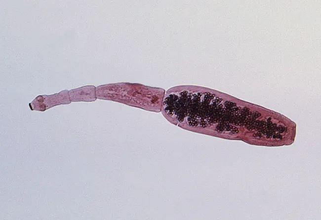
9. Echinococcus granulosus (adult worm, scolex + hooklets)
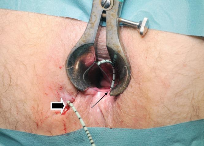
10. Anal Fistula — MRI tract & Seton placement
Complile all plus also make guess paper
~/attachments/d22a85cd-3600-4174-9f3e-1289593e3168/
~/attachments/
~/
~/conversation_history
~/conversation_history/media
Answer
Give me guess paper for paper one
Guess Paper — MS General Surgery, Paper I (Applied Basic Science / Basic Medical Science)
Frequency analysis (why these topics were picked)
| Topic | Appeared in |
|---|---|
| Shock / Sepsis / SIRS | B.J. (Q1), Dr. M.K. Shah (Q2) — 2/4 colleges |
| Wound healing / chronic ulcers | B.J. (Q3), Dr. M.K. Shah (Q3) — 2/4 colleges |
| Trauma — primary/secondary survey, damage control surgery | Dr. M.K. Shah (Q4A), Smt. N.H.L. (Q4, EDH) — 2/4 colleges |
| Hernia anatomy/repair (inguinal + ventral) | B.J. (Q5c), Dr. M.K. Shah (Q4B), Smt. N.H.L. (Q1b) — 3/4 colleges |
| ERAS protocols | Dr. M.K. Shah (Q5a), Narendra Modi (Q5a) — 2/4 colleges |
| TPN | Dr. M.K. Shah (Q5b), Smt. N.H.L. (Q5c) — 2/4 colleges |
| Informed consent / medicolegal | Smt. N.H.L. (Q5a) — recurring theme across Papers I & II in general |
| Surgical infection / OPSI / SSI | Narendra Modi (Q1, Q4c) — high-yield standalone |
GUESS PAPER — PAPER I (Applied Basic Science)
Reserve / backup topics (asked only once so far but classic exam favorites — revise these too)
- Surgical infections: pathogenesis, host immune response, antimicrobial stewardship
- Complicated appendicitis — appendicular abscess and mass management
- Principles of disaster surgery, mass casualty triage
- Pheochromocytoma — preoperative preparation and surgical management
- Skin bank and skin substitutes
- Mycetoma — epidemiology, pathogenesis, clinical features, management
- Fournier's gangrene
- Modified Kuppuswamy scale / socioeconomic status in surgical patients
- Thyroglossal cyst / pharyngeal pouch (branchial anomalies)
- Rectus abdominis anatomy and sheaths
- Pancreas surgical anatomy and cystic lesions (mucinous cystadenoma)
- Total gut irrigation
- Management of biofilm formation in surgical patients
- Hyperbaric oxygen therapy
- Functional constipation / Visual Analogue Scale for pain
With full answers
Surviving Sepsis Campaign 2021 guidelines septic shock management bundle
"septic shock" AND management
Guess Paper — Paper I (Applied Basic Science) — Full Model Answers
Q1. Shock: Classification, Pathophysiology and Management of Septic Shock (20 marks)
| Type | Mechanism | Examples | Hemodynamics |
|---|---|---|---|
| Hypovolaemic | Loss of intravascular volume | Haemorrhage, burns, GI losses | Low CO, high SVR, low CVP |
| Cardiogenic | Pump failure | MI, arrhythmia, valve rupture | Low CO, high SVR, high CVP |
| Obstructive | Mechanical obstruction to flow | Tension pneumothorax, cardiac tamponade, massive PE | Low CO, high SVR, high CVP |
| Distributive | Loss of vascular tone/maldistribution | Septic, anaphylactic, neurogenic shock | High/normal CO, low SVR |
- Massive nitric-oxide-mediated vasodilatation and loss of vasomotor tone -> profound distributive shock
- Endothelial injury and capillary leak -> third-spacing, relative hypovolaemia
- Microvascular thrombosis (activation of coagulation cascade, consumption of protein C) -> DIC and tissue ischaemia despite normal/high cardiac output
- Myocardial depression from circulating cytokines
- Mitochondrial dysfunction -> impaired oxygen utilisation even when delivery is adequate ("cytopathic hypoxia")
- Hour-1 bundle: measure lactate, obtain blood cultures before antibiotics, administer broad-spectrum antibiotics within 1 hour, begin rapid 30 mL/kg crystalloid (balanced solution preferred) for hypotension/lactate >4, start vasopressors if hypotensive during/after fluid resuscitation to keep MAP >=65 mmHg.
- Source control: identify and drain/debride/remove the septic focus as early as feasible (abscess drainage, necrotic tissue debridement, line removal).
- Vasopressor of choice: noradrenaline first-line; add vasopressin or adrenaline if inadequate; dobutamine for persistent myocardial dysfunction.
- Adjuncts: hydrocortisone (200 mg/day) if shock persists despite fluids and vasopressors; lung-protective ventilation if ARDS develops; glycaemic control; stress ulcer and DVT prophylaxis; nutrition within 24-48 hours.
- Reassessment: dynamic fluid responsiveness (passive leg raise, IVC variability) rather than fixed CVP targets; serial lactate clearance to guide resuscitation.
Q2. Wound Healing: Phases, Factors Affecting, and Management of Chronic Leg Ulcers (20 marks)
- Haemostasis (minutes): vessel spasm, platelet plug formation, activation of coagulation cascade forming a fibrin clot that acts as a scaffold.
- Inflammatory phase (0-3 days): neutrophils arrive first, clearing bacteria and debris; macrophages follow (day 2-3), releasing growth factors (PDGF, TGF-beta, VEGF) that drive the next phase.
- Proliferative phase (3 days-3 weeks): fibroblasts lay down type III collagen; angiogenesis forms granulation tissue; epithelial cells migrate across the wound bed (epithelialisation); myofibroblasts cause wound contraction.
- Remodelling/maturation phase (3 weeks-1 year+): type III collagen is replaced by stronger type I collagen, cross-linking increases tensile strength, and the scar matures, reaching about 80% of original skin strength.
- Local: infection, foreign body, poor blood supply/ischaemia, haematoma, mechanical stress/movement, radiation, repeated trauma, presence of necrotic tissue or slough.
- Systemic: age, diabetes mellitus (impaired neutrophil function, microangiopathy), malnutrition (protein, vitamin C, zinc deficiency), anaemia and hypoxia, smoking (nicotine-induced vasoconstriction), obesity, corticosteroids/immunosuppressants/chemotherapy, uraemia, jaundice, malignancy, connective tissue disorders (e.g. Ehlers-Danlos).
| Type | Key feature | Management principle |
|---|---|---|
| Venous ulcer | Gaiter area, shallow, irregular edge, associated lipodermatosclerosis/varicosities | Compression bandaging/stockings (after excluding arterial disease with ABPI), leg elevation, treat underlying venous reflux (endovenous ablation), wound dressings |
| Arterial (ischaemic) ulcer | Punched-out edge, painful, over pressure points/toes, absent pulses | Revascularisation (angioplasty/bypass) is priority; compression contraindicated |
| Neuropathic (diabetic) ulcer | Painless, over pressure points (heel, metatarsal heads), surrounding callus | Offloading, glycaemic control, debridement, treat infection, screen for PAD |
| Malignant (Marjolin's ulcer) | Ulcer in old scar/burn that fails to heal or changes character | Biopsy to exclude SCC, wide excision |
Q3. Trauma: Primary and Secondary Survey, Damage Control Surgery vs Early Total Care, and Extradural Haematoma Management (20 marks)
- Airway with cervical spine protection - look for obstruction, maintain in-line immobilisation.
- Breathing and ventilation - exclude tension pneumothorax, open pneumothorax, massive haemothorax, flail chest.
- Circulation with haemorrhage control - identify and control external haemorrhage, assess for internal bleeding (chest, abdomen, pelvis, long bones), obtain IV access, send for group & crossmatch.
- Disability - rapid neurological assessment (GCS, pupils).
- Exposure/Environment - fully expose the patient while preventing hypothermia.
- DCS is a staged approach for the physiologically deranged trauma patient (hypothermia, acidosis, coagulopathy - the "lethal triad"): (1) abbreviated laparotomy for haemorrhage/contamination control only (packing, vessel ligation/shunting, stapled bowel without anastomosis, temporary abdominal closure e.g. "Vac-Pac"/OPSITE sandwich); (2) transfer to ICU for physiological correction; (3) planned re-look and definitive repair once the patient is warm, coagulating and haemodynamically stable. - Bailey and Love's Short Practice of Surgery, 28th ed.
- ETC involves definitive single-stage repair of all injuries at the index operation and is reserved for physiologically stable patients who can tolerate a prolonged procedure. In unstable, multiply injured patients, ETC increases the risk of the lethal triad and death; DCS improves survival in this subgroup.
Q4(a). Applied Surgical Anatomy of the Inguinal Canal and Myopectineal Orifice of Fruchaud (10 marks)
Q4(b). Principles of Lichtenstein Repair, Laparoscopic (TEP/TAPP) Repair, and Posterior Component Separation (10 marks)
Q5. Short Notes (5 marks each)
Yes
mkdir -p /tmp/workspace/surgery-exam/content && ls -la /tmp/workspace/surgery-exam/
~/surgery-exam/content/guess_paper1.md
# GUESS PAPER — Paper I (Applied Basic Science / Basic Medical Science)
*Compiled from cross-college pattern analysis (B.J. Medical College, Dr. M.K. Shah Medical College, Smt. N.H.L. Municipal Medical College, Narendra Modi Medical College)*
**Maximum Marks: 100 | Time: 3 Hours**
---
## Q1. Shock: Classification, Pathophysiology and Management of Septic Shock (20 marks)
**Definition:** Shock is a state of acute circulatory failure resulting in inadequate tissue oxygen delivery relative to metabolic demand, leading to cellular hypoxia, anaerobic metabolism and, if uncorrected, multi-organ dysfunction.
**Classification (4 major types):**
| Type | Mechanism | Examples | Hemodynamics |
|---|---|---|---|
| Hypovolaemic | Loss of intravascular volume | Haemorrhage, burns, GI losses | Low CO, high SVR, low CVP |
| Cardiogenic | Pump failure | MI, arrhythmia, valve rupture | Low CO, high SVR, high CVP |
| Obstructive | Mechanical obstruction to flow | Tension pneumothorax, cardiac tamponade, massive PE | Low CO, high SVR, high CVP |
| Distributive | Loss of vascular tone/maldistribution | Septic, anaphylactic, neurogenic shock | High/normal CO, low SVR |
**Pathophysiology of septic shock:** Septic shock is a subset of sepsis with circulatory, cellular and metabolic abnormalities that carry a higher mortality risk than sepsis alone. Pathogen-associated molecular patterns (PAMPs, e.g. endotoxin) are recognised by pattern recognition receptors (TLR-4) on macrophages, triggering release of TNF-alpha, IL-1, IL-6 and other cytokines — the Systemic Inflammatory Response Syndrome (SIRS). This causes:
- Massive nitric-oxide-mediated vasodilatation and loss of vasomotor tone → profound distributive shock
- Endothelial injury and capillary leak → third-spacing, relative hypovolaemia
- Microvascular thrombosis (activation of coagulation cascade, consumption of protein C) → DIC and tissue ischaemia despite normal/high cardiac output
- Myocardial depression from circulating cytokines
- Mitochondrial dysfunction → impaired oxygen utilisation even when delivery is adequate ("cytopathic hypoxia")
**Clinical features:** Fever or hypothermia, tachycardia, tachypnoea, warm peripheries early (warm shock) progressing to cold, mottled skin (cold shock), altered sensorium, oliguria, hypotension refractory to fluids requiring vasopressors.
**Diagnosis and scoring:** Sepsis-3 criteria define sepsis as life-threatening organ dysfunction (SOFA score increase ≥2) caused by a dysregulated host response to infection; septic shock = sepsis with persisting hypotension requiring vasopressors to maintain MAP ≥65 mmHg plus serum lactate >2 mmol/L despite adequate fluid resuscitation. qSOFA (altered mentation, RR≥22, SBP≤100) is used for rapid bedside screening outside ICU.
**Management (per Surviving Sepsis Campaign 2021 guidelines):**
1. **Hour-1 bundle:** measure lactate, obtain blood cultures before antibiotics, administer broad-spectrum antibiotics within 1 hour, begin rapid 30 mL/kg crystalloid (balanced solution preferred) for hypotension/lactate >4, start vasopressors if hypotensive during/after fluid resuscitation to keep MAP ≥65 mmHg.
2. **Source control:** identify and drain/debride/remove the septic focus as early as feasible (abscess drainage, necrotic tissue debridement, line removal).
3. **Vasopressor of choice:** noradrenaline first-line; add vasopressin or adrenaline if inadequate; dobutamine for persistent myocardial dysfunction.
4. **Adjuncts:** hydrocortisone (200 mg/day) if shock persists despite fluids and vasopressors; lung-protective ventilation if ARDS develops; glycaemic control; stress ulcer and DVT prophylaxis; nutrition within 24-48 hours.
5. **Reassessment:** dynamic fluid responsiveness (passive leg raise, IVC variability) rather than fixed CVP targets; serial lactate clearance to guide resuscitation.
*Source: Bailey and Love's Short Practice of Surgery, 28th ed.; Surviving Sepsis Campaign 2021 (Evans et al., Crit Care Med 2021).*
---
## Q2. Wound Healing: Phases, Factors Affecting, and Management of Chronic Leg Ulcers (20 marks)
**Phases of wound healing:**
1. **Haemostasis (minutes):** vessel spasm, platelet plug formation, activation of coagulation cascade forming a fibrin clot that acts as a scaffold.
2. **Inflammatory phase (0-3 days):** neutrophils arrive first, clearing bacteria and debris; macrophages follow (day 2-3), releasing growth factors (PDGF, TGF-beta, VEGF) that drive the next phase.
3. **Proliferative phase (3 days-3 weeks):** fibroblasts lay down type III collagen; angiogenesis forms granulation tissue; epithelial cells migrate across the wound bed (epithelialisation); myofibroblasts cause wound contraction.
4. **Remodelling/maturation phase (3 weeks-1 year+):** type III collagen is replaced by stronger type I collagen, cross-linking increases tensile strength, and the scar matures, reaching about 80% of original skin strength.
**Types of healing:** primary intention (clean incised wound, edges apposed), secondary intention (wound left open to granulate and contract, e.g. abscess cavity, pressure ulcer) and tertiary intention/delayed primary closure (initially left open, closed later once infection risk subsides).
**Factors affecting wound healing:**
- *Local:* infection, foreign body, poor blood supply/ischaemia, haematoma, mechanical stress/movement, radiation, repeated trauma, presence of necrotic tissue or slough.
- *Systemic:* age, diabetes mellitus (impaired neutrophil function, microangiopathy), malnutrition (protein, vitamin C, zinc deficiency), anaemia and hypoxia, smoking (nicotine-induced vasoconstriction), obesity, corticosteroids/immunosuppressants/chemotherapy, uraemia, jaundice, malignancy, connective tissue disorders (e.g. Ehlers-Danlos).
**Chronic leg ulcers — types and management:**
| Type | Key feature | Management principle |
|---|---|---|
| Venous ulcer | Gaiter area, shallow, irregular edge, associated lipodermatosclerosis/varicosities | Compression bandaging/stockings (after excluding arterial disease with ABPI), leg elevation, treat underlying venous reflux (endovenous ablation), wound dressings |
| Arterial (ischaemic) ulcer | Punched-out edge, painful, over pressure points/toes, absent pulses | Revascularisation (angioplasty/bypass) is priority; compression contraindicated |
| Neuropathic (diabetic) ulcer | Painless, over pressure points (heel, metatarsal heads), surrounding callus | Offloading, glycaemic control, debridement, treat infection, screen for PAD |
| Malignant (Marjolin's ulcer) | Ulcer in old scar/burn that fails to heal or changes character | Biopsy to exclude SCC, wide excision |
General principles: control infection (culture-directed antibiotics), debridement of slough/necrotic tissue, optimise the wound bed (moist wound healing, appropriate dressings — hydrocolloids, alginates, negative pressure wound therapy), correct the underlying cause, optimise nutrition and glycaemic control, and consider skin grafting for large ulcers once a healthy granulating bed is achieved.
---
## Q3. Trauma: Primary and Secondary Survey, Damage Control Surgery vs Early Total Care, and Extradural Haematoma Management (20 marks)
**Primary survey (ATLS <ABCDE> approach):**
- **A**irway with cervical spine protection
- **B**reathing and ventilation — exclude tension pneumothorax, open pneumothorax, massive haemothorax, flail chest
- **C**irculation with haemorrhage control — identify/control external haemorrhage, assess internal bleeding, IV access, group & crossmatch
- **D**isability — GCS, pupils
- **E**xposure/Environment — full exposure while preventing hypothermia
**Secondary survey:** head-to-toe examination once primary survey complete, full AMPLE history, complete physical exam, appropriate imaging. Tertiary survey repeated to detect missed injuries.
**Damage control surgery (DCS) vs Early Total Care (ETC):**
- DCS is a staged approach for the physiologically deranged trauma patient (hypothermia, acidosis, coagulopathy — the "lethal triad"): (1) abbreviated laparotomy for haemorrhage/contamination control only (packing, vessel ligation/shunting, stapled bowel without anastomosis, temporary abdominal closure e.g. "Vac-Pac"/OPSITE sandwich); (2) transfer to ICU for physiological correction; (3) planned re-look and definitive repair once stable.
- ETC involves definitive single-stage repair at index operation, reserved for physiologically stable patients.
**Significance of time:** the "golden hour" concept — early haemorrhage control and resuscitation reduce preventable trauma deaths.
**Venous lactate in resuscitation:** reflects tissue hypoperfusion/anaerobic metabolism; correlates with injury severity/mortality better than BP alone. Serial lactate clearance (>10-20% fall) guides adequacy of resuscitation.
**Extradural (epidural) haematoma:** Results from rupture of middle meningeal artery (classically with temporal bone fracture) or venous sinus injury. Classic pattern: transient LOC → lucid interval → rapid deterioration (contralateral hemiparesis, ipsilateral fixed dilated pupil) as intracranial compensation (Monro-Kellie doctrine) is exhausted. Only ~1/3 show classic "talk and die" pattern. CT: biconvex/lentiform hyperdense collection limited by dural attachments at suture lines. **Management:** urgent neurosurgical transfer; craniotomy and clot evacuation for deteriorating/comatose patients or large haematomas; conservative management with serial imaging for small stable haematomas. Excellent prognosis if evacuated promptly.
*Source: Bailey and Love's Short Practice of Surgery, 28th ed., p.387.*
---
## Q4(a). Applied Surgical Anatomy of the Inguinal Canal and Myopectineal Orifice of Fruchaud (10 marks)
The inguinal canal is an oblique intermuscular passage, ~4 cm long, from the deep inguinal ring (transversalis fascia, ~2-3 cm above and lateral to femoral pulse) to the superficial inguinal ring (V-shaped defect in external oblique aponeurosis). Boundaries: **roof** — conjoint tendon (internal oblique + transversus abdominis); **floor** — inguinal (Poupart's) ligament, reinforced medially by lacunar ligament; **anterior wall** — external oblique aponeurosis; **posterior wall** — transversalis fascia, reinforced medially by conjoint tendon. Transmits spermatic cord (round ligament in females), ilioinguinal nerve, genital branch of genitofemoral nerve. Inferior epigastric vessels lie medial to the deep ring, separating direct (medial) from indirect (lateral) hernia defects.
**Myopectineal orifice of Fruchaud:** a single, oval musculo-aponeurotic weak area bordered superiorly by arching internal oblique/transversus, medially by rectus sheath, laterally by iliopsoas, inferiorly by pectineal (Cooper's) ligament. The inguinal ligament divides it into an upper (inguinal) compartment — indirect and direct hernias — and lower (femoral) compartment — femoral hernias. Since all three hernia types emerge through this single weak area, adequate repair (open preperitoneal or laparoscopic TEP/TAPP) must place mesh large enough to cover the entire orifice — the anatomical rationale for posterior/preperitoneal mesh repairs having lower recurrence than simple anterior tissue repairs.
---
## Q4(b). Principles of Lichtenstein Repair, Laparoscopic (TEP/TAPP) Repair, and Posterior Component Separation (10 marks)
**Lichtenstein (open tension-free mesh) repair:** External oblique aponeurosis opened, spermatic cord mobilised, hernia sac reduced/ligated. Flat polypropylene mesh placed over the posterior wall (floor) of the inguinal canal, overlapping 2-3 cm beyond pubic tubercle medially and beyond deep ring laterally, sutured to conjoint tendon superiorly and inguinal ligament inferiorly, slit for spermatic cord laterally. Tension-free, low recurrence (<2%), gold-standard open technique.
**Laparoscopic repair (TEP/TAPP):** Both reduce the hernia sac and place large (≥10x15 cm) mesh in the preperitoneal plane covering the myopectineal orifice — Hesselbach's triangle, deep ring, femoral canal — extending ≥5 cm lateral to deep ring. **TEP:** extraperitoneal space developed with balloon dissector without entering peritoneal cavity. **TAPP:** peritoneal cavity entered first, peritoneal flap raised over defect, peritoneum closed over mesh afterward. Versus open: less acute/chronic pain, faster return to activity, fewer wound complications; advantageous for bilateral/recurrent hernias; longer learning curve.
**Posterior component separation (Transversus Abdominis Release, TAR):** For large/complex ventral hernias. Posterior rectus sheath divided medial to linea semilunaris, transversus abdominis muscle divided, entering retromuscular/preperitoneal plane developed laterally to psoas, connecting bilaterally in midline retrorectus space. Allows medial advancement of rectus muscles for tension-free midline closure, creates large retromuscular space for wide uninterrupted mesh, avoids wide subcutaneous flaps (skin necrosis/seroma risk) of anterior (Ramirez) component separation.
---
## Q5. Short Notes (5 marks each)
**(a) Enhanced Recovery After Surgery (ERAS) protocols:** Multimodal, evidence-based perioperative pathway to reduce surgical stress response and accelerate recovery. Key elements: preop counselling, carbohydrate loading, avoidance of mechanical bowel prep, goal-directed intraoperative fluids, minimally invasive/short-acting anaesthesia, avoidance of routine NG tubes/drains, multimodal opioid-sparing analgesia, early mobilisation, early oral feeding within 24 hours. Reduces complications, LOS and cost without increasing readmission.
**(b) Total Parenteral Nutrition (TPN):** *Indications:* prolonged inability to tolerate enteral feeding >5-7 days (ileus, high-output fistula, short bowel, severe pancreatitis, obstruction). *Methods:* dedicated central venous catheter; "all-in-one" bag with dextrose, amino acids, lipids, electrolytes, vitamins, trace elements. *Complications:* catheter-related (pneumothorax, line sepsis, thrombosis), metabolic (hyperglycaemia, refeeding syndrome, electrolyte derangement, EFA deficiency), hepatobiliary (fatty liver, cholestasis, acalculous cholecystitis), gut mucosal atrophy. Enteral feeding always preferred when gut is usable.
**(c) Informed consent in surgical practice:** Voluntary agreement to treatment after adequate information disclosure. Requires: capacity, voluntariness, disclosure (diagnosis, nature/purpose of procedure, material risks/benefits, alternatives). Documented in writing for invasive procedures; procedure-specific; withdrawable any time. Emergency exception: doctrine of necessity/best interests. Inadequate consent = negligence/battery, leading cause of medicolegal litigation.
**(d) Overwhelming Post-Splenectomy Infection (OPSI):** Rare, fulminant sepsis in asplenic patients from encapsulated organisms (S. pneumoniae, H. influenzae type b, N. meningitidis) normally cleared by splenic macrophages. Highest risk in first 2-3 years post-splenectomy and in children; mortality up to 50-70% untreated. **Prevention:** vaccination (pneumococcal 23-valent, Hib, meningococcal) with 5-year pneumococcal booster; prophylactic penicillin V in children; patient education for urgent care with fever; splenic preservation/partial splenectomy preferred over total splenectomy when feasible.
*Source: Current Surgical Therapy, 14th ed.*
Yes
~/surgery-exam/content
You are writing textbook-quality model exam answers for MS General Surgery postgraduate exam papers (Indian medical college format). You have access to query_library_db (medical textbook library with FTS5 search over paragraphs_fts and headings_fts), web_search, and pubmed_search for grounding. Read /skills/readonly/medical-library/SKILL.md first if unsure how to query the library. DEPTH/LENGTH STANDARD (strictly follow): - 20 marks ≈ 600-800 words, use tables where helpful - 14-15 marks ≈ 450-600 words - 10-13 marks ≈ 300-500 words - 6-8 marks ≈ 200-300 words - 5 marks ≈ 150-250 words - 4 marks ≈ 130-180 words Write in clean markdown with clear headers per question/college. No meta-commentary, no "as an AI" type language. Cite textbook sources inline like "- Bailey and Love's Short Practice of Surgery, 28th ed." where content is drawn from a specific book. YOUR TASK: Write full model answers for THREE separate exam papers below. Use query_library_db to look up topics in the medical textbook library (Bailey & Love, Current Surgical Therapy, Sabiston, Schwartz's Principles of Surgery, etc. — search via paragraphs_fts/headings_fts) and web_search/pubmed_search for any recent-guideline topics. Work through these efficiently — you have limited tool budget, so batch your library searches in parallel where possible, and rely on established surgical knowledge to supplement where the library doesn't have a direct match. === PAPER 1: ROUND 1 — "Paper IV: Modern Trends & Recent Advances in General Surgery" (3 colleges, write ALL as one compiled document with a header per college) === **B.J. Medical College — Paper IV:** Q1: Discuss the recent advances in Surgery for all types of Hernia with its merits and demerits (20) Q2: Discuss the recent advances in the management of varicose veins of lower limb (20) Q3: Discuss the recent advances in management of CBD injury (20) Q4: (a) Write in detail about MIPH - Minimally Invasive Parathyroidectomy/Hepatectomy [use surgical context: Minimally Invasive Parathyroid/Hepatobiliary surgery] (10) (b) NOTES - Natural Orifice Transluminal Endoscopic Surgery (10) Q5 short notes (5 each): (a) Use of Robot in Surgery (b) MRCP - Magnetic Resonance Cholangiopancreatography (c) Staplers in Gastrointestinal Surgery (d) Use of 3D printing in Surgery **Narendra Modi Medical College — Paper IV:** Q1: Discuss tissue and molecular diagnosis in surgery. Explain biopsy techniques, immunohistochemistry, molecular pathology and their role in precision medicine (20) Q2: Discuss the principles of organ transplantation including donor selection, recipient selection, organ allocation, immunology, immunosuppression and ethical issues (20) Q3: (a) Robotic surgery: Principles, applications, advantages, limitations and future directions (14) (b) Nutritional deficiency after bariatric surgery (6) Q4 notes: (a) Principle of tissue engineering and regenerative medicine (7) (b) Endoscopic management of salivary gland disorders (sialendoscopy) (7) (c) Recent advance in perioperative optimization, including prehabilitation (6) Q5 short notes (5 each): (a) Transanal minimally invasive surgery (TAMIS) (b) Indocyanine green (ICG) applications in surgery (c) Vacuum assisted biopsy (d) Cancer Registry **Smt. N.H.L. Municipal Medical College — Paper 4 (Recent Advances):** Q1: Recent advances in Varicose Veins management (20) Q2: Enumerate different energy sources used in surgery, their principles, advantages and disadvantages (20) Q3: (a) OT set up for Bariatric Surgery (10) (b) Magnet therapy in Endoscopy (10) Q4: (a) Fallacies in E-FAST (Extended Focused Assessment with Sonography for Trauma) (10) (b) Tissue engineering scaffold (10) Q5: (a) Compatibility testing for Transplant Surgery (10) (b) Principles of Ventral Hernia Repair (10) === PAPER 2: ROUND 2 — "Paper I: Applied Basic Science" (4 colleges — NOTE: skip B.J. Medical College Paper I, it is already done separately. Write ONLY Dr. M.K. Shah, Smt N.H.L., and Narendra Modi versions) === **Dr. M.K. Shah Medical College — Paper I:** Q1: Describe Epidemiology, Pathogenesis, Clinical features, Spread, Diagnosis, Differential Diagnosis, and management of Mycetoma (20) Q2: Define Sepsis. Describe the pathophysiology and systemic inflammatory response associated with sepsis. Discuss clinical features, diagnosis, scoring systems and management of Septic Shock (20) Q3: Describe the process of normal wound healing. Discuss the phases of healing, factors affecting wound healing. Discuss in detail management of Leg Ulcers (20) Q4: (A) Discuss in detail the Magnitude of Problem of TRAUMA, Primary Survey, Secondary Survey, Damage Control Surgery versus Early Total Care, Significance of time in the outcome, Venous lactate in resuscitation of TRAUMA (10) (B) Describe applied surgical anatomy of Inguinal region. Discuss importance of Myopectineal orifice of Fruchaud. Discuss the guiding principles of Open Lichtenstein and Laparoscopic inguinal hernia repair (10) Q5 short notes (5 each): (A) ERAS protocols (B) TPN: indications, methods and complications (C) Fournier's Gangrene (D) Ultrasonic Coagulator Dissector Scalpel: Principle and Applications **Smt. N.H.L. Municipal Medical College — Paper 1 (Basic medical science as applied to subject):** Q1: (a) Anatomy, blood supply, innervation of rectus abdominis muscle and its sheaths (10) (b) Indications and technique of posterior component separation in ventral hernia repair (10) Q2: (a) Modified Kuppuswamy scale (10) (b) Clinical importance of socio-economic status in surgical patients (10) Q3: (a) Surgical anatomy, blood supply, innervation and lymphatic drainage of Pancreas (10) (b) Classify cystic lesions of Pancreas and write in detail about management of Mucinous Cystadenoma of Pancreas (10) Q4: Describe in detail about different clinical presentations of Head injury and write in detail about management of Extra Dural Hematoma (20) Q5 short notes (5 each): (a) Informed Consent (b) Methods to do Surgical AUDIT (c) Total Parenteral Nutrition (d) Management of Biofilm formation in general surgical patients **Narendra Modi Medical College — Paper I (Basic Medical Science):** Q1: Discuss Surgical infections including their pathogenesis, host immune response, principles of antimicrobial stewardship and recent advance in their prevention and management (20) Q2: Discuss the management of complicated appendicitis including Appendicular abscess and appendicular mass (20) Q3: Discuss the principles of disaster surgery, mass casualty triage, resource allocation, and hospital disaster preparedness (20) Q4 notes: (a) Pheochromocytoma: Preoperative preparation and surgical management (7) (b) Skin Bank and Skin Substitutes (7) (c) OPSI (6) Q5 short notes (5 each): (a) Enhanced Recovery after surgery (ERAS) (b) Hyperbaric Oxygen therapy (c) Functional Constipation (d) Visual Analogue Scale (VAS) for pain assessment === PAPER 3: GUESS PAPER II — "Principles & Practice of Surgery" (a predicted exam paper, write full model answers) === Q1 (20): Liver hydatid cyst — life cycle of Echinococcus granulosus, clinical features, diagnosis, and management Q2 (20): 70-year-old male with abdominal distension and obstipation for 5 days (large bowel obstruction) — causes, differential diagnosis, investigations, and management Q3: (a) Surgical anatomy of liver, blood supply, and lymphatic drainage (10) (b) Grading of liver trauma (AAST scale) and management of high-grade liver injury (10) Q4: (a) Etiopathogenesis and management of Deep Vein Thrombosis (10) (b) Legal aspects of surgical practice — informed consent, medical negligence, documentation, consumer protection (10) Q5 short notes (5 each): (a) GIST (Gastrointestinal Stromal Tumor) (b) Hinchey classification for diverticulitis (c) Child-Pugh and MELD scoring systems (d) Techniques and indications for enteral tube feeding OUTPUT INSTRUCTIONS: 1. Write full model answers for all three papers above. 2. Save each paper as a SEPARATE markdown file using the write_file tool at these exact paths: - /tmp/workspace/surgery-exam/content/round1_paper4.md - /tmp/workspace/surgery-exam/content/round2_paper1.md - /tmp/workspace/surgery-exam/content/guess_paper2.md 3. Each file should start with a top-level markdown header naming the paper, then sub-headers per college, then Q1, Q2 etc. 4. In your FINAL response to me (the orchestrator), do NOT repeat the full text. Just confirm the three file paths were written successfully and give a one-line summary of each file's word count/scope.
You are writing textbook-quality model exam answers for MS General Surgery postgraduate exam papers (Indian medical college format). You have access to query_library_db (medical textbook library with FTS5 search over paragraphs_fts and headings_fts), web_search, and pubmed_search for grounding. Read /skills/readonly/medical-library/SKILL.md first if unsure how to query the library. DEPTH/LENGTH STANDARD (strictly follow): - 20 marks ≈ 600-800 words, use tables where helpful - 14-15 marks ≈ 450-600 words - 10-13 marks ≈ 300-500 words - 6-8 marks ≈ 200-300 words - 5 marks ≈ 150-250 words - 4 marks ≈ 130-180 words Write in clean markdown with clear headers per question/college. No meta-commentary, no "as an AI" type language. Cite textbook sources inline like "- Bailey and Love's Short Practice of Surgery, 28th ed." where content is drawn from a specific book. YOUR TASK: Write full model answers for FOUR separate exam papers below. Use query_library_db to look up topics in the medical textbook library (Bailey & Love, Current Surgical Therapy, Sabiston, Schwartz's Principles of Surgery, etc. — search via paragraphs_fts/headings_fts) and web_search/pubmed_search for any recent-guideline topics. Work through these efficiently — you have limited tool budget, so batch your library searches in parallel where possible, and rely on established surgical knowledge to supplement where the library doesn't have a direct match. === PAPER 1: ROUND 3 — "Paper II: Principles & Practice of Surgery" (3 colleges) === **B.J. Medical College — Paper II:** Q1: Write down the life cycle of causative organism of liver Hydatid cyst. How will you manage a case of Liver Hydatid cyst (20) Q2: Differential diagnosis of posterior triangle neck swelling (20) Q3: How will you manage a case of blunt abdominal injury with suspected splenic injury (20) Q4: (a) Diagnosis and management of GIST — Gastrointestinal Stromal Tumor (10) (b) Classification of CBD (common bile duct) injury (Strasberg classification) (10) Q5 short notes (5 each): (a) Lucid interval (b) Propylthiouracil (c) Radiological scoring system for breast lump (BI-RADS) (d) Urinary diversion **Smt. N.H.L. Municipal Medical College — Paper 2:** Q1: (a) Anatomy, blood supply, lymphatic drainage of Stomach (10) (b) Management of Stage 2 Gastric Malignancy (10) Q2: 70yr male, abdominal distension, obstipation 5 days — large bowel obstruction — differential diagnoses and management (20) Q3: (a) Surgical anatomy, blood supply, lymphatic drainage of Liver (10) (b) Classify grades of Liver trauma (AAST); management of Grade 4 liver trauma (10) Q4: (a) Clinical presentations of GI NeuroEndocrine tumors (5) (b) DOTA scan (5) (c) Management of NET including recent advances (10) Q5 short notes (5 each): (a) Pylephlebitis (b) Hinchey Classification for Colonic Diverticulosis (c) Intestinal Duplication Cyst (d) Glasgow-Blatchford Bleeding Score (GBS) for Upper GI Haemorrhage **Narendra Modi Medical College — Paper II:** Q1: Discuss the various techniques and indications for the establishment of enteral tube feeding (20) Q2: Discuss the legal aspects of surgical practice, including medical negligence, documentation, informed consent, medicolegal issues and consumer protection (20) Q3: (a) Describe DVT including its etiopathogenesis, management and prevention (14) (b) Surgical anatomy of Inguinal canal and its clinical importance (6) Q4 notes: (a) Advanced vessel sealing devices (7) (b) Principle and treatment of perianal fistula surgery (7) (c) Preoperative management of patients receiving anticoagulants and antiplatelet drugs (6) Q5 short notes (5 each): (a) Hungry Bone Syndrome (b) ABCD approach to nutritional assessment (c) Interventional radiology (d) Child-Pugh and MELD scoring system === PAPER 2: ROUND 4 — "Paper III: Principles and Practices of Operative Surgery" (3 colleges) === **B.J. Medical College — Paper III:** Q1: Write down the life cycle of causative organism of amoebic liver abscess. How will you manage a case of amoebic liver abscess (20) Q2: How will you manage a case of lower limb arterial embolism (20) Q3: How will you manage a case of acid poisoning (corrosive/acid ingestion injury) (20) Q4: (a) Diagnosis and management of Urethral injury (10) (b) Management of Carcinoma Breast Stage 2A (10) Q5 short notes (5 each): (a) Pneumothorax (b) Glasgow Coma Scale (c) Scoring system for pancreatitis (Ranson's/BISAP/APACHE II) (d) Classification of ano-rectal fistula (Parks classification) **Smt. N.H.L. Municipal Medical College — Paper 3:** Q1: (a) Classify Oral ulcers and write about their characteristics (8) (b) Management of malignant ulcers over anterior 2/3rd of Tongue (12) Q2: (a) Management of Locally Advanced Breast Cancer (15) (b) Operative steps of Modified Radical Mastectomy (5) Q3: Classify Testicular Tumors and write about their management. Describe operative steps of Retroperitoneal Lymph node dissection in brief (20) Q4: (a) Aetiopathogenesis of Urolithiasis (7) (b) Various treatment modalities of Renal Pelvic Stones (13) Q5 short notes (4 each): (a) Ectopia Vesicae (b) Operative Steps of Hemithyroidectomy (c) Sialadenitis (d) Hypersplenism (e) Perianal Fistula **Narendra Modi Medical College — Paper III:** Q1: Discuss the basic surgical skills required for a postgraduate resident, including operating room safety and ergonomics (20) Q2: Describe Laparoscopic Cholecystectomy. Discuss its complications and the management of bile duct injury (20) Q3: (a) Discuss surgical anatomy of the thyroid gland. Briefly describe the steps of left hemithyroidectomy and discuss complications of thyroid surgery (14) (b) PIRS — Percutaneous Internal Ring Suturing (6) Q4 notes: (a) Diagnosis and management of obstructive uropathy (7) (b) Oncoplastic breast surgery (7) (c) Diagnosis and certification of Brain Death (6) Q5 short notes (5 each): (a) Saphenous vein graft (b) Counselling of patients undergoing prostatectomy (c) Balloon assisted enteroscopy (d) Blood transfusion in the perioperative period === PAPER 3: GUESS PAPER III — "Principles and Practices of Operative Surgery" (a predicted exam paper, write full model answers) === Q1 (20): Describe Laparoscopic Cholecystectomy — indications, technique, complications, and management of bile duct injury (Strasberg classification) Q2 (20): Surgical anatomy of thyroid gland; steps of hemithyroidectomy; complications of thyroid surgery Q3 (20): Management of carcinoma breast — locally advanced breast cancer and Stage 2A, multimodality treatment, oncoplastic approaches, operative steps of Modified Radical Mastectomy Q4: (a) Classify testicular tumors, their management, and operative steps of RPLND (10) (b) Diagnosis and management of obstructive uropathy (10) Q5 short notes (5 each): (a) Glasgow Coma Scale (b) Diagnosis and certification of brain death (c) Perianal fistula (Parks classification and treatment) (d) Balloon assisted enteroscopy === PAPER 4: GUESS PAPER IV — "Modern Trends & Recent Advances in General Surgery" (a predicted exam paper, write full model answers) === Q1 (20): Recent advances in management of varicose veins of lower limb Q2 (20): Recent advances in surgery for hernia (inguinal and ventral) with merits/demerits, including mesh technology and component separation Q3 (20): Principles of organ transplantation — donor/recipient selection, organ allocation, immunosuppression, compatibility testing, ethical issues Q4: (a) Robotic surgery — principles, applications, advantages, limitations (10) (b) Tissue engineering and regenerative medicine / scaffolds (10) Q5 short notes (5 each): (a) NOTES — Natural Orifice Transluminal Endoscopic Surgery (b) Indocyanine Green (ICG) applications in surgery (c) 3D printing in surgery (d) E-FAST (Extended Focused Assessment with Sonography for Trauma) OUTPUT INSTRUCTIONS: 1. Write full model answers for all four papers above. 2. Save each paper as a SEPARATE markdown file using the write_file tool at these exact paths: - /tmp/workspace/surgery-exam/content/round3_paper2.md - /tmp/workspace/surgery-exam/content/round4_paper3.md - /tmp/workspace/surgery-exam/content/guess_paper3.md - /tmp/workspace/surgery-exam/content/guess_paper4.md 3. Each file should start with a top-level markdown header naming the paper, then sub-headers per college, then Q1, Q2 etc. 4. In your FINAL response to me (the orchestrator), do NOT repeat the full text. Just confirm the four file paths were written successfully and give a one-line summary of each file's word count/scope.
recent advances endovenous thermal ablation varicose veins guidelines 2023 2024
recent advances inguinal hernia mesh robotic ventral hernia repair component separation 2023
indocyanine green fluorescence imaging applications general surgery
~/surgery-exam/content/round3_paper2.md
# Round 3 — Paper II: Principles & Practice of Surgery — Model Answers
---
# B.J. Medical College — Paper II
## Q1. Life cycle of the causative organism of Liver Hydatid Cyst and its management (20 marks)
### Causative organism
*Echinococcus granulosus* (occasionally *E. multilocularis* causing alveolar disease) — a small tapeworm (3-6 mm) of the family Taeniidae.
### Life cycle
- **Definitive host**: Dog (and other canids) — adult tapeworm lives in the small intestine.
- **Intermediate host**: Sheep, cattle, goat, pig, and accidentally **man**.
- **Cycle**:
1. The adult worm in the dog's gut sheds eggs (containing oncospheres) in faeces, contaminating soil, water, and vegetables.
2. Intermediate host (sheep) or man ingests eggs → oncospheres released in duodenum by gastric/duodenal juices → penetrate intestinal mucosa → enter portal venous system.
3. Most are trapped in the **liver** (70%); some pass on to **lungs** (20%), and a few reach other organs (spleen, kidney, brain, bone) via systemic circulation.
4. In the organ, the oncosphere develops into a **hydatid cyst** — a fluid-filled cyst with an outer laminated membrane and inner germinal layer that buds off **brood capsules and protoscolices**.
5. Cycle completes when the dog eats infected offal (viscera of sheep) containing protoscolices, which attach to the dog's intestinal wall and mature into adult worms in ~6 weeks.
- Man is a **dead-end accidental host** — does not transmit disease further.
### Structure of the cyst
- **Pericyst** (host-derived fibrous reaction)
- **Ectocyst/laminated layer** (acellular, permits nutrient exchange)
- **Endocyst/germinal layer** — produces brood capsules, daughter cysts, protoscolices, and hydatid sand.
### Clinical features
Often asymptomatic for years; dull right upper quadrant pain, palpable hepatomegaly/mass, or presentation with complications — secondary infection (liver abscess), rupture into biliary tree (biliary colic, obstructive jaundice, cholangitis), rupture into peritoneum (anaphylaxis, secondary peritoneal hydatidosis), or rupture into pleura/lung.
### Diagnosis
- **Ultrasound** — cystic lesion, may show daughter cysts, "cyst within cyst," floating membranes ("water-lily sign"), wall calcification. **Gharbi classification** (CE1–CE5) used for staging (WHO-IWGE classification).
- **CT/MRI** — better delineates daughter cysts, biliary communication, calcification.
- **Serology** — ELISA/indirect haemagglutination for Echinococcus antibodies (Casoni test historically used, now largely abandoned).
- Eosinophilia may be present.
- **Percutaneous aspiration is contraindicated** as a diagnostic test unless PAIR protocol followed, due to anaphylaxis risk.
### Management
**1. Medical therapy**
- **Albendazole** 10-15 mg/kg/day in 2 divided doses, given for 1-3 months (or several cycles of 4 weeks with 2-week gaps); used as sole treatment for small (<5 cm) CE1/CE3a cysts, as peri-operative adjunct (2-4 weeks before and 4 weeks after surgery/PAIR to reduce recurrence and risk of secondary echinococcosis from spillage), and in inoperable/disseminated disease.
**2. PAIR (Puncture-Aspiration-Injection-Reaspiration)**
- Ultrasound-guided percutaneous puncture, aspiration of cyst fluid, injection of scolicidal agent (hypertonic saline 20%, or absolute alcohol) for 15-20 minutes, then re-aspiration.
- Indicated for CE1 and CE3a cysts; combined with albendazole cover; contraindicated if cyst communicates with biliary tree (risk of sclerosing cholangitis) or is superficial (risk of rupture/peritoneal spillage).
**3. Surgery** — mainstay for large, complicated, multiloculated, or biliary-communicating cysts (CE2, CE3b).
- **Principles**: avoid spillage of cyst contents (anaphylaxis, secondary hydatidosis); use scolicidal agents (hypertonic saline) packed around the operative field; wide exposure.
- **Conservative surgery**: cyst deroofing/partial cystectomy, evacuation of contents, obliteration of residual cavity by omentoplasty or capitonnage.
- **Radical surgery**: total pericystectomy or formal hepatic resection (anatomical segmentectomy/lobectomy) — lower recurrence, used for peripheral, solitary cysts in fit patients.
- Laparoscopic approach increasingly used in specialised centres.
- If biliary communication/obstructive jaundice: pre-operative ERCP with sphincterotomy, or intra-operative CBD exploration/T-tube drainage.
- Peri-operative **albendazole** cover mandatory to reduce recurrence risk.
### Complications to watch for
Anaphylaxis (intraoperative rupture), biliary fistula, recurrence, secondary bacterial infection, secondary peritoneal echinococcosis.
- Bailey and Love's Short Practice of Surgery, 28th ed.; Maingot's Abdominal Operations; WHO-IWGE Guidelines.
---
## Q2. Differential diagnosis of a posterior triangle neck swelling (20 marks)
### Anatomical boundaries (recall)
Posterior triangle: anteriorly by posterior border of sternocleidomastoid, posteriorly by anterior border of trapezius, inferiorly by middle third of clavicle; roof by investing layer of deep cervical fascia; floor by prevertebral fascia over scalenes/levator scapulae.
### Systematic differential diagnosis
| Category | Examples |
|---|---|
| **Lymph nodal** | Reactive lymphadenitis (TB, pyogenic), tuberculous cold abscess, lymphoma, metastatic nodes (nasopharynx, scalp, thyroid) |
| **Congenital** | Cystic hygroma (cystic, brilliantly transilluminant, seen in infants), branchial cyst (usually anterior but can extend), dermoid cyst, cervical rib (bony, pulsatile transmission) |
| **Vascular** | Subclavian artery aneurysm, external jugular vein phlebectasia/thrombophlebitis, cervical rib with subclavian aneurysm |
| **Neurogenic** | Neurofibroma/schwannoma of brachial plexus or accessory nerve, Pancoast tumour extending into the triangle |
| **Muscular/soft tissue** | Lipoma, sebaceous cyst, pharyngeal pouch (rare posterior extension) |
| **Skeletal** | Cervical rib, clavicular swelling (fracture callus, tumour), Sprengel's shoulder |
| **Others** | Subclavian/supraclavicular lipoma, Virchow's node (left supraclavicular — Troisier's sign, from GI malignancy), suprasternal/omohyoid tenderness (omohyoid sling), pancoast tumour, TB abscess pointing from apex of lung |
### Clinical approach
- **History**: duration, growth rate, pain, constitutional symptoms (fever, night sweats, weight loss — TB/lymphoma), occupational/contact history (TB), primary malignancy elsewhere.
- **Examination**: site (upper vs lower part of triangle), consistency, mobility, fluctuation, transillumination, pulsatility, relation to sternocleidomastoid/trapezius, cough impulse (cervical rib), fixity to skin/deep structures, lymph node areas, abdominal/chest/ENT examination (search for primary), left supraclavicular node (Troisier's sign — think stomach/oesophageal Ca), respiratory system (Pancoast tumour).
### Investigations
- FNAC — first-line for any neck node/swelling.
- USG neck ± Doppler (for vascular lesions).
- Contrast CT/MRI neck and chest.
- Chest X-ray/CT thorax (cervical rib, Pancoast, TB).
- Excision biopsy if FNAC inconclusive or lymphoma suspected (needs architecture).
- Mantoux/AFB/CBNAAT for suspected TB.
- Upper GI endoscopy if Virchow's node suspicious.
### Management
Directed at the aetiology — antitubercular therapy for cold abscess/TB nodes, antibiotics for pyogenic lymphadenitis, excision for cystic hygroma/branchial anomalies/lipoma/neurofibroma, oncological workup and multidisciplinary treatment for lymphoma or metastatic disease, vascular repair for aneurysms, and first rib resection for symptomatic cervical rib.
- Bailey and Love's Short Practice of Surgery, 28th ed.; SRB's Manual of Surgery.
---
## Q3. Management of blunt abdominal injury with suspected splenic injury (20 marks)
### Primary survey (ATLS)
Airway with C-spine control, Breathing, **Circulation** (two large-bore IV lines, group & crossmatch, controlled fluid resuscitation with crystalloids ± blood, permissive hypotension until haemorrhage controlled), Disability, Exposure. Identify haemorrhagic shock class from vitals.
### Focused assessment
- **E-FAST** at bedside — free fluid in Morrison's pouch/splenorenal recess/pelvis; also excludes pneumothorax/haemothorax.
- If haemodynamically **unstable** despite resuscitation with positive FAST → **emergency laparotomy** — no time for CT.
- If **haemodynamically stable/responds to resuscitation** → **contrast-enhanced CT abdomen** (gold standard) to grade injury and detect associated injuries (look for "blush" indicating active bleeding/pseudoaneurysm).
### AAST Splenic Organ Injury Scale (2018 revision, abbreviated)
| Grade | Findings |
|---|---|
| I | Subcapsular haematoma <10% SA; capsular laceration <1 cm depth |
| II | Subcapsular haematoma 10-50% SA; laceration 1-3 cm depth not involving trabecular vessel |
| III | Haematoma >50% SA or expanding; laceration >3 cm or involving trabecular vessels |
| IV | Laceration involving segmental/hilar vessels producing >25% devascularisation; active bleeding contained within splenic capsule |
| V | Shattered spleen; hilar vascular injury with devascularisation |
Presence of contrast blush (pseudoaneurysm/AV fistula/active bleed) upgrades management regardless of grade.
### Management strategy
**1. Non-operative management (NOM)** — treatment of choice in haemodynamically stable patients (success rate 80-90%), especially children.
- Criteria: haemodynamic stability, no peritonitis, no other indication for laparotomy, availability of ICU monitoring, blood bank support, and facility for angioembolisation and emergency surgery if needed.
- Admission to ICU/HDU, serial abdominal examination, serial Hb, bed rest, NPO initially.
- **Splenic artery angioembolisation** — for Grade III-V injuries with contrast blush/active bleed, or in NOM failure; proximal (main splenic artery) or selective embolisation preserves splenic tissue and immune function.
**2. Operative management** — indicated for haemodynamic instability, peritonitis, failed NOM, other associated injuries requiring laparotomy, or unavailability of angioembolisation/ICU support.
- **Splenic conservation techniques**: direct pressure, topical haemostatics (Surgicel, fibrin glue), electrocautery/argon beam, mesh splenorrhaphy, partial splenectomy, ligation of segmental vessels.
- **Splenectomy** for Grade V/shattered spleen, hilar injury, uncontrolled haemorrhage, or unstable patient — via midline laparotomy, control of splenic pedicle first (splenic artery ligation), mobilisation of spleen with division of splenocolic, splenorenal, splenophrenic ligaments, ligation of short gastric vessels, removal, meticulous haemostasis, drain if needed.
### Post-splenectomy care
- **OPSI (Overwhelming Post-Splenectomy Infection)** prevention: vaccination against encapsulated organisms — **pneumococcus, Haemophilus influenzae type b, meningococcus** — ideally 2 weeks post-splenectomy (or pre-op if elective); annual influenza vaccine.
- Prophylactic penicillin in children up to 5 years/2 years post splenectomy.
- Patient education card, prompt antibiotics for febrile illness.
- Monitor for thrombocytosis (platelet count), consider antiplatelet if >1 million/µL with risk factors.
- Bailey and Love's Short Practice of Surgery, 28th ed.; ATLS 10th ed.; Current Surgical Therapy, 14th ed.
---
## Q4(a). Diagnosis and management of GIST (10 marks)
**Gastrointestinal Stromal Tumour (GIST)** — most common mesenchymal tumour of GI tract, arising from the **interstitial cells of Cajal**; driven by gain-of-function mutation in **c-KIT (CD117)** (~85%) or **PDGFRA** genes.
**Sites**: stomach (60%) > small intestine (30%) > colorectum/oesophagus (rest).
**Clinical features**: often incidental; may present with vague abdominal pain, GI bleed (mucosal ulceration), palpable mass, anaemia; rarely acute abdomen from rupture/obstruction.
**Diagnosis**:
- CT abdomen — well-defined exophytic/submucosal enhancing mass, may show necrosis/ulceration.
- Endoscopy with EUS — submucosal lesion; EUS-guided FNA/core biopsy for tissue diagnosis (avoid percutaneous biopsy — risk of seeding/haemorrhage; preferred only if unresectable/need for neoadjuvant confirmation).
- **Immunohistochemistry**: CD117 (KIT) positive in ~95%, DOG1 positive (more sensitive/specific), CD34 positive in ~70%; SMA/desmin/S100 usually negative (differentiates from leiomyoma/schwannoma).
- Mitotic count and tumour size determine risk stratification (**Miettinen/Fletcher NIH consensus criteria**).
- PET-CT for staging/response assessment (GISTs are FDG-avid).
**Management**:
- **Surgery** — complete surgical resection with negative margins (R0) is the treatment of choice for localised, resectable GIST; wedge/segmental resection, no need for formal lymphadenectomy (lymphatic spread is rare); avoid tumour rupture (upstages disease).
- **Imatinib mesylate** (tyrosine kinase inhibitor, targets KIT/PDGFRA):
- **Neoadjuvant** — for large/borderline resectable tumours to downsize and permit organ-preserving surgery (esp. GE junction/rectal GIST).
- **Adjuvant** — for high-risk tumours (based on size, mitotic count, site, rupture — Miettinen risk criteria) for **3 years** post resection, shown to improve recurrence-free and overall survival.
- **Palliative/first-line** for metastatic/unresectable disease — response monitored by PET/CT (Choi criteria).
- Second line: **Sunitinib**; third line: **Regorafenib** for imatinib/sunitinib-resistant disease.
- Follow-up with periodic CT for recurrence surveillance.
- Sabiston Textbook of Surgery; Robbins & Kumar Basic Pathology.
## Q4(b). Strasberg classification of CBD injury (10 marks)
Developed by Strasberg et al. to classify all bile duct injuries occurring chiefly during laparoscopic cholecystectomy, superseding the Bismuth classification (which only covered strictures).
| Type | Description |
|---|---|
| **A** | Bile leak from cystic duct stump or a minor duct in the liver bed (duct of Luschka); biliary continuity intact |
| **B** | Occlusion (clipping) of an aberrant right sectoral hepatic duct — usually asymptomatic unless it drains significant liver volume |
| **C** | Transection of an aberrant right sectoral duct without ligation → bile leak, duct not in continuity with main biliary tree |
| **D** | Lateral injury to a major bile duct (<50% circumference) — leak, biliary continuity preserved |
| **E** | Circumferential injury to the main bile duct (equivalent to Bismuth strictures), subdivided by level: |
| E1 | Transection >2 cm from hilar confluence |
| E2 | Transection <2 cm from confluence |
| E3 | Transection at the confluence, right and left ducts still in communication |
| E4 | Separation of right and left hepatic ducts (confluence destroyed) |
| E5 | Injury involving an aberrant right hepatic duct together with a stricture of the common hepatic duct |
**Clinical relevance/management correlation**:
- Type A/D leaks — ERCP with sphincterotomy ± stenting, or percutaneous drainage; usually settle without major reconstruction.
- Type B — observation if asymptomatic; resection of atrophic segment if symptomatic cholangitis.
- Type C — reimplantation/hepaticojejunostomy of the aberrant duct.
- Type E injuries — early recognition and referral to hepatobiliary centre; Roux-en-Y **hepaticojejunostomy** is the standard reconstructive procedure; the higher the E grade, the more complex the reconstruction (bilateral duct anastomoses in E4).
- Early recognition intra-operatively (on-table cholangiogram) and conversion to open surgery with expert hepatobiliary input improves outcome; delayed repair after sepsis control is preferred if injury recognised post-operatively.
- Bailey and Love's Short Practice of Surgery, 28th ed.; Sabiston Textbook of Surgery.
---
## Q5. Short Notes (5 marks each)
### (a) Lucid interval
The symptom-free period following a head injury during which the patient is conscious/orientated, before subsequent deterioration in consciousness. Classically seen in **extradural (epidural) haematoma** due to rupture of the **middle meningeal artery** — initial concussion with brief loss of consciousness, recovery to normal (lucid interval of minutes to hours), followed by progressive headache, vomiting, deteriorating GCS, and lateralising signs (dilated pupil, contralateral hemiparesis) as the haematoma expands and herniates the uncus. Recognising the lucid interval is critical — any head injury patient must be observed/monitored even if initially seems well; urgent CT scan and neurosurgical evacuation (craniotomy) are required once deterioration is noted, as delay increases mortality.
### (b) Propylthiouracil (PTU)
An **antithyroid drug** (thionamide) that inhibits **thyroid peroxidase**, blocking iodine organification and coupling of iodotyrosines, thus reducing thyroid hormone synthesis. Uniquely also inhibits peripheral conversion of T4 to T3 (5'-deiodinase). Used in **thyrotoxicosis** — preferred over methimazole in **first trimester of pregnancy** and in **thyroid storm** (rapid onset of action, blocks peripheral conversion). Dose 100-150 mg 8th hourly, titrated. Side effects: agranulocytosis, hepatotoxicity (black-box warning — reserved for specific indications), rash, arthralgia, ANCA-associated vasculitis (rare). Requires regular monitoring of LFT and WBC count.
### (c) BI-RADS — radiological scoring for breast lump
**Breast Imaging-Reporting and Data System**, developed by the American College of Radiology to standardise mammography/USG/MRI reporting and guide management.
| Category | Meaning | Action |
|---|---|---|
| 0 | Incomplete | Further imaging needed |
| 1 | Negative | Routine screening |
| 2 | Benign | Routine screening |
| 3 | Probably benign (<2% malignancy) | Short-interval (6-month) follow-up |
| 4 | Suspicious (2-95%) — subdivided 4a/4b/4c | Biopsy |
| 5 | Highly suggestive of malignancy (>95%) | Biopsy/treatment planning |
| 6 | Biopsy-proven malignancy | Treatment planning |
Ensures uniform communication between radiologist and clinician and drives the decision for tissue diagnosis (core biopsy/FNAC).
### (d) Urinary diversion
Diversion of urinary flow away from the normal pathway, temporary or permanent, used after cystectomy, in neurogenic bladder, obstructive uropathy, or trauma.
**Types**:
- **Temporary**: nephrostomy tube, ureteric stent (DJ stent), suprapubic catheter, cutaneous ureterostomy.
- **Permanent — non-continent**: **Ileal conduit** (Bricker's operation, most common), cutaneous ureterostomy.
- **Permanent — continent cutaneous**: Indiana pouch, Mitrofanoff (appendicovesicostomy).
- **Orthotopic neobladder**: ileal/colonic reservoir anastomosed to native urethra, allowing volitional voiding — best functional and cosmetic outcome, requires intact urethra/sphincter and good renal function.
Choice depends on renal function, patient's manual dexterity/cognitive status, tumour extent (urethral involvement precludes neobladder), and patient preference. Complications: stomal stenosis, pyelonephritis, metabolic acidosis (hyperchloraemic), stone formation, and long-term risk of malignancy at ureterocolic anastomosis site.
- Bailey and Love's Short Practice of Surgery, 28th ed.; Smith's General Urology.
---
# Smt. N.H.L. Municipal Medical College — Paper 2
## Q1(a). Anatomy, blood supply, lymphatic drainage of Stomach (10 marks)
**Anatomy**: J-shaped organ with cardia, fundus, body, antrum, pylorus; lesser and greater curvatures; anterior and posterior surfaces. Lies in the epigastrium/left hypochondrium, related anteriorly to left lobe of liver, anterior abdominal wall; posteriorly forms anterior wall of stomach bed (pancreas, spleen, left kidney, splenic artery — via lesser sac).
**Blood supply**:
- **Lesser curvature**: left gastric artery (from coeliac trunk) + right gastric artery (from hepatic artery proper).
- **Greater curvature**: right gastroepiploic artery (from gastroduodenal) + left gastroepiploic artery (from splenic artery).
- **Fundus**: short gastric vessels from splenic artery.
- Venous drainage mirrors arterial supply, draining into the **portal venous system** (left/right gastric veins directly into portal vein; gastroepiploic veins into superior mesenteric/splenic vein).
**Lymphatic drainage** (basis of D1/D2 lymphadenectomy in gastric cancer surgery) — 4 zones per Japanese Gastric Cancer Association, draining ultimately to coeliac nodes → thoracic duct:
1. Left gastric nodes (lesser curvature, upper stomach)
2. Right gastric/suprapyloric nodes (lesser curvature, lower stomach)
3. Right gastroepiploic/subpyloric nodes (greater curvature, distal stomach)
4. Left gastroepiploic/pancreaticosplenic nodes (greater curvature, proximal stomach/fundus)
All ultimately drain to **coeliac axis nodes** (station 9) → **cisterna chyli/thoracic duct**. Skip metastasis and Virchow's node (left supraclavicular) are clinically relevant.
## Q1(b). Management of Stage 2 Gastric malignancy (10 marks)
Stage II gastric cancer (AJCC 8th, roughly T2-3N1 or T1N2 etc., no distant metastasis) requires **multimodality treatment** with curative intent.
- **Staging workup**: upper GI endoscopy + biopsy, EUS (T/N staging), contrast CT chest-abdomen-pelvis, staging laparoscopy (to exclude peritoneal metastasis in locally advanced disease), tumour markers (CEA, CA19-9) for baseline.
- **Perioperative chemotherapy**: **FLOT regimen** (5-FU, leucovorin, oxaliplatin, docetaxel) preferred where tolerated — neoadjuvant + adjuvant, per MAGIC/FLOT4 trial data; alternative is **peri-operative ECF/ECX** or upfront surgery followed by **adjuvant chemoradiotherapy** (INT-0116/MacDonald protocol) or **adjuvant S-1/capecitabine-oxaliplatin (CLASSIC trial)** depending on region/resectability.
- **Surgery** — mainstay: **radical (sub)total gastrectomy** with **D2 lymphadenectomy** (removal of perigastric + named 2nd tier nodal stations along named vessels), ensuring 5 cm proximal/distal margins (frozen section as needed).
- Distal tumours: subtotal gastrectomy with Billroth II/Roux-en-Y reconstruction.
- Proximal/diffuse tumours: total gastrectomy with Roux-en-Y oesophagojejunostomy.
- **Adjuvant therapy** post-surgery based on pathological staging and whether neoadjuvant given.
- **Follow-up**: clinical review, endoscopy, CT surveillance for recurrence; nutritional support (B12 supplementation post total gastrectomy).
- MDT (surgical oncologist, medical oncologist, radiation oncologist, dietitian) approach recommended.
- Sabiston Textbook of Surgery; NCCN Gastric Cancer Guidelines.
---
## Q2. 70-year male, abdominal distension, obstipation for 5 days — Large bowel obstruction: differentials and management (20 marks)
### Differential diagnosis of large bowel obstruction (LBO) in elderly
| Cause | Clue |
|---|---|
| **Colorectal carcinoma** (most common, ~60%) | Progressive symptoms, altered bowel habit, weight loss, anaemia |
| **Sigmoid volvulus** | Sudden onset, elderly/chronic constipation/psychiatric-institutionalised, coffee-bean sign on X-ray |
| **Diverticular stricture/Hinchey complication** | Prior diverticulitis history, LIF pain |
| **Caecal volvulus** | Younger, more acute |
| **Pseudo-obstruction (Ogilvie's syndrome)** | Post-op/elderly bedridden, no mechanical cause on contrast study |
| **Faecal impaction** | Elderly, bed-bound, hard stool on PR |
| **Hernia with colonic obstruction** | Groin/incisional swelling |
| **Extrinsic compression** | Pelvic/ovarian malignancy, metastatic nodal mass |
| **Anastomotic/inflammatory stricture** | Prior surgery, IBD |
### Clinical evaluation
History: pain (colicky, central/lower abdomen), progressive distension, absolute constipation (obstipation), late vomiting (faeculent if complete), previous bowel habit change, PR bleed, weight loss.
Examination: distended tympanitic abdomen, visible peristalsis, tenderness (look for peritonism suggesting ischaemia/perforation — surgical emergency), hernial orifices, **digital rectal examination** (mandatory — may reveal rectal growth, empty rectum, blood).
### Investigations
- Erect/supine X-ray abdomen — dilated colon with haustration, "coffee bean" sign in sigmoid volvulus.
- **CT abdomen-pelvis with contrast** — investigation of choice: confirms level and cause of obstruction, "transition point," pneumatosis/free air (perforation/ischaemia), staging if malignant.
- Blood: CBC, electrolytes (dehydration), renal function, group & crossmatch, lactate (ischaemia marker).
- Colonoscopy (once stabilised, if no complete obstruction/perforation) for confirming and biopsying a mass, or as therapeutic (decompression of volvulus, stenting).
### Management
**1. Resuscitation** — NBM, NG tube decompression, IV fluids, correct electrolyte imbalance, urinary catheter for monitoring, antibiotics if signs of sepsis/perforation.
**2. Assess for complications** (strangulation, perforation, peritonitis, closed-loop obstruction with competent ileocaecal valve — risk of caecal perforation) → these mandate **emergency laparotomy**.
**3. Cause-specific management**:
- **Sigmoid volvulus (uncomplicated)**: rigid/flexible sigmoidoscopic detorsion and flatus tube decompression; elective sigmoid colectomy in the same admission (high recurrence otherwise) once bowel prepped.
- **Obstructing carcinoma (left-sided)**:
- If fit and no perforation: **Hartmann's procedure** (resection + end colostomy) or resection with **on-table lavage and primary anastomosis** in selected stable patients; **self-expanding metal stent (SEMS)** as a bridge to elective surgery in selected patients or as palliation in unresectable/metastatic disease/high surgical risk.
- Right-sided obstructing lesion: **right hemicolectomy with primary ileocolic anastomosis** (small bowel-to-colon anastomosis is generally safe even in unprepped bowel).
- **Ogilvie's syndrome**: exclude mechanical cause (gastrograffin enema/CT), correct electrolytes, stop offending drugs (opioids, anticholinergics), neostigmine trial (with cardiac monitoring) if caecal diameter >12 cm/no response, colonoscopic decompression if neostigmine fails, surgery (caecostomy/colectomy) as last resort if perforation risk.
- **Faecal impaction**: manual evacuation, enemas.
**4. Postoperative care**: ICU/HDU if elderly with comorbidities, DVT prophylaxis, early mobilisation, oncological follow-up if malignant.
- Bailey and Love's Short Practice of Surgery, 28th ed.; Current Surgical Therapy 14e.
---
## Q3(a). Surgical anatomy, blood supply, lymphatic drainage of Liver (10 marks)
**Anatomy**: largest solid organ, occupies right hypochondrium/epigastrium; **Couinaud's segmental anatomy** divides liver into 8 functionally independent segments based on portal pedicles and hepatic venous drainage (segment I = caudate; II-IVb = left lobe; V-VIII = right lobe), independent of the anatomical right/left lobe division by the falciform ligament. The **Cantlie's line** (gallbladder fossa to IVC) is the true anatomical plane dividing right and left hemilivers, following the middle hepatic vein.
**Blood supply** — dual:
- **Portal vein** (75% of flow, nutrient-rich, low oxygen) — formed by union of SMV and splenic vein.
- **Hepatic artery proper** (25% of flow, high oxygen) — from coeliac trunk via common hepatic artery.
- Both enter at the porta hepatis together with the bile duct (portal triad), branching per Couinaud segment.
- **Venous outflow**: right, middle, and left **hepatic veins** draining into the IVC; segment I drains directly into IVC via short veins.
**Lymphatic drainage**: superficial lymphatics drain to hepatic (porta hepatis) nodes → coeliac nodes; some drain along hepatic veins to nodes around IVC → posterior mediastinal/right paracardial nodes. Deep lymphatics follow portal triads to porta hepatis nodes.
## Q3(b). AAST liver injury grading; management of Grade IV liver trauma (10 marks)
**AAST-OIS Liver Injury Scale (2018 revision)** — summarized:
| Grade | Description |
|---|---|
| I | Subcapsular haematoma <10% SA; capsular tear <1 cm depth |
| II | Subcapsular haematoma 10-50% SA; laceration 1-3 cm depth, <10 cm length |
| III | Subcapsular haematoma >50% SA/ruptured/expanding; laceration >3 cm depth |
| IV | Parenchymal disruption involving 25-75% of a hepatic lobe or 1-3 Couinaud segments |
| V | Parenchymal disruption >75% of lobe or >3 segments; juxtahepatic venous injury (retrohepatic IVC/major hepatic veins) |
| VI | Hepatic avulsion (usually fatal) |
Vascular contrast blush on CT upgrades risk of NOM failure regardless of grade.
**Management of Grade IV liver trauma**:
- **Haemodynamically stable** → **Non-operative management** in a monitored HDU/ICU setting is preferred even for high-grade injuries provided no other indication for laparotomy: serial haemoglobin, clinical monitoring, bed rest, blood availability; **angioembolisation** for contrast blush/active bleeding — very effective for parenchymal (non-major-vessel) bleeding.
- **Haemodynamically unstable/peritonitis/failed NOM** → **emergency laparotomy**:
- Damage-control principles: **perihepatic packing** with laparotomy pads, temporary abdominal closure, correct coagulopathy/hypothermia/acidosis in ICU, planned re-look at 24-48 hours for pack removal — preferred strategy for Grade IV-V injuries in unstable patients.
- **Pringle manoeuvre** (temporary occlusion of hepatoduodenal ligament — hepatic artery + portal vein) to control inflow bleeding and assess for hepatic vs retrohepatic venous source (persistent bleeding despite Pringle suggests hepatic vein/retrohepatic IVC injury).
- Direct suture ligation of bleeding vessels within the laceration ("finger-fracture" technique), selective hepatic artery ligation for uncontrolled arterial bleeding, argon beam/topical haemostatics for surface ooze.
- Anatomical resection/lobectomy reserved for extensively devascularised segments — associated with high mortality in trauma setting, generally avoided acutely in favour of damage control.
- Post-op imaging to check for bile leak/abscess/missed vascular injury; delayed angioembolisation for pseudoaneurysm detected later.
- Bailey and Love's Short Practice of Surgery, 28th ed.; Schwartz's Principles of Surgery, 11th ed.
---
## Q4. GI Neuroendocrine Tumours (20 marks)
### (a) Clinical presentations of GI NETs (5 marks)
- **Functional (hormone-secreting)**:
- **Carcinoid syndrome** (from midgut NETs with liver metastases) — flushing, diarrhoea, wheezing, right-sided cardiac valvulopathy (carcinoid heart disease); precipitated by tyramine-rich food/alcohol/stress.
- **Insulinoma** — Whipple's triad (hypoglycaemic symptoms, low glucose, relief with glucose).
- **Gastrinoma** (Zollinger-Ellison syndrome) — refractory/recurrent peptic ulcers, diarrhoea.
- **VIPoma** — WDHA syndrome (watery diarrhoea, hypokalaemia, achlorhydria).
- **Glucagonoma** — necrolytic migratory erythema, diabetes, weight loss.
- **Non-functional**: incidental finding, mass effect, obstruction, bleeding, or found during workup of liver metastases of unknown primary.
- **Appendiceal NET**: often incidental at appendicectomy for "appendicitis."
- **Rectal NET**: incidental at colonoscopy, submucosal nodule.
### (b) DOTA scan (5 marks)
**⁶⁸Ga-DOTATATE/DOTANOC PET-CT** — a somatostatin-receptor–based functional imaging modality. NETs overexpress **somatostatin receptors (SSTR2)**; DOTA-conjugated peptides labelled with Gallium-68 bind these receptors, enabling highly sensitive/specific whole-body localisation of primary and metastatic NET lesions — superior to conventional Octreotide (¹¹¹In-pentetreotide) scintigraphy in resolution and sensitivity. Used for: initial staging, localising occult primary in metastatic NET, assessing eligibility for **peptide receptor radionuclide therapy (PRRT)**, and post-treatment surveillance.
### (c) Management of NET including recent advances (10 marks)
- **Localised, resectable disease**: surgical resection is curative treatment of choice — appendicectomy (if <1-2 cm, no mesoappendix invasion) vs right hemicolectomy (if >2 cm/base involvement/high grade); segmental small bowel resection with mesenteric lymphadenectomy for midgut NET; pancreaticoduodenectomy/enucleation for pancreatic NET depending on size/location.
- **Somatostatin analogues** (Octreotide LAR, Lanreotide) — control hormonal symptoms (carcinoid syndrome) and have anti-proliferative effect (PROMID, CLARINET trials) — first-line for unresectable/metastatic well-differentiated NET.
- **Liver-directed therapy** for hepatic metastases: resection if feasible, otherwise transarterial (chemo)embolisation, radiofrequency ablation.
- **Targeted therapy**: **Everolimus** (mTOR inhibitor) and **Sunitinib** (multi-TKI) for progressive pancreatic NET.
- **Peptide Receptor Radionuclide Therapy (PRRT)** — ¹⁷⁷Lu-DOTATATE (Lutathera) — recent major advance; for SSTR-positive, progressive, well-differentiated metastatic NET (NETTER-1 trial showed significant PFS benefit) — now standard second-line option.
- **Interferon-α** — occasionally used for symptom control.
- **Chemotherapy** (streptozocin/5-FU, or capecitabine-temozolomide) for higher-grade or pancreatic NETs.
- Liver transplantation in highly selected metastatic NET confined to liver.
- Multidisciplinary NET tumour board approach with Ki-67/grading (G1/G2/G3) guiding treatment intensity.
- Sabiston Textbook of Surgery; NCCN NET Guidelines; NETTER-1 trial (Strosberg et al., NEJM 2017).
---
## Q5. Short Notes (5 marks each)
### (a) Pylephlebitis
Septic thrombophlebitis of the **portal venous system**, usually complicating intra-abdominal sepsis (appendicitis, diverticulitis, pancreatitis) via spread through mesenteric venous tributaries. Presents with high fever, rigors, RUQ pain, jaundice, hepatomegaly; may progress to hepatic abscesses and portal vein thrombosis. Diagnosis by contrast CT abdomen (thrombus in portal vein/SMV, may show gas, associated abscesses) and blood cultures (often *E. coli*, *Bacteroides*). Treatment: prolonged broad-spectrum IV antibiotics (covering anaerobes/gram-negatives) for 4-6 weeks, anticoagulation in selected cases to prevent extension/enable recanalisation, drainage of primary source and any hepatic abscess, treatment of underlying cause (e.g., appendicectomy).
### (b) Hinchey classification for colonic diverticulitis
Grades complicated acute diverticulitis by CT findings, guiding management:
- **Stage I** — pericolic/mesenteric abscess (phlegmon)
- **Stage II** — pelvic/distant intra-abdominal or retroperitoneal abscess
- **Stage III** — generalised purulent peritonitis (perforated abscess, no faecal communication)
- **Stage IV** — generalised faecal peritonitis (free perforation)
Modified Hinchey (Wasvary) adds **Stage 0** (mild clinical diverticulitis) and subdivides Stage I into Ia (phlegmon) and Ib (confined abscess <5 cm). Management: Stage 0/Ia — outpatient antibiotics; Ib/II — IV antibiotics ± percutaneous drainage of abscess >4 cm; III/IV — emergency surgery (Hartmann's procedure or resection with primary anastomosis ± diverting stoma, or laparoscopic lavage in selected Stage III).
### (c) Intestinal duplication cyst
Congenital anomaly — a cystic or tubular structure attached to the mesenteric border of the alimentary tract, sharing a common muscular wall and blood supply with the adjacent bowel, lined by GI mucosa (often ectopic gastric mucosa). Most common in **ileum**. May present in infancy/childhood with abdominal mass, obstruction (intussusception/volvulus), or GI bleeding (from ectopic gastric mucosa causing peptic ulceration). Diagnosis: USG/CT/MRI showing cystic structure with "gut signature" (echogenic mucosa, hypoechoic muscularis on USG). Treatment: **surgical excision** — segmental resection of duplication along with adjoining normal bowel (due to shared blood supply) with primary anastomosis; mucosal stripping for long tubular duplications sharing extensive common wall to preserve bowel length.
### (d) Glasgow-Blatchford Bleeding Score (GBS) for Upper GI Haemorrhage
A validated clinical risk-scoring tool for triaging suspected acute UGIB, calculated from clinical/laboratory parameters **without endoscopy**: blood urea, haemoglobin (sex-specific), systolic BP, pulse ≥100/min, presence of melaena, syncope, hepatic disease, cardiac failure. Score ranges 0-23.
- **Score 0-1** — very low risk; safe for outpatient management/early discharge.
- **Higher scores** — predict need for blood transfusion, endoscopic/surgical intervention, and mortality; score ≥7 has high sensitivity for need for intervention.
Used to identify low-risk patients suitable for outpatient care, and to triage high-risk patients for urgent admission/early endoscopy (within 24 hours), thereby optimising resource use. Superior to the Rockall score for pre-endoscopy risk stratification.
- Bailey and Love's Short Practice of Surgery, 28th ed.; Current Surgical Therapy 14e; Rosen's Emergency Medicine.
---
# Narendra Modi Medical College — Paper II
## Q1. Techniques and indications for establishment of enteral tube feeding (20 marks)
### Rationale
"If the gut works, use it" — enteral nutrition is preferred over parenteral as it maintains gut mucosal integrity, prevents bacterial translocation, is cheaper, more physiological, and has fewer septic complications.
### Indications
- Inability to swallow safely but functioning gut: stroke/neurological dysphagia, head and neck cancer, maxillofacial trauma/surgery, oesophageal obstruction (proximal to a stricture being bypassed), prolonged mechanical ventilation/ICU patients, severe anorexia/hypercatabolic states (major burns, trauma, sepsis) unable to meet needs orally, altered consciousness, and as supplementation in malnourished pre-operative patients to optimise nutrition.
- Contraindications: complete mechanical intestinal obstruction distal to feeding point, severe ileus, high-output enterocutaneous fistula, uncontrolled peritonitis, severe haemodynamic instability/bowel ischaemia, and severe uncontrolled GI bleeding.
### Techniques (short-term vs long-term access)
**Short-term (<4-6 weeks)**:
- **Nasogastric (NG) tube** — simplest, bedside placement, confirm position (aspirate pH <5.5, or X-ray); risk of aspiration if impaired airway reflexes/high gastric residuals.
- **Nasojejunal (NJ) tube** — placed endoscopically/fluoroscopically past the pylorus; preferred in gastroparesis, severe pancreatitis, high aspiration risk.
- **Orogastric tube** — in patients with base-of-skull fracture/facial trauma where NG is contraindicated.
**Long-term (>4-6 weeks) — enterostomal access**:
- **Percutaneous Endoscopic Gastrostomy (PEG)** — endoscopically guided, "pull" or "push" technique, under sedation; most common long-term method (e.g., stroke, motor neuron disease, head-neck cancer).
- **Percutaneous Endoscopic Jejunostomy (PEJ)/PEG-J** — for patients needing post-pyloric feeding.
- **Surgical/open gastrostomy** (Stamm, Witzel) — when endoscopic access not feasible (e.g., oesophageal obstruction preventing scope passage).
- **Laparoscopic gastrostomy/jejunostomy**.
- **Feeding jejunostomy** — commonly fashioned at laparotomy for upper GI cancer surgery (e.g., oesophagectomy, gastrectomy) to allow early post-operative enteral feeding while anastomosis heals; needle-catheter jejunostomy is a low-morbidity option.
- **Radiologically inserted gastrostomy (RIG)** — fluoroscopy-guided, alternative when endoscopy not feasible.
### Feeding protocols
- Confirm tube position before starting feeds (chest X-ray for NJ/PEJ, aspirate pH, auscultation is unreliable alone).
- Start with low rate (e.g., 20-30 ml/hr), advance to goal over 48-72 hours as tolerated; use feeding pump for controlled continuous or bolus/intermittent regimens.
- Elevate head end 30-45° to reduce aspiration risk; monitor gastric residual volumes.
- Use polymeric/semi-elemental/elemental formulae as per gut function; disease-specific formulae (renal, hepatic, diabetic, immune-modulating) where indicated.
- Monitor for **refeeding syndrome** in malnourished patients — start low, correct electrolytes (phosphate, potassium, magnesium) and thiamine before/during initiation.
### Complications
Tube blockage/dislodgement, aspiration pneumonia, diarrhoea, stomal leakage/infection, buried bumper syndrome (PEG), metabolic derangement, refeeding syndrome.
- Sabiston Textbook of Surgery; ASPEN/ESPEN Enteral Nutrition Guidelines.
---
## Q2. Legal aspects of surgical practice (20 marks)
### Medical negligence
Defined as a breach of duty of care by a doctor causing injury to the patient. Tested by the **Bolam test** (standard of ordinary skilled practitioner in that field) and refined by **Bolitho test** (that standard must also be logically defensible). Elements required to prove negligence: **duty of care existed, breach of duty, causation (breach caused harm), and damage** resulted. Consumer Protection Act and Indian Medical Council (Professional Conduct) Regulations, 2002 govern accountability. Both civil (compensation) and criminal (Section 106 BNS, earlier Sec 304A IPC, for death by negligence — requires "gross negligence"/recklessness, per **Jacob Mathew vs State of Punjab (2005)** judgement) liability may arise.
### Informed consent
Must be **voluntary, informed (nature, risks, benefits, alternatives, prognosis explained in a language the patient understands), and given by a person with legal capacity**. Types: implied (routine examination), expressed (written, for any invasive procedure/surgery/anaesthesia/blood transfusion/HIV testing/research/photography for teaching). For minors (<18 yrs)/mentally incompetent — consent from parent/legal guardian. Therapeutic privilege and emergency exception (life-threatening situation, consent presumed) exist. Consent must be taken by the operating surgeon, not delegated to a junior/non-medical staff.
### Documentation
Meticulous, contemporaneous, legible medical records are the surgeon's primary legal defence — must include history, examination, investigations, consent forms, operative notes (indication, findings, procedure, complications), post-op orders, discharge summary. Records must be preserved as per MCI regulations (3 years for outpatient, longer for medico-legal/surgical cases) and provided to patient on request within 72 hours.
### Medicolegal issues
- **Medico-legal cases (MLC)** — registration mandatory in cases of assault, RTA, poisoning, burns, suspicious death — police intimation required.
- **Res ipsa loquitur** — "the thing speaks for itself," applied when negligence is obvious (e.g., retained surgical instrument, wrong-site surgery).
- **Vicarious liability** — hospital/senior liable for negligence of employed staff acting within scope.
- **Doctrine of informed refusal** — patient's right to refuse treatment must be respected and documented.
- **Death certification, dying declaration, and preservation of evidence** in trauma/assault cases.
### Consumer Protection Act (COPRA), 1986/2019
Since **Indian Medical Association vs V.P. Shantha (1995)**, medical services (except free services in government/charitable hospitals for certain categories) fall under "service" as defined by COPRA, making doctors/hospitals liable to consumer fora (District, State, National Commission) for deficiency in service — patients can seek compensation for negligence without needing to prove criminal intent, with a lower burden of proof and faster redress than civil courts.
### Practical safeguards for the surgeon
Maintain updated skills/CME, adhere to protocols/checklists (WHO Surgical Safety Checklist), obtain proper written informed consent, meticulous documentation, timely referral when beyond one's competence, professional indemnity insurance, and open disclosure of complications to patients/relatives.
- Bailey and Love's Short Practice of Surgery, 28th ed. (Medico-legal chapter); Indian case law (Jacob Mathew, IMA vs V.P. Shantha).
---
## Q3(a). DVT — etiopathogenesis, management, prevention (14 marks)
### Etiopathogenesis — Virchow's Triad
1. **Stasis** — immobility, prolonged surgery/bed rest, long travel, cardiac failure, varicose veins, pregnancy/pelvic mass compression.
2. **Endothelial injury** — trauma, surgery (esp. pelvic/orthopaedic), central venous catheters, prior DVT.
3. **Hypercoagulability** — malignancy, pregnancy/oestrogen therapy, thrombophilias (Factor V Leiden, protein C/S deficiency, antithrombin III deficiency, antiphospholipid syndrome), sepsis, nephrotic syndrome.
Risk stratification models: **Caprini score** (surgical patients), **Padua score** (medical patients), **Wells' score** (clinical probability of established DVT).
### Clinical features
Unilateral limb swelling, pain, calf tenderness, warmth, dilated superficial veins, low-grade fever, Homan's sign (unreliable/not recommended). May be entirely asymptomatic until presenting with **pulmonary embolism** (dyspnoea, chest pain, tachycardia, hypoxia, sudden death in massive PE).
### Diagnosis
- **D-dimer** — high sensitivity, low specificity; useful to rule out DVT in low pre-test probability (Wells score) patients.
- **Compression Doppler ultrasound** — investigation of choice; non-compressibility of vein confirms thrombus.
- CT pulmonary angiography if PE suspected; V/Q scan alternative.
- Thrombophilia workup in unprovoked/recurrent/young patients.
### Management
- **Anticoagulation** — mainstay: **Low molecular weight heparin** (bridging) or directly **Direct Oral Anticoagulants (DOACs — rivaroxaban, apixaban)** now first-line for most patients; **warfarin** (INR 2-3) if DOAC contraindicated; duration — 3 months for provoked DVT, longer/indefinite for unprovoked/recurrent/active malignancy (LMWH preferred in cancer-associated thrombosis, though DOACs increasingly used).
- **Compression stockings** and early ambulation to reduce post-thrombotic syndrome.
- **Thrombolysis** (catheter-directed) considered in massive iliofemoral DVT with limb-threatening ischaemia (phlegmasia cerulea dolens).
- **IVC filter** — for patients with contraindication to anticoagulation or recurrent PE despite adequate anticoagulation.
- Treat underlying cause (malignancy workup if unprovoked in older patient).
### Prevention
- **Mechanical**: early mobilisation, graduated compression stockings, intermittent pneumatic compression devices — used in all surgical patients, mandatory when anticoagulant prophylaxis contraindicated (active bleeding, recent neurosurgery).
- **Pharmacological**: prophylactic LMWH (e.g., enoxaparin 40 mg SC OD) or unfractionated heparin, started pre-operatively/post-operatively based on bleeding risk, continued through hospital stay and often extended (e.g., major cancer/orthopaedic surgery) for 2-4 weeks post-discharge.
- Risk assessment (Caprini score) for every surgical admission to individualise prophylaxis.
- Bailey and Love's Short Practice of Surgery, 28th ed.; ACCP Antithrombotic Guidelines.
## Q3(b). Surgical anatomy of inguinal canal and clinical importance (6 marks)
The inguinal canal is an oblique intermuscular passage in the anterior abdominal wall, extending from the **deep (internal) inguinal ring** (mid-inguinal point, above femoral pulse, in transversalis fascia, lateral to inferior epigastric vessels) to the **superficial (external) inguinal ring** (a triangular defect in external oblique aponeurosis, above pubic tubercle), running parallel and just above the inguinal ligament.
**Boundaries**:
- **Anterior wall**: external oblique aponeurosis (+ conjoint tendon medially).
- **Posterior wall**: transversalis fascia (+ conjoint tendon of internal oblique/transversus abdominis medially — the "shutter mechanism").
- **Roof**: arching fibres of internal oblique and transversus abdominis.
- **Floor**: inguinal ligament (+ lacunar ligament medially).
**Contents**: spermatic cord (in males)/round ligament (females), ilioinguinal nerve, genital branch of genitofemoral nerve.
**Clinical importance**:
- Site of **indirect inguinal hernia** (through deep ring, lateral to inferior epigastric vessels, follows the canal — congenital, patent processus vaginalis) vs **direct inguinal hernia** (through posterior wall weakness in Hesselbach's triangle, medial to inferior epigastric vessels — acquired, weak conjoint tendon).
- Landmark for hernia repair (Lichtenstein mesh repair, laparoscopic TAPP/TEP), **relevant for testicular descent anomalies**, hydrocele, varicocele surgery, and as an approach for femoral vessel exposure.
- Ilioinguinal nerve at risk during hernia surgery — injury causes groin numbness/chronic pain.
- Bailey and Love's Short Practice of Surgery, 28th ed.; SRB's Manual of Surgery.
---
## Q4. Notes (7+7+6 = 20 marks)
### (a) Advanced vessel sealing devices (7 marks)
Modern energy devices enable haemostatic vessel division without suture ligation, reducing operative time/blood loss, central to minimally invasive surgery.
- **Monopolar/bipolar electrocautery** — basic; bipolar safer (current confined between jaws).
- **Ultrasonic devices (Harmonic scalpel)** — mechanical vibration (55,500 Hz) denatures protein/coagulates via frictional heat, minimal lateral thermal spread, simultaneous cut + coagulate, can seal vessels up to 5 mm.
- **Advanced bipolar/vessel sealing systems (LigaSure)** — combines pressure + bipolar energy to fuse vessel walls (collagen/elastin denaturation), reliably seals vessels up to 7 mm, used widely in thyroidectomy, colorectal, hepatobiliary surgery.
- **Ultrasonic + advanced bipolar hybrid devices (Thunderbeat, Sonicision)** — combine cutting and sealing in a single device.
- Advantages: reduced blood loss, shorter operative time, no need for clips/sutures, reduced smoke/lateral thermal injury (newer generation) compared to monopolar diathermy.
- Limitations: cost, thermal spread to adjacent structures (nerve — recurrent laryngeal nerve during thyroidectomy) if used carelessly, device malfunction.
### (b) Principle and treatment of perianal fistula surgery (7 marks)
Principle (per **Parks classification** — inter-, trans-, supra-, extra-sphincteric) — **Goodsall's rule** predicts internal opening location from external opening. Goals: eradicate sepsis, obliterate tract, preserve continence, prevent recurrence.
- **Fistulotomy/fistulectomy** — for simple low inter-/trans-sphincteric fistula (involving minimal sphincter) — laying open the tract, allows healing by secondary intention; gold standard for simple fistula, low recurrence, minimal continence risk.
- **Seton placement** (loose or cutting) — for high/complex fistula involving significant sphincter; loose seton drains sepsis/allows staged definitive treatment; cutting seton gradually divides sphincter with fibrosis to minimise incontinence.
- **LIFT (Ligation of Intersphincteric Fistula Tract)** — sphincter-sparing, ligates tract in intersphincteric plane.
- **Advancement flap (endorectal/anodermal)** — covers internal opening, sphincter-sparing.
- **Fibrin glue/fistula plug** — sphincter-preserving but higher recurrence.
- **VAAFT (Video-Assisted Anal Fistula Treatment)** — endoscopic visualisation and fulguration of tract.
- Management of Crohn's-related fistula differs — combines seton drainage with medical therapy (biologics).
### (c) Pre-operative management of patients on anticoagulants/antiplatelets (6 marks)
- **Risk assessment**: balance thrombotic risk (indication for the drug — mechanical valve, AF with high CHA2DS2-VASc, recent VTE/stent) against bleeding risk of the planned surgery.
- **Warfarin**: stop 5 days pre-op, check INR (<1.5 for surgery); **bridging with therapeutic LMWH** in high thrombotic-risk patients, stopping LMWH 24 hrs before surgery; restart warfarin post-op once haemostasis secure.
- **DOACs** (rivaroxaban, apixaban, dabigatran): stop 24-72 hours pre-op depending on renal function and bleeding risk of procedure (longer hold for high-bleeding-risk surgery/renal impairment); generally no bridging needed given short half-life.
- **Antiplatelets**: **Aspirin** — usually continued for most surgeries (low bleeding risk procedures) given cardiovascular protection, stopped only for high-bleeding-risk/neurosurgical procedures 5-7 days pre-op.
- **Clopidogrel/P2Y12 inhibitors**: stop 5-7 days pre-op if elective; for patients with recent coronary stents (within 6 weeks bare-metal/6-12 months drug-eluting), defer elective surgery or continue dual antiplatelet with cardiology input given high stent-thrombosis risk.
- Emergency surgery on anticoagulants: reversal agents — Vitamin K/FFP/PCC for warfarin, idarucizumab for dabigatran, andexanet alfa for factor Xa inhibitors, platelet transfusion/desmopressin for antiplatelet effect.
- Multidisciplinary discussion (surgeon, cardiologist, haematologist/anaesthetist) individualised to patient risk.
- Current Surgical Therapy, 14th ed.; ACC/AHA Perioperative Guidelines.
---
## Q5. Short Notes (5 marks each)
### (a) Hungry Bone Syndrome
Severe, prolonged **hypocalcaemia** (with hypophosphataemia, hypomagnesaemia) occurring after **parathyroidectomy** for severe hyperparathyroidism (esp. long-standing secondary/tertiary renal hyperparathyroidism or large adenomas with osteitis fibrosa cystica), due to sudden withdrawal of PTH's bone-resorptive effect while remineralisation of previously PTH-depleted bone causes massive calcium/phosphate influx into bone. Presents with tetany, perioral tingling, Chvostek's/Trousseau's sign, cardiac arrhythmia (prolonged QT). Risk factors: high pre-op ALP/PTH, large adenoma, radiological evidence of bone disease. Management: aggressive IV calcium gluconate infusion (may need days-weeks), oral calcium and high-dose calcitriol (activated Vitamin D) supplementation, monitoring serum calcium/phosphate/magnesium closely in the immediate post-operative period.
### (b) ABCD approach to nutritional assessment
A structured framework for peri-operative nutritional assessment:
- **A — Anthropometry**: weight, height, BMI, % weight loss (>10% in 6 months = significant), mid-arm circumference, triceps skinfold thickness.
- **B — Biochemical**: serum albumin, prealbumin (transthyretin — shorter half-life, more sensitive to acute change), transferrin, total lymphocyte count, CRP.
- **C — Clinical**: history (dietary intake, GI symptoms, chronic disease, functional status — Subjective Global Assessment/SGA), physical signs of deficiency (muscle wasting, oedema, skin/hair changes).
- **D — Dietary**: dietary history/recall, nutrient intake assessment, food frequency.
Used to identify at-risk/malnourished surgical patients (validated tools: NRS-2002, MUST) so pre-operative nutritional optimisation (oral supplements, enteral/parenteral feeding) can reduce post-operative morbidity.
### (c) Interventional radiology (IR)
Minimally invasive, image-guided (fluoroscopy/USG/CT) diagnostic and therapeutic procedures performed by radiologists, increasingly integral to modern surgical practice. Applications in general surgery: **angioembolisation** (trauma — splenic/hepatic/pelvic bleeding, GI bleed, post-partum haemorrhage), **percutaneous drainage** of abscess/collections, **biliary interventions** (PTC, biliary drainage/stenting), **nephrostomy**, **TIPS** (transjugular intrahepatic portosystemic shunt) for variceal bleeding/refractory ascites, **vascular access** (central lines, ports), **tumour ablation** (RFA/microwave for HCC, RCC, lung nodules), **vena cava filter placement**, and **vertebroplasty**. Advantages: lower morbidity, shorter hospital stay, local anaesthesia often sufficient, repeatable; complementary to (and often replaces) open surgical intervention in high-risk patients.
### (d) Child-Pugh and MELD scoring systems
**Child-Pugh score** — assesses severity of chronic liver disease/cirrhosis and predicts surgical risk; 5 parameters each scored 1-3: **bilirubin, albumin, INR/PT, ascites, encephalopathy**. Total score 5-15 → **Class A (5-6)**, **B (7-9)**, **C (10-15)** — correlates with peri-operative mortality (Class C surgery carries prohibitive risk for elective procedures).
**MELD (Model for End-stage Liver Disease)** score — objective, uses **bilirubin, creatinine, INR** (± sodium in MELD-Na) in a logarithmic formula; primarily used for **organ allocation priority in liver transplantation** and predicts 3-month mortality; also used to estimate peri-operative risk for non-transplant surgery in cirrhotics (higher MELD = higher mortality for abdominal surgery). Both scores guide decisions on surgical candidacy, need for pre-operative optimisation, and referral for transplantation.
- Bailey and Love's Short Practice of Surgery, 28th ed.; Schwartz's Principles of Surgery.
~/surgery-exam/content/round1_paper4.md
# ROUND 1 — Paper IV: Modern Trends & Recent Advances in General Surgery
---
# B.J. Medical College — Paper IV
## Q1. Recent advances in surgery for all types of hernia — merits and demerits (20 marks)
Hernia surgery has evolved from pure-tissue repairs to a mesh- and minimal-access-dominated field, with robotics now the fastest-growing modality.
**Inguinal hernia**
- *Lichtenstein tension-free mesh repair* remains the open gold standard — low recurrence (<2%), easy to learn. Demerit: chronic groin pain (inguinodynia) in 10–12%.
- *Laparoscopic TEP (totally extraperitoneal)* and *TAPP (transabdominal preperitoneal)*: exploit the myopectineal orifice of Fruchaud to cover all potential hernia sites with one mesh. Merits — less pain, faster return to work, better for bilateral/recurrent hernia. Demerits — steep learning curve, general anaesthesia needed, higher cost, risk of visceral/vascular injury during peritoneal dissection (TAPP), CO₂-related complications.
- *Robotic TAPP (r-TAPP)*: articulated instruments, 3D vision, easier intracorporeal suturing of peritoneal flap; ideal for complex/recurrent groin hernias. Demerits — high cost, docking time, lack of haptic feedback.
- *Endoscopic totally extraperitoneal repair with balloon dissection* has standardised the extraperitoneal space creation.
**Ventral/incisional hernia**
- *Laparoscopic IPOM (intraperitoneal onlay mesh) ± defect closure*: quicker recovery; demerit — mesh-bowel contact requiring costly composite/anti-adhesive meshes, seroma.
- *Robotic transabdominal retromuscular repair, eTEP (enhanced-view totally extraperitoneal), and robotic/laparoscopic component separation (Rives–Stoppa, TAR – transversus abdominis release)*: restore the linea alba, permit retromuscular mesh placement even for large defects while minimising mesh-viscera contact and wound complications versus open component separation. Demerits — long operative time, need for advanced skill, cost.
- *Sublay (retromuscular) mesh* is now favoured over onlay/inlay for lowest recurrence.
**Hiatus and other hernias**
- Laparoscopic hiatal hernia repair with crural repair ± mesh cruroplasty and fundoplication is now standard; robotic approach aids suturing in obese patients.
**Newer mesh technology**: lightweight macroporous polypropylene, self-gripping (non-fixation) meshes reducing chronic pain, biologic/bioabsorbable meshes for contaminated fields, and 3D-anatomically contoured meshes.
| Technique | Merits | Demerits |
|---|---|---|
| Lichtenstein (open) | Simple, cheap, LA possible | Chronic pain, longer recovery |
| Laparoscopic TEP/TAPP | Less pain, quick return to work, good for bilateral | Learning curve, GA, cost |
| Robotic TAPP/TAR | Precision suturing, ergonomic | Very high cost, docking time |
| eTEP/TAR ventral repair | Wide myofascial release, sublay mesh, low wound morbidity | Long OT time, expertise-dependent |
| Self-gripping/lightweight mesh | Less pain, less foreign-body reaction | Cost, migration in some series |
- Bailey and Love's Short Practice of Surgery, 28th ed.
- Sabiston Textbook of Surgery, 21st ed.
- Current Surgical Therapy, 14th ed.
## Q2. Recent advances in management of varicose veins of lower limb (20 marks)
**Pathophysiology recap**: superficial venous reflux through incompetent saphenofemoral/saphenopopliteal junctions or perforators causes venous hypertension.
**Diagnostic advances**
- Duplex ultrasound is now mandatory pre-procedure — maps reflux, vein diameter, tortuosity.
- Air plethysmography for functional assessment in select cases.
**Endovenous thermal ablation** (first-line per NICE/SVS guidelines)
- *Endovenous Laser Ablation (EVLA)*: laser fibre delivers thermal energy causing endothelial destruction and fibrosis. Tumescent LA, day-care, >90% occlusion rates.
- *Radiofrequency Ablation (RFA/VNUS ClosureFast)*: segmental heating (120°C) of vein wall; similar efficacy to EVLA, less bruising.
- Merits: no groin incision, minimal scarring, rapid recovery, comparable long-term efficacy to open surgery (5-yr occlusion ~85–90%).
- Demerits: cost, equipment-dependent, risk of skin burn, endovenous heat-induced thrombosis (EHIT), unsuitable for very tortuous/superficial veins.
**Non-thermal, non-tumescent techniques**
- *Mechanochemical ablation (ClariVein)*: rotating wire + sclerosant, avoids tumescent anaesthesia, useful near nerves.
- *Cyanoacrylate glue embolization (VenaSeal)*: adhesive occludes vein; no tumescent LA, minimal pain; demerit — glue-induced phlebitis, hypersensitivity, cost, unclear very-long-term data.
**Ultrasound-guided foam sclerotherapy (UGFS)**: for perforators, recurrent/residual veins, telangiectasia; office-based, cheap; demerit — higher recurrence than thermal ablation, risk of visual disturbance/rare stroke via right-to-left shunt.
**Surgery** — conventional high ligation and stripping (Trendelenburg operation) with multiple stab avulsions/Muller's phlebectomy still used where endovenous therapy unavailable or vein anatomy unsuitable; increasingly reserved for very large/tortuous veins.
**Perforator management**: *SEPS (Subfascial Endoscopic Perforator Surgery)* has largely replaced Linton's open procedure for incompetent perforators in venous ulceration.
**Adjuncts**: transdermal laser/IPL for telangiectasia; compression therapy and venoactive drugs (micronized purified flavonoid fraction) as conservative/adjunct measures; early ablation now recommended even in active venous ulcers (EVRA trial) to hasten healing.
- Bailey and Love's Short Practice of Surgery, 28th ed. (Endovenous glue, Varicose veins)
- SVS/AVF 2022 Clinical Practice Guidelines on venous disease
## Q3. Recent advances in management of CBD injury (20 marks)
CBD injury occurs most often during laparoscopic cholecystectomy (0.3–0.5%).
**Classification** (guides treatment): *Strasberg classification* (A–E, with E1–E5 subtypes for major duct injuries) is the most widely used; Bismuth classification grades level of stricture for reconstruction planning; the Amsterdam and Hannover classifications add clinical detail (bile leak vs stricture, vascular injury).
**Recent diagnostic advances**
- Intraoperative *near-infrared fluorescence cholangiography with indocyanine green (ICG)* delineates biliary anatomy in real time, reducing inadvertent injury — a major recent addition to "critical view of safety."
- Routine adoption of the *Critical View of Safety (CVS)* by SAGES/SSAT as a safe cholecystectomy standard.
- MRCP has become the first-line non-invasive imaging for suspected injury/stricture; combined with contrast-enhanced CT for associated vascular injury and collections.
- ERCP now serves both diagnostic and primary therapeutic role for low-grade leaks (Strasberg A/D) via sphincterotomy and stenting.
**Management advances**
- *Early recognition and multidisciplinary referral* to a hepatobiliary centre improves outcomes; immediate repair by the non-specialist surgeon is discouraged.
- *Minor injuries (Strasberg A, C, D)*: percutaneous/endoscopic drainage + ERCP stenting; laparoscopic clip/repair for cystic duct leaks.
- *Major injuries (Strasberg E)*: after controlling sepsis/drainage and optimizing nutrition, *Roux-en-Y hepaticojejunostomy* remains the definitive repair of choice, ideally at 6–8 weeks (delayed repair) unless early clean transection allows immediate repair by an expert.
- *Vascular injury assessment* (right hepatic artery is commonly co-injured) is now routine by CT angiography, as it changes prognosis and repair strategy.
- Minimally invasive reconstruction: laparoscopic and robotic hepaticojejunostomy are being reported from high-volume centres with comparable outcomes to open repair.
- *Percutaneous transhepatic biliary drainage (PTBD)* and balloon dilatation for post-repair strictures; long-term transhepatic stenting protocols reduce re-stricture rates.
- Liver transplantation is a rare last resort for irreversible secondary biliary cirrhosis.
- Litigation/documentation and use of a structured "bile duct injury pathway" with tertiary-centre referral is now recommended as a quality/patient-safety measure.
| Strasberg type | Injury | Preferred management |
|---|---|---|
| A | Cystic duct/duct of Luschka leak | ERCP stent/drainage |
| B/C | Aberrant right sectoral duct occlusion/leak | Drainage ± hepaticojejunostomy |
| D | Lateral CBD injury | Primary repair over T-tube or ERCP stent |
| E1–E5 | Major duct transection/stricture at various levels | Roux-en-Y hepaticojejunostomy |
- Sabiston Textbook of Surgery, 21st ed. — Bile Duct Injury
- Current Surgical Therapy, 14th ed. — Classification of Biliary Injuries
- Bailey and Love's Short Practice of Surgery, 28th ed.
## Q4(a). MIPH — Minimally Invasive Parathyroidectomy / Hepatobiliary Surgery (10 marks)
**Minimally Invasive Parathyroidectomy (MIP)**
- Preoperative localisation (Sestamibi scan, 4D-CT, ultrasound) permits focused, image-guided exploration instead of bilateral neck exploration.
- Techniques: *Minimally Invasive Video-Assisted Parathyroidectomy (MIVAP)*, endoscopic parathyroidectomy, and *focused open parathyroidectomy* via small (2–2.5 cm) incision under local/regional anaesthesia.
- *Intraoperative parathormone (ioPTH) monitoring* (Miami criterion: >50% fall at 10 min post-excision) confirms cure and avoids need for bilateral exploration.
- Merits: smaller scar, less pain, day-care surgery, lower recurrent laryngeal nerve/hypocalcaemia risk from limited dissection.
- Demerits: misses multiglandular disease if localisation is wrong; not suitable for MEN syndromes or reoperative necks.
**Minimally Invasive Hepatectomy**
- *Laparoscopic and robotic liver resection* now performed for benign lesions, HCC and selected metastases, following the Southampton/Louisville consensus guidelines.
- Techniques: laparoscopic segmentectomy/lobectomy using intraoperative ultrasound for parenchymal transection planes, CUSA/harmonic dissection, and stapler-assisted control of major pedicles/hepatic veins.
- *ICG fluorescence guidance* delineates tumour margins and segmental boundaries (positive/negative staining technique).
- Merits: less blood loss, shorter hospital stay, fewer wound complications, comparable oncological margins/survival to open resection in matched series.
- Demerits: cost, learning curve, difficulty controlling major intraoperative haemorrhage, limited for posterosuperior segments (VII, VIII) and major vascular resections.
- Sabiston Textbook of Surgery, 21st ed.; Current Surgical Therapy, 14th ed.
## Q4(b). NOTES — Natural Orifice Transluminal Endoscopic Surgery (10 marks)
NOTES accesses the peritoneal or thoracic cavity through a natural orifice (mouth, anus, vagina) with a flexible endoscope, avoiding any abdominal wall incision.
**Principle**: entry through stomach (transgastric), colon (transcolonic), vagina (transvaginal) or bladder, followed by closure of the visceral opening, then intra-abdominal procedures (cholecystectomy, appendicectomy, peritoneoscopy, tubal ligation) using flexible endoscopic platforms.
**Applications reported**: transvaginal cholecystectomy, transgastric peritoneoscopy/appendicectomy, NOTES-assisted sleeve gastrectomy components, diagnostic staging in GI malignancy.
**Hybrid evolution**: Pure NOTES has largely evolved into *hybrid NOTES* (combined with 1–2 laparoscopic ports for safety) and into *TAMIS/POEM* platforms which retain the "no external scar" philosophy for specific organs (rectum, oesophagus).
**Merits**: scarless abdomen (cosmesis), potentially less parietal pain, reduced wound infection/hernia risk, feasible under conscious sedation in select cases.
**Demerits**: difficult triangulation and instrument control with flexible platforms, risk of visceral closure leak/peritonitis, limited working space, prolonged operative time, lack of dedicated instrumentation, steep learning curve, and unresolved training/credentialing pathways — hence NOTES remains largely investigational/limited to high-volume centres rather than routine practice.
- Bailey and Love's Short Practice of Surgery, 28th ed.; Sabiston Textbook of Surgery, 21st ed.
## Q5. Short notes (5 marks each)
**(a) Use of Robot in Surgery**
Robotic platforms (da Vinci Xi/SP, Hugo RAS, Versius) provide 3D magnified vision, EndoWrist articulation with 7 degrees of freedom, tremor filtration, and ergonomic console control. Applications span urology (radical prostatectomy — the pioneering application), general surgery (cholecystectomy, hernia — TAPP/TAR, colorectal resection, bariatric surgery, hepatobiliary/pancreatic resections), gynaecology and thoracic surgery. Merits: precision suturing in confined spaces, reduced surgeon fatigue, superior in obese/narrow pelvis surgery. Demerits: very high capital/consumable cost, no haptic (tactile) feedback, long docking/set-up time, need for dedicated training, and limited access in resource-poor settings.
- Bailey and Love's Short Practice of Surgery, 28th ed.; Current Surgical Therapy, 14th ed.
**(b) MRCP — Magnetic Resonance Cholangiopancreatography**
A non-invasive MR technique exploiting the long T2 signal of static bile/pancreatic fluid to generate high-contrast images of the biliary and pancreatic ductal system without contrast or radiation. Indications: choledocholithiasis, biliary strictures/CBD injury, congenital anomalies (choledochal cyst, pancreas divisum), IPMN/cystic pancreatic lesion characterisation, preoperative mapping before hepatobiliary surgery. Advantages over ERCP: non-invasive, no sedation/radiation, no risk of pancreatitis; disadvantage: purely diagnostic (no therapeutic capability), lower resolution for subtle small stones, motion artefact, cost/availability.
**(c) Staplers in Gastrointestinal Surgery**
Linear and circular staplers apply titanium/polymer staple rows (commonly triple/multi-row with progressive height) to achieve rapid, secure closure or anastomosis. Types: linear cutter (GIA) for bowel division/side-to-side anastomosis, circular stapler (EEA) for oesophagojejunal/colorectal anastomosis, TA (linear non-cutting) for closure of stumps. Advantages: speed, consistency, reduced contamination, feasible in deep pelvis/thoracic cavity via minimal access. Disadvantages: cost, staple-line leak/bleeding, need for correct tissue thickness selection, malfunction risk, and learning curve for circular stapler anastomotic technique.
**(d) Use of 3D Printing in Surgery**
Additive manufacturing from CT/MRI-derived DICOM data creates patient-specific anatomical models. Applications: preoperative planning of complex liver/renal/orthopaedic resections, custom cutting guides, patient-specific implants (cranioplasty plates, mandibular reconstruction), surgical training/simulation models, and bioprinting of tissue scaffolds (emerging). Merits: improved spatial understanding, reduced operative time, patient education/consent, individualized implant fit. Demerits: cost and time for printing, need for specialised software/expertise, regulatory issues for implantable printed devices, and limited durability of some bioprinted materials.
---
# Narendra Modi Medical College — Paper IV
## Q1. Tissue and molecular diagnosis in surgery: biopsy techniques, immunohistochemistry, molecular pathology and role in precision medicine (20 marks)
**Biopsy techniques**
- *FNAC (fine needle aspiration cytology)*: rapid, minimally invasive, cytological diagnosis (thyroid, breast, lymph node); limited architecture assessment.
- *Core needle/Tru-cut biopsy*: preserves tissue architecture, allows IHC and molecular testing; standard for breast, liver, prostate lesions.
- *Vacuum-assisted biopsy (Mammotome)*: larger volume sample, therapeutic excision of benign lesions.
- *Incisional/excisional biopsy*: open surgical sampling for soft tissue/bone tumours where core biopsy is non-diagnostic.
- *Image-guided biopsy* (USG/CT-guided) improves yield for deep-seated lesions.
- *Sentinel lymph node biopsy*: dye/radiocolloid mapping in breast cancer/melanoma to stage axilla/nodal basin with minimal morbidity.
- *Frozen section*: rapid intraoperative diagnosis to guide extent of resection/margin assessment.
**Immunohistochemistry (IHC)**
Detects specific antigens using labelled antibodies to characterise tumour lineage and guide therapy:
- Breast cancer: ER, PR, HER2 status determine hormonal/targeted therapy eligibility.
- GIST: CD117 (c-KIT), DOG1 positivity confirms diagnosis, guides imatinib therapy.
- Lymphoma subtyping: CD markers (CD20, CD3, CD15/30) differentiate B-cell, T-cell, Hodgkin lymphoma.
- Neuroendocrine tumours: chromogranin, synaptophysin, Ki-67 proliferation index for grading.
- Ki-67 index also used in breast cancer proliferative activity assessment.
**Molecular pathology**
- PCR-based mutation analysis: KRAS/NRAS/BRAF in colorectal cancer determines anti-EGFR therapy eligibility.
- FISH (fluorescence in situ hybridisation): HER2 amplification confirmation, ALK/ROS1 rearrangement in lung cancer.
- Next-generation sequencing (NGS) panels: multi-gene profiling (BRCA1/2, TP53, PIK3CA) guiding targeted therapy and familial cancer risk assessment.
- Liquid biopsy: circulating tumour DNA/cells from blood for minimal residual disease monitoring and recurrence surveillance — an emerging non-invasive molecular tool.
- Gene expression profiling (Oncotype DX, MammaPrint) in breast cancer stratifies recurrence risk and chemotherapy benefit.
**Role in precision medicine**
Combining histomorphology with IHC/molecular signature allows individualised therapy selection (e.g., trastuzumab for HER2+ breast cancer, imatinib for CD117+ GIST, EGFR-TKIs for mutant lung cancer), prognostication, minimal residual disease detection, and identification of hereditary cancer syndromes for family screening — shifting surgical oncology from "one size fits all" resection/adjuvant protocols toward biomarker-driven, individualized management, including neoadjuvant strategy selection and surgical extent (e.g., nipple-sparing feasibility, extent of nodal dissection).
- Robbins & Kumar Basic Pathology; Bailey and Love's Short Practice of Surgery, 28th ed.; Sabiston Textbook of Surgery, 21st ed.
## Q2. Principles of organ transplantation: donor/recipient selection, allocation, immunology, immunosuppression, ethics (20 marks)
**Donor selection**
- *Living donor*: healthy, ABO/HLA-compatible, normal organ function, free of transmissible disease/malignancy, thorough psychosocial evaluation to exclude coercion/commercial motives.
- *Deceased donor*: brain-dead donor after formal brain-death certification (apnoea test, absent reflexes, confirmatory tests) or donation after circulatory death (DCD); donor work-up excludes sepsis, transmissible malignancy, HIV/hepatitis unless specific exception protocols.
- Extended-criteria donors (older age, mild comorbidity) increasingly used given organ shortage, with risk-adjusted allocation.
**Recipient selection**
Based on disease severity/prognosis without transplant (e.g., MELD score for liver, eGFR/dialysis dependence for kidney), absence of absolute contraindications (active malignancy, uncontrolled sepsis, severe irreversible extra-organ disease, active substance abuse/non-compliance), and psychosocial support assessment.
**Organ allocation**
Governed by national transplant organisations (in India — National Organ and Tissue Transplant Organisation, NOTTO/state SOTTOs) using principles of: medical urgency, waiting time, ABO/HLA matching, geographic zoning, and utility/equity balance. Deceased donor organs allocated per matching algorithms (e.g., calculated panel-reactive antibody, crossmatch compatibility).
**Immunology**
- ABO compatibility mandatory (except ABO-incompatible protocols with desensitization).
- HLA typing (Class I — A, B; Class II — DR) — degree of mismatch affects rejection risk.
- Crossmatch (complement-dependent cytotoxicity/flow cytometry) detects donor-specific antibodies — positive crossmatch is generally a contraindication unless desensitized.
- Rejection types: hyperacute (preformed antibody — now rare due to crossmatch), acute cellular/antibody-mediated (T-cell/B-cell mediated, treatable), chronic rejection (fibrosis/vasculopathy, largely irreversible).
**Immunosuppression**
- Induction: anti-thymocyte globulin, IL-2 receptor antagonists (basiliximab), or alemtuzumab.
- Maintenance: calcineurin inhibitors (tacrolimus/cyclosporine), antiproliferatives (mycophenolate mofetil), mTOR inhibitors (sirolimus), corticosteroids — combined in tailored regimens balancing rejection risk vs infection/malignancy risk.
- Newer strategies: steroid-minimisation/withdrawal protocols, belatacept (costimulation blockade), and biomarker-guided personalised immunosuppression.
**Ethical issues**
- Informed consent and voluntariness (especially for living donors), prohibition of organ commerce (Transplantation of Human Organs Act, India), equitable allocation irrespective of socioeconomic status, brain-death certification safeguards, presumed vs explicit consent debates for deceased donation, and management of ABO/HLA-incompatible or paired exchange programs. Xenotransplantation and use of organs from executed prisoners raise additional unresolved ethical questions.
- Sabiston Textbook of Surgery, 21st ed.; Schwartz's Principles of Surgery, 11th ed.
## Q3(a). Robotic surgery: principles, applications, advantages, limitations, future directions (14 marks)
**Principle**: A surgeon-controlled console translates hand movements into scaled, tremor-filtered motion of wristed instruments (7 degrees of freedom) mounted on robotic arms, with a stereoscopic 3D camera providing magnified vision; telemanipulation replaces direct hand-instrument contact.
**Components**: surgeon console, patient-side cart with robotic arms, high-definition 3D vision cart; newer platforms (da Vinci Xi/SP, Hugo RAS, Versius, Senhance) add single-port capability, modular arms, and haptic-feedback development.
**Applications**: urology (radical prostatectomy, partial nephrectomy — most established use), general surgery (cholecystectomy, TAPP/TAR hernia repair, colorectal resection with low pelvic dissection, bariatric surgery, hepatobiliary and pancreatic resections), gynaecological oncology, cardiothoracic (mitral valve repair, lobectomy), and head-neck (transoral robotic surgery).
**Advantages**: 3D magnified view, wristed instruments enabling suturing/dissection in confined spaces (pelvis, deep mediastinum) superior to laparoscopy, tremor elimination, ergonomic seated console reducing surgeon fatigue, potentially reduced blood loss and conversion rates in complex minimally invasive resections.
**Limitations**: very high acquisition and per-case consumable cost, large footprint/docking time, absence of true haptic/tactile feedback (surgeon relies on visual cues), need for dedicated training/credentialing, longer operative times especially during learning curve, and dependence on technical/engineering support.
**Future directions**: single-port and miniaturised platforms, haptic feedback integration, AI-assisted image guidance and autonomous suturing modules, augmented-reality overlay of imaging (ICG fluorescence, tumour margins), tele-surgery/tele-mentoring over 5G networks enabling remote expert-assisted procedures in underserved areas, and cost-reduction through competing indigenous platforms (e.g., SSI Mantra in India).
- Bailey and Love's Short Practice of Surgery, 28th ed.; Current Surgical Therapy, 14th ed.
## Q3(b). Nutritional deficiency after bariatric surgery (6 marks)
Malabsorptive/restrictive procedures (Roux-en-Y gastric bypass, sleeve gastrectomy, biliopancreatic diversion) predispose to deficiencies due to reduced intake, bypass of absorptive sites (duodenum/proximal jejunum), and reduced intrinsic factor/acid secretion.
| Nutrient | Deficiency consequence | Note |
|---|---|---|
| Iron | Microcytic anaemia | Common after RYGB (duodenal bypass) |
| Vitamin B12 | Macrocytic anaemia, neuropathy | Reduced intrinsic factor/acid |
| Folate | Megaloblastic anaemia | Poor intake |
| Calcium & Vitamin D | Metabolic bone disease, secondary hyperparathyroidism | Bypass of duodenum |
| Thiamine (B1) | Wernicke's encephalopathy, beriberi | Especially with persistent vomiting — surgical emergency |
| Fat-soluble vitamins (A, D, E, K) | Night blindness, coagulopathy, osteomalacia | More with malabsorptive procedures (BPD) |
| Protein | Hypoalbuminemia, oedema, hair loss | Especially malabsorptive procedures |
Lifelong multivitamin/mineral supplementation, periodic biochemical surveillance, and prompt IV thiamine in any post-bariatric vomiting patient before glucose administration are essential preventive/management strategies.
- Sabiston Textbook of Surgery, 21st ed. (Nutritional Optimization in Bariatric Patients)
## Q4. Notes
**(a) Principles of tissue engineering and regenerative medicine (7 marks)**
Tissue engineering combines three elements — scaffold, cells, and signalling/growth factors — to regenerate functional tissue. Scaffolds (biodegradable synthetic polymers like PLA/PGA, or biologic collagen/decellularised matrices) provide a 3D template for cell attachment and guide architecture as they degrade. Cell sources include autologous, allogeneic, and stem cells (mesenchymal, induced pluripotent). Growth factors/cytokines direct differentiation and vascularisation. Applications: skin substitutes for burns, cartilage/bone regeneration, tissue-engineered vascular grafts and urinary bladder, and hernia mesh biologics. Regenerative medicine extends this to stem-cell therapies and in-situ tissue regeneration without ex-vivo scaffolds. Challenges: vascularisation of larger constructs, immune rejection of allogeneic components, and regulatory/cost barriers.
**(b) Endoscopic management of salivary gland disorders — sialendoscopy (7 marks)**
Sialendoscopy uses semi-rigid/flexible micro-endoscopes (<1.5 mm) introduced through the natural salivary papilla to visualise Wharton's/Stensen's duct systems. Indications: sialolithiasis (stones <4 mm removable directly with baskets/forceps; larger stones fragmented by intracorporeal lithotripsy or combined with a small transoral incision — "combined approach"), duct strictures (balloon dilatation, stent placement), and recurrent juvenile parotitis (therapeutic saline/steroid irrigation). Advantages: gland-preserving (avoids sialoadenectomy and facial nerve risk), day-care procedure, repeatable. Limitations: cost of equipment, risk of duct perforation/false passage, limited for very proximal or large (>7-8mm) stones needing open surgery.
**(c) Recent advances in perioperative optimization, including prehabilitation (6 marks)**
Enhanced Recovery After Surgery (ERAS) protocols bundle evidence-based perioperative measures — minimal fasting with carbohydrate loading, avoidance of routine bowel prep/drains, multimodal opioid-sparing analgesia, early mobilisation and oral feeding, and goal-directed fluid therapy — to reduce complications and length of stay. *Prehabilitation* extends this to the preoperative period: structured exercise training, nutritional optimisation (protein/immunonutrition supplementation), correction of anaemia, smoking/alcohol cessation, and psychological preparation, particularly in frail or oncological patients before major surgery, improving physiological reserve and postoperative recovery trajectory. Multidisciplinary "surgical school" and tele-prehabilitation programs are recent extensions.
## Q5. Short notes (5 marks each)
**(a) Transanal Minimally Invasive Surgery (TAMIS)**
A single-port access platform inserted transanally, insufflated with CO₂, through which standard or robotic laparoscopic instruments excise rectal lesions (early rectal cancer, large polyps not amenable to endoscopic resection) via full-thickness local excision. Combines the visualization advantage of TEM (transanal endoscopic microsurgery) with cheaper, more widely available single-port laparoscopic equipment. Advantages: organ preservation, low morbidity, avoids abdominal incision; limitation: only for selected early-stage/low-risk lesions, risk of inadequate margins/incomplete staging for more advanced tumours, and specimen fragmentation risk.
**(b) Indocyanine green (ICG) applications in surgery**
ICG is a near-infrared fluorescent dye bound to plasma proteins, detected by special camera systems. Applications: assessment of anastomotic perfusion in colorectal/bariatric surgery (reducing leak rates), sentinel lymph node mapping (breast, gastric, melanoma), biliary tree visualization/"fluorescence cholangiography" during cholecystectomy, delineation of liver tumour margins and segmental boundaries in hepatectomy, and free-flap perfusion assessment in reconstructive surgery. Advantages: real-time, low allergy risk, inexpensive dye; limitations: reduced signal in obese/cirrhotic tissue, need for special imaging equipment, and qualitative rather than fully quantitative perfusion assessment.
**(c) Vacuum-assisted biopsy**
A percutaneous, image-guided (stereotactic/ultrasound) technique using a vacuum-powered probe to obtain multiple large-volume contiguous cores through a single insertion, primarily for breast lesions. Allows complete removal of small benign lesions (fibroadenoma) therapeutically and reliable sampling of microcalcifications/architectural distortion for diagnosis, reducing sampling error compared to core biopsy. Limitations: cost, occasional haematoma, and inability to substitute for surgical excision in atypical/borderline lesions requiring larger margins.
**(d) Cancer Registry**
A systematic, population-based or hospital-based data collection system recording incidence, treatment, and outcome of cancer cases. Hospital-based registries aid institutional audit and research; population-based registries (in India, coordinated under the National Cancer Registry Programme, ICMR) provide incidence/mortality trends, guide public health policy, resource allocation, and screening program evaluation, and enable international comparison of cancer burden. Essential for planning cancer control programs and evaluating early detection initiatives.
---
# Smt. N.H.L. Municipal Medical College — Paper 4 (Recent Advances)
## Q1. Recent advances in Varicose Veins management (20 marks)
*(Content as detailed under B.J. Medical College Q2 above — duplicate coverage expected across colleges.)*
Modern management is duplex-ultrasound-driven and endovenous-first. **Endovenous thermal ablation** — Endovenous Laser Ablation (EVLA) and Radiofrequency Ablation (RFA) — has replaced open stripping as first-line therapy for truncal (great/small saphenous) reflux, performed under tumescent local anaesthesia as day-care procedures with occlusion rates >90% and quicker return to work; drawbacks include cost, EHIT, and skin burns if tumescence inadequate. **Non-thermal non-tumescent (NTNT) techniques** — mechanochemical ablation (ClariVein) and cyanoacrylate glue embolization (VenaSeal) — avoid tumescent anaesthesia and nerve injury risk, useful near the SPJ/tibial nerve, but carry cost and glue-phlebitis/hypersensitivity concerns with limited long-term data. **Ultrasound-guided foam sclerotherapy** remains useful for perforators, reticular veins, and recurrences, cheaply performed in-office, though with higher recurrence than thermal methods and rare risk of visual disturbance/neurological symptoms. **Subfascial Endoscopic Perforator Surgery (SEPS)** has replaced open Linton's procedure for incompetent perforators in venous ulcer disease, reducing wound complications. Conventional high ligation-stripping and stab avulsion/phlebectomy persist for large tortuous veins or where endovenous equipment is unavailable. The EVRA trial established that early endovenous ablation (within 2 weeks) in active venous leg ulcers significantly speeds healing compared to compression alone, changing ulcer-care pathways. Adjuncts include graduated compression stockings, venoactive drugs, and transdermal laser for telangiectasia. Overall trend: minimally invasive, office-based, ultrasound-guided techniques with surgery reserved for select anatomic patterns.
- Bailey and Love's Short Practice of Surgery, 28th ed.; SVS/AVF Clinical Practice Guidelines 2022.
## Q2. Energy sources used in surgery — principles, advantages, disadvantages (20 marks)
| Energy source | Principle | Advantages | Disadvantages |
|---|---|---|---|
| Monopolar electrosurgery | High-frequency AC current flows from active electrode through tissue to a remote return pad, generating heat via tissue resistance (cut = continuous low-voltage waveform, coagulation = intermittent high-voltage) | Cheap, versatile, widely available, effective cutting and coagulation | Risk of stray current injury, capacitive coupling, return-pad burns, unpredictable lateral thermal spread |
| Bipolar electrosurgery | Current passes only between two electrode tips (forceps jaws) through grasped tissue, no return pad needed | Safer (no distant burn risk), precise coagulation of small vessels | Cannot cut effectively, slower, limited to grasped tissue |
| Advanced bipolar (vessel-sealing, e.g. LigaSure) | Combines pressure + bipolar energy to denature collagen/elastin, fusing vessel walls up to 7mm | Reliable haemostasis of larger vessels without clips/sutures, reduces smoke/lateral spread vs monopolar | Cost of disposable generators/instruments, thermal spread still possible near vital structures |
| Ultrasonic energy (Harmonic scalpel) | Mechanical ultrasonic vibration (55,500 Hz) of blade causes protein hydrogen bond disruption/coagulation and cavitation cutting, no electrical current through patient | Simultaneous cut+coagulate, minimal smoke, less lateral thermal spread than monopolar, no electrical hazard | Slower on larger vessels, blade fragility, cost, learning curve for tissue tension technique |
| Laser (CO2, Nd:YAG, diode) | Coherent monochromatic light absorbed by tissue chromophores causing vaporisation/coagulation depending on wavelength | Precise, bloodless field for select applications (skin, airway, endovenous ablation) | Expensive equipment, eye/fire hazard, plume, limited depth control |
| Argon plasma coagulation | Ionised argon gas conducts monopolar current in a non-contact spray for superficial coagulation | Rapid superficial haemostasis (GI bleeding, liver surface), non-contact reduces sticking | Only superficial effect, gas embolism risk if misused, cannot cut |
| Cryotherapy/cryoablation | Rapid freeze-thaw cycles (argon/nitrogen-based probes) cause cell membrane rupture and ischaemic necrosis | Useful for tumour ablation (liver, prostate, renal), minimal bleeding | Slow, expensive probes, risk of adjacent structure injury from ice-ball extension |
| Radiofrequency ablation (tumour) | Alternating current at radiofrequency generates resistive heating causing coagulative necrosis around an electrode | Effective for small HCC/liver metastases, minimally invasive percutaneous/laparoscopic | Incomplete ablation near vessels ("heat-sink effect"), risk of tract seeding, limited to lesions <3-5cm |
| CUSA (Cavitron Ultrasonic Surgical Aspirator) | Ultrasonic vibration fragments low-density parenchymal cells while sparing higher-density vessels/ducts, simultaneously aspirated | Precise parenchyma-sparing dissection (liver, brain, pancreas) preserving vessels/ducts | Slow, costly, needs separate haemostasis for exposed vessels |
**General trend**: modern surgery combines multiple energy modalities (e.g., ultrasonic dissection + advanced bipolar sealing) tailored to tissue type, aiming to minimise thermal spread, smoke, and collateral injury while maximising haemostatic efficiency, especially in minimal-access and robotic platforms.
- Bailey and Love's Short Practice of Surgery, 28th ed. (Ultrasonic energy devices); Fischer's Mastery of Surgery, 8th ed.
## Q3(a). OT set-up for Bariatric Surgery (10 marks)
**Patient positioning**: reverse Trendelenburg (anti-Trendelenburg) with split-leg or supine position, footboard/anti-slide devices, and secure strapping given steep angulation needed for upper abdominal access in obese patients; reinforced bariatric-rated operating table (capacity ≥250 kg).
**Equipment**: longer-shaft laparoscopic instruments and trocars (bariatric-length, 15 cm+) to reach through thick abdominal wall; high-flow insufflator to maintain pneumoperitoneum against increased intra-abdominal pressure; high-definition/4K laparoscopic tower; liver retractor (Nathanson) for left lobe elevation.
**Staff/team**: bariatric-trained anaesthesiologist familiar with difficult airway (STOP-BANG assessment), OT technicians familiar with bariatric-specific instruments, dietician/physician input pre- and postoperatively.
**Anaesthesia set-up**: video laryngoscope and difficult airway cart readily available, ramped positioning for intubation, DVT prophylaxis (mechanical + pharmacological) applied before induction, careful drug dosing for altered pharmacokinetics.
**Ancillary equipment**: circular/linear staplers with appropriate cartridge (green/blue loads for thick tissue), leak testing setup (methylene blue/endoscopy), warming devices/forced-air warmers given prolonged surgery, and calibration tube/bougie for sleeve gastrectomy.
**Safety checks**: bariatric-specific WHO checklist addition (verify equipment weight rating, DVT prophylaxis, stapler cartridge selection), fire-safety awareness with electrosurgery given increased tissue vascularity, and postoperative high-dependency unit availability for monitoring given higher cardiopulmonary risk.
## Q3(b). Magnet therapy in Endoscopy (10 marks)
Magnetic compression anastomosis (magnamosis) uses paired self-aligning rare-earth (Nd-FeB) magnets, delivered endoscopically/percutaneously to opposing luminal walls; the magnets compress intervening tissue causing pressure necrosis and simultaneously creates a sutureless/staple-free anastomosis or fistula tract as tissue heals in apposition, then the magnets slough off after transmural fusion.
**Applications**: recanalisation of oesophageal/colonic atresia or complete anastomotic strictures without open surgery, creation of biliary-enteric or gastro-jejunal bypass tracts in malignant obstruction (magnetic compression biliary drainage), management of long-gap oesophageal atresia in neonates (magnet-assisted lengthening/anastomosis), and closure of fistulae by compression.
**Advantages**: avoids formal anastomotic suturing/major surgery in high-risk/frail patients, minimally invasive delivery (endoscopic/percutaneous), reduced leak risk as anastomosis forms gradually with ingrowth of granulation tissue, feasible under fluoroscopic/endoscopic guidance repeatedly if needed.
**Disadvantages**: requires precise magnet alignment (imaging guidance essential), risk of pressure necrosis extending to adjacent structures if malpositioned, slow process (days to weeks) needing repeated imaging follow-up, limited long-term stricture data, and device/technique availability restricted to specialised centres. Emerging technology, increasingly reported in bariatric (magnetic gastric bypass anastomosis) and paediatric surgery.
## Q4(a). Fallacies in E-FAST (10 marks)
E-FAST (Extended Focused Assessment with Sonography for Trauma) screens for haemoperitoneum, haemopericardium, and pneumo/haemothorax in trauma resuscitation, but has recognised limitations:
- **Operator-dependent**: accuracy heavily depends on sonographer experience; steep learning curve for novices.
- **Body habitus**: obesity, subcutaneous emphysema, or extensive dressings/bandages degrade image quality.
- **Small-volume haemoperitoneum**: FAST can miss <200–250 mL of free fluid, giving false negatives in early presentation.
- **Cannot characterize solid organ injury**: detects free fluid, not the specific organ injury, grade, or ongoing bleeding — a negative FAST does not exclude solid organ injury without free fluid (e.g., contained subcapsular haematoma, retroperitoneal injury).
- **Retroperitoneal structures poorly visualised**: pancreas, duodenum, retroperitoneal great vessels, and some renal injuries are frequently missed.
- **Bowel and mesenteric injury**: hollow viscus/mesenteric injuries are notoriously under-detected by FAST.
- **Pelvic fluid ambiguity**: physiological fluid (ascites, ruptured ovarian cyst, menstrual fluid) can mimic haemoperitoneum causing false positives.
- **Pneumothorax assessment limitations**: absent lung sliding is sensitive but non-specific (also seen in pleurodesis, main-stem intubation, apnoea); cannot reliably size/quantify pneumothorax.
- **Cardiac views**: obesity, subcutaneous air, or pericardial clot (rather than free fluid) may be missed on FAST for haemopericardium.
- **Serial/dynamic nature**: a single negative FAST is a snapshot; repeat FAST or CT is needed if clinical suspicion persists, especially in haemodynamically stable patients — FAST should never be used in isolation to "clear" the abdomen definitively.
Hence E-FAST is a rapid triage/screening adjunct in unstable patients, not a substitute for CT (in stable patients) or diagnostic laparotomy/exploration when clinical suspicion is high despite a negative scan.
- ATLS 10th ed. principles; Bailey and Love's Short Practice of Surgery, 28th ed.
## Q4(b). Tissue engineering scaffold (10 marks)
A scaffold is the 3D structural framework that supports cell attachment, proliferation, and organized tissue regeneration while gradually degrading as native extracellular matrix is deposited.
**Ideal properties**: biocompatibility (non-immunogenic), biodegradability at a rate matching tissue regeneration, adequate porosity/pore-interconnectivity for cell migration and nutrient/oxygen diffusion, mechanical strength matching native tissue, and surface chemistry promoting cell adhesion.
**Types of scaffold materials**:
- *Natural biologic*: collagen, chitosan, alginate, decellularised extracellular matrix (e.g., decellularised dermis/small intestinal submucosa) — good bioactivity but variable mechanical strength and batch variability.
- *Synthetic polymers*: polylactic acid (PLA), polyglycolic acid (PGA), PLGA copolymers — tunable degradation and mechanical properties, but lower inherent bioactivity.
- *Ceramic/composite*: hydroxyapatite, tricalcium phosphate for bone scaffolds — osteoconductive.
- *Hydrogels*: for soft tissue/cartilage, cell-laden 3D bioprinted constructs.
**Fabrication techniques**: electrospinning (nanofibrous scaffolds), freeze-drying/lyophilisation, salt-leaching, and 3D bioprinting allowing patient-specific geometry with computer-aided design.
**Applications**: dermal substitutes for burns (Integra), cartilage/bone regeneration, vascular grafts, nerve conduits, and experimental bladder/tracheal tissue-engineered constructs.
**Limitations**: achieving adequate vascularisation of thick scaffolds, immune response to residual biologic material, mismatch of degradation rate with tissue ingrowth, and regulatory/cost hurdles for clinical translation.
## Q5(a). Compatibility testing for Transplant Surgery (10 marks)
**ABO blood grouping**: mandatory first step; ABO-incompatible transplant requires desensitisation protocols (plasmapheresis, rituximab) — routine ABO compatibility avoids hyperacute rejection from preformed isoagglutinins.
**HLA typing**: Class I (HLA-A, -B, -C) and Class II (HLA-DR, -DQ) matching by serological or molecular (PCR-SSP/SSO) methods; degree of mismatch correlates with graft survival, most critical in kidney transplantation.
**Crossmatch testing**:
- *Complement-dependent cytotoxicity (CDC) crossmatch*: donor lymphocytes + recipient serum + complement — cell lysis indicates preformed donor-specific antibody (positive = contraindication in standard protocol).
- *Flow cytometry crossmatch*: more sensitive detection of low-level donor-specific antibodies.
- *Virtual crossmatch*: using solid-phase Luminex-based single antigen bead testing to predict compatibility without live cells, increasingly used for deceased-donor allocation speed.
**Panel-reactive antibody (PRA)/calculated PRA**: quantifies recipient sensitisation from prior transplants, transfusions, or pregnancy, affecting donor pool availability.
**Additional testing**: donor-recipient size/organ compatibility, infectious disease screening (HIV, hepatitis B/C, CMV, EBV status matching for immunosuppression planning), and ABO-incompatible/HLA-incompatible paired kidney exchange programs as alternatives when direct match unavailable.
**Post-transplant monitoring**: serial donor-specific antibody titres and biopsy-proven rejection surveillance to detect antibody-mediated rejection early.
## Q5(b). Principles of Ventral Hernia Repair (10 marks)
**Preoperative optimisation**: weight reduction, smoking cessation, glycaemic control, and treatment of chronic cough/constipation to reduce recurrence and wound complication risk; abdominal wall CT for large/complex hernias to assess loss of domain; consideration of preoperative botulinum toxin injection into lateral oblique muscles or progressive pneumoperitoneum for large hernias with loss of domain.
**Principles of repair**:
- Restore normal anatomy — approximate the linea alba/midline whenever feasible (reduces bulge, restores physiological muscle function), using component separation techniques (anterior — Ramirez; posterior — transversus abdominis release/TAR) for large defects with myofascial retraction.
- Tension-free reinforcement with mesh in almost all but the smallest defects — mesh placement plane (onlay, inlay, retrorectus/sublay, or intraperitoneal) determined by defect size, contamination, and available planes; sublay/retromuscular placement has the lowest recurrence and infection rates.
- Wide mesh overlap (minimum 3-5 cm beyond defect margins) to reduce recurrence.
- Minimise mesh-viscus contact using composite/anti-adhesive meshes when intraperitoneal placement unavoidable.
- Manage contamination appropriately (CDC wound class) — biologic or biosynthetic mesh preferred in contaminated/infected fields; synthetic mesh reserved for clean fields.
- Minimally invasive approaches (laparoscopic/robotic IPOM, eTEP, robotic TAR) reduce wound morbidity while achieving similar myofascial closure principles.
- Adequate postoperative abdominal binder support, avoidance of heavy strain, and management of seroma/wound complications complete the principle-based approach.
- Sabiston Textbook of Surgery, 21st ed. (Approaches to Ventral Hernia Repair); Current Surgical Therapy, 14th ed. (Component Separation)
~/surgery-exam/content/round2_paper1.md
# ROUND 2 — Paper I: Applied Basic Science
---
# Dr. M.K. Shah Medical College — Paper I
## Q1. Mycetoma: epidemiology, pathogenesis, clinical features, spread, diagnosis, differential diagnosis, management (20 marks)
**Definition**: Mycetoma is a chronic granulomatous, suppurative and progressively destructive subcutaneous infection of skin, subcutaneous tissue, fascia and bone, caused by true fungi (eumycetoma) or filamentous bacteria/actinomycetes (actinomycetoma), characterised by a triad of tumefaction, sinus tracts, and discharge containing grains.
**Epidemiology**: Endemic in the "mycetoma belt" (between latitudes 15°S and 30°N) — Sudan (highest burden), India, Mexico, and other tropical/subtropical countries. Affects young adult males (agricultural workers, barefoot labourers) most commonly, feet ("Madura foot") being the classic site due to thorn-prick inoculation. *Actinomycetoma* (Nocardia, Actinomadura, Streptomyces) predominates in India/Mexico; *eumycetoma* (Madurella mycetomatis, Madurella grisea) predominates in Sudan/Africa.
**Pathogenesis**: Traumatic subcutaneous inoculation of soil/vegetation-derived organism (thorn prick, splinter) → organism proliferates as compact colonies ("grains") within tissue → chronic granulomatous inflammatory response with neutrophilic abscess formation around grains → progressive fibrosis, sinus tract formation to skin surface discharging grains, and slow contiguous spread to deeper fascia, muscle, and bone (osteolysis) with characteristic sparing of neurovascular bundles.
**Clinical features**: Painless, slowly progressive subcutaneous swelling over months to years; multiple sinuses discharging serosanguinous fluid containing grains (black grains — eumycetoma/Madurella; white/yellow — actinomycetoma; red — Actinomadura pelletieri); woody induration of the foot/limb with deformity; regional lymphadenopathy uncommon early; disease may involve bone causing pathological fracture in late stages; systemic symptoms rare unless secondary bacterial infection.
**Spread**: Local contiguous spread along fascial planes to adjacent muscle and bone is typical; haematogenous/lymphatic dissemination is rare (unlike actinomycosis), which helps differentiate mycetoma from malignancy and other systemic mycoses.
**Diagnosis**:
- Clinical triad (swelling, sinuses, grains) is often sufficient.
- Direct microscopy of grains (crushed in saline/KOH) — differentiate fungal (broad hyphae) from actinomycotic (fine filaments) grains.
- Culture of grains on Sabouraud's agar (fungal) or specific bacteriologic media (actinomycetes) for species identification and antimicrobial sensitivity.
- Histopathology: grains surrounded by neutrophilic/granulomatous reaction (Grocott/PAS stain for fungal elements).
- Imaging: plain X-ray shows "dot-in-circle" sign on MRI (pathognomonic), bone erosion/periosteal reaction; ultrasound shows characteristic hyperechoic grains with cavities.
- Fine needle aspiration cytology (FNAC) of grains as a minimally invasive diagnostic option.
**Differential diagnosis**: chronic osteomyelitis, tuberculosis (lupus vulgaris/skeletal TB), botryomycosis, soft tissue sarcoma, filariasis/elephantiasis, chronic venous ulcer with secondary infection, and actinomycosis.
**Management**:
- *Actinomycetoma*: responds well to prolonged combination antimicrobial therapy — e.g., Welsh regimen (amikacin + cotrimoxazole in cycles), or streptomycin/dapsone/cotrimoxazole combinations; surgery reserved for extensive disease.
- *Eumycetoma*: relatively poor response to antifungals (itraconazole, ketoconazole, terbinafine) given as prolonged courses (months to years); surgery (wide local excision with medical therapy, or amputation in advanced bony destruction/failed medical therapy) often required as fungal grains are less accessible to antifungal penetration.
- Combined medical-surgical approach, physiotherapy for limb function, and early diagnosis to avoid amputation are emphasised.
- Bailey and Love's Short Practice of Surgery, 28th ed.; GOLDMAN-CECIL Medicine.
## Q2. Sepsis, pathophysiology, SIRS, septic shock — clinical features, diagnosis, scoring, management (20 marks)
**Definition (Sepsis-3, 2016)**: Sepsis is life-threatening organ dysfunction caused by a dysregulated host response to infection, quantified by an increase in SOFA score ≥2 points. Septic shock is sepsis with persisting hypotension requiring vasopressors to maintain MAP ≥65 mmHg and serum lactate >2 mmol/L despite adequate fluid resuscitation.
**Pathophysiology/SIRS**: Pathogen-associated molecular patterns (PAMPs, e.g., endotoxin/LPS) and damage-associated molecular patterns activate pattern-recognition receptors (Toll-like receptors) on innate immune cells, triggering massive release of pro-inflammatory cytokines (TNF-α, IL-1, IL-6) — the Systemic Inflammatory Response Syndrome (SIRS). This causes widespread endothelial activation, increased capillary permeability, complement and coagulation cascade activation (leading to disseminated intravascular coagulation), microvascular thrombosis, and maldistribution of blood flow. A compensatory anti-inflammatory response (CARS) and immune paralysis follow, predisposing to secondary infection. The net effect is tissue hypoperfusion, mitochondrial dysfunction, and progressive multi-organ dysfunction syndrome (MODS).
**Clinical features**: fever or hypothermia, tachycardia, tachypnoea, altered mental status, oliguria, mottled/cold extremities in shock, and evidence of the underlying source (abdominal, pulmonary, urinary, wound).
**Diagnosis and scoring systems**:
- SIRS criteria (historic): ≥2 of temperature >38°C or <36°C, HR >90/min, RR >20/min or PaCO2<32, WBC >12,000 or <4,000.
- qSOFA (quick bedside screen): RR ≥22/min, altered mentation (GCS<15), SBP ≤100 mmHg — ≥2 positive flags high-risk patients for escalation.
- SOFA score: quantifies dysfunction across respiratory, coagulation, hepatic, cardiovascular, CNS, and renal systems — used to define sepsis (ΔSOFA≥2).
- Lactate level: marker of tissue hypoperfusion and resuscitation response; serial clearance predicts outcome.
- Blood cultures (before antibiotics if feasible), procalcitonin as an adjunct biomarker, and source-specific imaging.
**Management (Surviving Sepsis Campaign bundle)**:
- *Within 1 hour*: measure lactate, obtain blood cultures, administer broad-spectrum antibiotics, begin rapid crystalloid resuscitation (30 mL/kg for hypotension/lactate ≥4), start vasopressors (norepinephrine first-line) if hypotensive despite/during fluids to maintain MAP ≥65 mmHg.
- Source control: prompt identification and control of the infective focus (drainage of abscess, debridement of necrotic tissue, removal of infected device/line) — a surgical priority that must not be delayed.
- Antimicrobial de-escalation once culture/sensitivity available.
- Supportive care: lung-protective ventilation if ARDS develops, renal replacement therapy for renal failure, glycaemic control, stress ulcer/DVT prophylaxis, and corticosteroids (hydrocortisone) for refractory vasopressor-dependent shock.
- Nutritional support (early enteral feeding when tolerated) and reassessment of the resuscitation bundle at 6 hours.
- Sabiston Textbook of Surgery, 21st ed.; Surviving Sepsis Campaign Guidelines 2021; Mulholland & Greenfield's Surgery.
## Q3. Normal wound healing, phases, factors affecting healing, management of leg ulcers (20 marks)
**Phases of wound healing**:
1. *Haemostasis* (immediate): vasoconstriction, platelet plug, fibrin clot formation.
2. *Inflammatory phase* (0–5 days): neutrophil then macrophage infiltration, debridement of debris/bacteria, release of growth factors (PDGF, TGF-β) initiating repair.
3. *Proliferative phase* (5 days–3 weeks): fibroblast proliferation and collagen (type III) synthesis, angiogenesis, granulation tissue formation, and epithelialisation from wound margins.
4. *Remodelling/maturation phase* (weeks–months to 1-2 years): type III collagen replaced by type I, cross-linking increases tensile strength, wound contraction via myofibroblasts; final scar attains ~80% of original tissue strength.
**Factors affecting wound healing**:
- *Local*: infection/foreign body, poor blood supply/ischaemia, inadequate debridement, mechanical tension/movement, radiation, local malignancy.
- *Systemic*: diabetes mellitus, malnutrition/hypoproteinemia, vitamin C/zinc deficiency, anaemia/hypoxia, corticosteroid/immunosuppressive therapy, smoking, advanced age, uraemia, and jaundice.
**Management of leg ulcers** (with venous ulcer as the prototype, differentiated from arterial/neuropathic/diabetic ulcers):
*Assessment*: detailed history (venous vs arterial risk factors), ankle-brachial pressure index (ABPI) mandatory before compression (ABPI <0.8 suggests arterial component — compression contraindicated/modified), duplex ultrasound for venous reflux mapping, wound swab if infected, and biopsy if atypical/non-healing (to exclude Marjolin's ulcer/malignant transformation).
*Conservative treatment*:
- Graduated compression bandaging/stockings (mainstay for venous ulcers, ABPI permitting) reduces venous hypertension and promotes healing.
- Regular wound debridement (surgical, larval, or enzymatic) to remove slough/biofilm.
- Moist wound dressings (hydrocolloid, foam, alginate) selected per exudate level; topical negative pressure (VAC) therapy for large/exudative wounds.
- Leg elevation, exercise, and weight reduction to improve venous return.
- Treatment of infection with systemic antibiotics if cellulitis present (routine antibiotic use without infection is discouraged).
*Definitive/surgical treatment*: correction of underlying venous reflux (endovenous ablation or SEPS for perforators) — the EVRA trial confirms early ablation accelerates healing and reduces recurrence even with active ulceration; split-skin grafting for large chronic ulcers after adequate granulation; management of associated lymphoedema; and long-term compression to prevent recurrence.
*Arterial ulcers*: managed by revascularisation (angioplasty/bypass) rather than compression; *diabetic/neuropathic ulcers*: offloading, glycaemic control, and infection/vascular assessment.
- Bailey and Love's Short Practice of Surgery, 28th ed. (Wound Healing, Venous Leg Ulcer)
## Q4(A). Magnitude of trauma, primary/secondary survey, damage control vs early total care, significance of time, venous lactate in resuscitation (10 marks)
**Magnitude**: Trauma is a leading cause of death and disability-adjusted life years globally in persons <40 years, with road traffic injuries a major contributor in India; the "trimodal death distribution" (immediate, early, late deaths) underlies trauma system design.
**Primary survey (ABCDE)**: Airway with cervical spine protection, Breathing and ventilation, Circulation with haemorrhage control, Disability (neurological — GCS, pupils), Exposure/Environment control — performed simultaneously with resuscitation, identifying and treating immediately life-threatening conditions first.
**Secondary survey**: Head-to-toe systematic examination once primary survey and resuscitation are underway/stable, including AMPLE history, complete physical exam, and adjunctive investigations (imaging, FAST/CT) to identify all injuries; performed only after the primary survey is completed and patient is stabilising.
**Damage Control Surgery (DCS) vs Early Total Care (ETC)**: In physiologically unstable patients (acidosis, hypothermia, coagulopathy — the "lethal triad"), DCS emphasizes abbreviated initial surgery (haemorrhage/contamination control, temporary abdominal closure) followed by ICU resuscitation and correction of physiology, with definitive fixation/reconstruction delayed 24-48+ hours later (staged approach). ETC (single definitive procedure) is reserved for physiologically stable patients able to tolerate prolonged surgery, avoiding a second anaesthetic/procedure. Choice is guided by injury severity, physiological parameters, and evolving trend towards individualized "damage control resuscitation" combining balanced blood product transfusion, permissive hypotension, and minimising crystalloid.
**Significance of time**: The "golden hour" concept emphasizes that early haemorrhage control and resuscitation within the first hour reduces mortality; delays in transport, diagnosis, or definitive care (source control) are directly associated with increased mortality, particularly in penetrating trauma and traumatic brain injury.
**Venous lactate in resuscitation**: Serves as a surrogate marker of tissue hypoperfusion/anaerobic metabolism; venous lactate correlates well with arterial lactate and avoids arterial puncture, useful for triage and monitoring resuscitation adequacy. Serial lactate clearance (>10-20%/2h) predicts survival and guides the endpoint of resuscitation better than vital signs alone, which can be normal despite ongoing occult hypoperfusion ("compensated shock").
- ATLS 10th ed.; Bailey and Love's Short Practice of Surgery, 28th ed. (Primary Survey)
## Q4(B). Applied anatomy of inguinal region, myopectineal orifice, Lichtenstein vs laparoscopic repair principles (10 marks)
**Applied anatomy**: The inguinal canal extends from the deep (internal) ring (above the mid-inguinal point, lateral to inferior epigastric vessels) to the superficial (external) ring (above the pubic tubercle), bounded by the external oblique aponeurosis anteriorly, conjoint tendon/transversalis fascia posteriorly, inguinal ligament inferiorly, and internal oblique/transversus abdominis arch superiorly. It transmits the spermatic cord (round ligament in females) and ilioinguinal/genitofemoral nerve branches.
**Myopectineal orifice of Fruchaud**: A single musculo-aponeurotic "weak area" of the groin, bounded by the internal oblique/transversus arch above, rectus sheath medially, iliopsoas laterally, and pectineal ligament (Cooper's ligament) below, divided by the inguinal ligament into an upper (inguinal, direct/indirect hernia) and lower (femoral hernia) compartment. This unifying concept explains why all groin hernias (direct, indirect, femoral) arise from the same anatomical defect, and underlies the rationale for prosthetic mesh repairs (open preperitoneal or laparoscopic) that cover the entire orifice rather than repairing individual defects, achieving lower recurrence than anatomical/tissue repairs addressing only one hernia type.
**Guiding principles**:
- *Open Lichtenstein repair*: anterior approach, tension-free onlay of a polypropylene mesh overlapping 2 cm beyond the pubic tubercle medially and the internal ring laterally, mesh slit around the cord with crossover, sutured to the inguinal ligament inferiorly and conjoint tendon/rectus sheath superiorly, avoiding tension and nerve entrapment — reduces recurrence to <2% compared with historical tissue repairs.
- *Laparoscopic (TEP/TAPP) repair*: posterior/preperitoneal approach placing a large mesh over the entire myopectineal orifice from within, based directly on Fruchaud's concept; requires careful dissection to identify the "triangle of doom" (external iliac vessels) and "triangle of pain" (lateral femoral cutaneous/genital nerves) to avoid vascular/nerve injury, adequate mesh overlap (fixation optional with self-fixating or large mesh), and complete reduction of the hernia sac/parietalisation of the cord structures.
- Sabiston Textbook of Surgery, 21st ed. (Myopectineal Orifice); Fischer's Mastery of Surgery, 8th ed.
## Q5. Short notes (5 marks each)
**(A) ERAS protocols**
Enhanced Recovery After Surgery is a multimodal, evidence-based perioperative care bundle aiming to reduce surgical stress response and expedite recovery: preoperative counselling and carbohydrate loading, avoidance of prolonged fasting and routine mechanical bowel preparation, minimally invasive surgical approach where feasible, avoidance of unnecessary drains/nasogastric tubes, opioid-sparing multimodal analgesia (regional blocks, NSAIDs), goal-directed intraoperative fluid therapy, early postoperative mobilisation, and early resumption of oral feeding. Benefits: reduced length of stay, lower complication rates, faster return of bowel function, without increasing readmission — validated extensively in colorectal, upper GI, and gynaecological surgery.
**(B) TPN: indications, methods, complications**
Total Parenteral Nutrition delivers complete nutrition (carbohydrate, lipid, protein, electrolytes, vitamins, trace elements) intravenously when the enteral route is unavailable or insufficient. Indications: prolonged ileus, high-output enterocutaneous fistula, short bowel syndrome, severe malabsorption, bowel obstruction, and severe acute pancreatitis with intolerance to enteral feeding. Methods: administered via a central venous catheter (subclavian/PICC) for hyperosmolar solutions, or peripherally (PPN) for short-term lower-osmolarity formulations; delivered via infusion pump with strict aseptic technique. Complications: catheter-related bloodstream infection, pneumothorax/central line insertion complications, metabolic derangements (hyperglycaemia, refeeding syndrome, electrolyte imbalance), hepatic steatosis/cholestasis, and catheter-related thrombosis.
**(C) Fournier's Gangrene**
A rapidly progressive necrotising fasciitis of the perineal, perianal, and genital region, typically polymicrobial (synergistic aerobic-anaerobic infection), arising from anorectal, urogenital, or cutaneous sources, common in diabetics and immunocompromised patients. Presents with severe pain disproportionate to findings, crepitus, foul-smelling discharge, and rapidly spreading erythema/necrosis, often with systemic sepsis. Management: aggressive resuscitation, broad-spectrum antibiotics, and emergency wide surgical debridement of all necrotic tissue (repeated debridements as needed), diversion colostomy if extensive anorectal involvement, and delayed reconstruction/skin grafting after control of sepsis; hyperbaric oxygen used adjunctively in some centres. High mortality (up to 20-30%) if treatment delayed.
**(D) Ultrasonic Coagulator Dissector Scalpel: Principle and Applications**
Uses a piezoelectric transducer to vibrate a titanium blade at ~55,500 Hz; mechanical energy denatures protein (disrupting hydrogen bonds) to form a coagulum that seals vessels, while simultaneous cavitation and friction allow tissue cutting — cutting/coagulation balance is controlled by blade sharpness, tissue tension, and power setting. No electrical current passes through the patient, minimising stray thermal injury and smoke production compared to monopolar diathermy. Applications: laparoscopic cholecystectomy, thyroidectomy, splenectomy, bowel mobilisation, and various open procedures needing simultaneous haemostatic dissection with minimal lateral thermal spread — particularly valued near nerves/vital structures.
---
# Smt. N.H.L. Municipal Medical College — Paper 1 (Basic Medical Science)
## Q1(a). Anatomy, blood supply, innervation of rectus abdominis and its sheaths (10 marks)
The rectus abdominis is a paired, vertical, strap-like muscle extending from the pubic crest/symphysis below to the xiphoid process and 5th-7th costal cartilages above, divided by 3-4 tendinous intersections and a midline linea alba, enclosed by the rectus sheath.
**Rectus sheath composition** (varies above/below the arcuate line):
- Above the costal margin: only anterior sheath (external oblique aponeurosis); muscle lies directly on costal cartilages posteriorly.
- Between costal margin and arcuate line (linea semicircularis of Douglas): anterior sheath = external oblique + anterior lamina of internal oblique aponeurosis; posterior sheath = posterior lamina of internal oblique + transversus abdominis aponeurosis.
- Below the arcuate line (~midway between umbilicus and pubis): all three aponeuroses (external oblique, internal oblique, transversus abdominis) pass anterior to the muscle; posteriorly only transversalis fascia lies behind the muscle — this transition is surgically important as the posterior sheath is deficient below the arcuate line, relevant to component separation and Spigelian hernia sites.
**Blood supply**: Superior epigastric artery (terminal branch of internal thoracic artery) supplies the upper muscle; inferior epigastric artery (branch of external iliac artery) supplies the lower muscle and anastomoses with the superior epigastric within the muscle substance/posterior rectus sheath — this rich anastomosis is exploited in the TRAM/DIEP flap for breast reconstruction. Segmental posterior intercostal and subcostal arteries contribute laterally.
**Innervation**: Anterior rami of the lower six thoracoabdominal nerves (T7-T12), which run in the neurovascular plane between internal oblique and transversus abdominis before piercing the posterior rectus sheath to supply the muscle segmentally and provide sensory supply to overlying skin — segmental innervation means transverse muscle-splitting incisions crossing multiple segments risk denervation, unlike vertical midline incisions.
**Surgical relevance**: The rectus sheath and its neurovascular anatomy underlie rectus sheath haematoma, TRAM flap harvesting, and abdominal wall reconstruction planning (component separation, TAR).
- Gray's Anatomy for Students; Sabiston Textbook of Surgery, 21st ed.
## Q1(b). Indications and technique of posterior component separation (TAR) in ventral hernia repair (10 marks)
**Indications**: Large/complex midline ventral or incisional hernias (typically defect width >8-10 cm) with significant myofascial retraction/loss of domain, recurrent hernias after failed prior repair, hernias requiring wide retromuscular mesh placement without adequate advancement by simple retrorectus (Rives-Stoppa) dissection alone, and cases needing a durable large-surface-area sublay mesh reconstruction.
**Technique — Posterior Component Separation via Transversus Abdominis Release (TAR)**:
1. Midline laparotomy/adhesiolysis and hernia sac reduction.
2. Retrorectus (Rives-Stoppa) plane developed bilaterally by incising the posterior rectus sheath medially and dissecting the plane between rectus muscle and posterior sheath toward the linea semilunaris.
3. At the linea semilunaris, the posterior lamina is incised and the transversus abdominis muscle fibres are divided (released), entering the plane between transversus abdominis and transversalis fascia — this "releases" the lateral myofascial unit, allowing medial mobilisation of the rectus complex to close the midline without tension.
4. Dissection continues laterally in the retromuscular/preperitoneal plane posterior to the transversus muscle (extending toward the retroperitoneum/psoas) to create a large pocket for wide mesh placement.
5. Posterior sheath/peritoneum closed primarily to separate mesh from viscera; a large synthetic (macroporous polypropylene) mesh is placed in the retromuscular plane with wide overlap; anterior rectus sheath/linea alba closed over the mesh restoring the midline; drains placed in the lateral retromuscular spaces.
**Advantages**: achieves durable midline restoration with sublay mesh placement even for very large defects, lower recurrence and mesh infection rates than bridging repairs, preserves blood supply/innervation to rectus muscle (unlike anterior component separation which sacrifices perforators), and is adaptable to laparoscopic/robotic (robotic TAR) approaches to reduce wound morbidity.
- Current Surgical Therapy, 14th ed. (Posterior Component Separation); Sabiston Textbook of Surgery, 21st ed.
## Q2(a). Modified Kuppuswamy scale (10 marks)
The Kuppuswamy Socio-Economic Status Scale (originally 1976, periodically updated/modified for inflation e.g. 2019 revision) is a composite scoring tool widely used in Indian community/clinical research to classify urban/rural families into socioeconomic strata, based on three domains:
| Domain | Scoring basis |
|---|---|
| Education (head of family) | Illiterate (1) to professional/postgraduate (7) |
| Occupation (head of family) | Unemployed (0) to profession (10) |
| Monthly family income | Graded income brackets (updated per current price index/CPI) scored 1–12 |
Total scores are summed and classified into five socioeconomic classes: Upper (I), Upper middle (II), Lower middle (III), Upper lower (IV), and Lower (V). The income component requires periodic updating according to All India Consumer Price Index to remain valid, which is the basis for "modified" versions published periodically.
**Use**: standardises socioeconomic classification for epidemiological/clinical research, hospital-based studies correlating disease outcomes with socioeconomic status, and health planning/resource allocation.
## Q2(b). Clinical importance of socio-economic status in surgical patients (10 marks)
- **Disease presentation**: Lower socioeconomic groups often present late with advanced disease (e.g., neglected hernias, advanced malignancy, complicated appendicitis) due to delayed healthcare-seeking, poor health literacy, and limited access.
- **Nutritional status**: Malnutrition/anaemia is more prevalent, impairing wound healing, increasing surgical site infection, and raising perioperative morbidity/mortality.
- **Comorbidity burden and screening**: Lower access to preventive screening (diabetes, hypertension) means undiagnosed comorbidities are frequently unmasked only at pre-anaesthetic evaluation, increasing perioperative risk.
- **Compliance and follow-up**: Financial constraints affect ability to afford investigations, medications, and follow-up visits, impacting outcomes of chronic disease management (e.g., ostomy care, wound dressing, adjuvant chemotherapy after cancer surgery).
- **Occupational factors**: Manual labourers may have different injury patterns (trauma, hernia from heavy lifting) and inability to afford prolonged work absence, affecting choice of treatment (e.g., preference for quicker-recovery minimally invasive options if affordable, or early return-to-work pressures leading to non-compliance with rest advice).
- **Health insurance/scheme utilization**: Awareness and utilization of government schemes (Ayushman Bharat/PMJAY) varies by socioeconomic strata, affecting access to costlier interventions (laparoscopic/robotic surgery, biologics, cancer therapy).
- **Public health planning**: Socioeconomic mapping guides targeted screening camps, subsidised surgical care programs, and resource allocation in government hospitals.
Recognition of socioeconomic status thus informs individualised counselling, discharge planning, and realistic treatment options tailored to the patient's ability to access care.
## Q3(a). Surgical anatomy, blood supply, innervation, lymphatic drainage of pancreas (10 marks)
**Anatomy**: Retroperitoneal organ extending transversely from the C-loop of duodenum (head, with uncinate process) across L1-L2 vertebral level through neck, body, to the tail reaching the splenic hilum; head lies anterior to IVC and right renal vessels, neck anterior to the superior mesenteric-portal vein confluence, body/tail anterior to splenic vein and left kidney.
**Blood supply**:
- Head: anterior and posterior pancreaticoduodenal arcades formed by superior pancreaticoduodenal branches (from gastroduodenal artery) and inferior pancreaticoduodenal branches (from superior mesenteric artery) — a dual supply making the head resistant to ischaemia but explaining bleeding challenges during pancreaticoduodenectomy.
- Body and tail: multiple branches of the splenic artery (dorsal pancreatic, great pancreatic/pancreatica magna, caudal pancreatic arteries).
- Venous drainage parallels arteries, draining into the portal venous system (splenic vein, superior mesenteric vein, and portal vein directly).
**Innervation**: Sympathetic fibres via splanchnic nerves (T6-T10) through the celiac plexus mediate pain (explaining classic epigastric pain radiating to back in pancreatitis/cancer) and inhibit secretion; parasympathetic (vagal) fibres stimulate exocrine/endocrine secretion.
**Lymphatic drainage**: Follows the arterial supply into peripancreatic nodal stations — pancreaticoduodenal, pyloric, superior and inferior pancreatic body/tail nodes, splenic hilar nodes, and eventually to celiac and superior mesenteric nodal groups — extensive lymphatic drainage explains the propensity for early nodal spread in pancreatic cancer and the rationale for regional lymphadenectomy during resection.
- Sabiston Textbook of Surgery, 21st ed.; Gray's Anatomy for Students.
## Q3(b). Classification of pancreatic cystic lesions and management of Mucinous Cystadenoma (10 marks)
**Classification of pancreatic cystic lesions**:
| Category | Examples |
|---|---|
| Non-neoplastic | Pseudocyst (most common overall), simple/retention cyst, lymphoepithelial cyst |
| Neoplastic — mucinous | Mucinous cystic neoplasm (MCN/mucinous cystadenoma-carcinoma), Intraductal papillary mucinous neoplasm (IPMN — main duct, branch duct, mixed) |
| Neoplastic — serous | Serous cystadenoma (microcystic/oligocystic) |
| Neoplastic — other | Solid pseudopapillary neoplasm, cystic neuroendocrine tumour, cystic degeneration of adenocarcinoma |
**Mucinous Cystic Neoplasm (Mucinous Cystadenoma)**: Occurs almost exclusively in middle-aged women, typically in the body/tail of pancreas, presenting as a large, unilocular or multilocular cyst with a thick fibrous capsule, lined by mucin-producing columnar epithelium with a characteristic ovarian-type stroma (pathognomonic), and no communication with the pancreatic duct (distinguishing it from branch-duct IPMN).
**Investigations**: contrast CT/MRI (MRCP) showing a septated cystic lesion without ductal communication; endoscopic ultrasound with fine-needle aspiration for cyst fluid analysis (elevated CEA suggests mucinous lesion; amylase typically low, unlike pseudocyst) and cytology.
**Management**: Because MCN harbours malignant potential (mucinous cystadenocarcinoma) and cannot be reliably distinguished from malignant transformation preoperatively based on imaging/cytology alone, **surgical resection is recommended for all MCN** regardless of size in fit patients — typically **distal pancreatectomy with or without splenectomy** for body/tail lesions (most common location), or pancreaticoduodenectomy for head lesions. Enucleation may be considered for small, clearly benign-appearing lesions away from the main duct in selected cases. Laparoscopic/robotic distal pancreatectomy is now the preferred minimally invasive approach in appropriate cases. Regular surveillance is NOT recommended for MCN (unlike small branch-duct IPMN) given malignant potential — resection is the standard of care once diagnosed.
- Sabiston Textbook of Surgery, 21st ed.; Current Surgical Therapy, 14th ed.
## Q4. Clinical presentations of head injury; management of Extradural Haematoma (20 marks)
**Clinical presentations of head injury** (spectrum by severity, GCS-based):
- *Mild head injury* (GCS 13-15): brief/no loss of consciousness, headache, vomiting, amnesia; most resolve without sequelae but require observation for deterioration.
- *Moderate* (GCS 9-12): definite loss of consciousness/confusion, focal neurological deficit possible; requires CT imaging and hospital observation.
- *Severe* (GCS ≤8): coma, requires airway protection, urgent CT, ICU/neurosurgical management.
- *Concussion*: transient neurological dysfunction without structural imaging abnormality.
- *Diffuse axonal injury*: prolonged coma disproportionate to CT findings, due to shear injury at grey-white matter junction, corpus callosum, brainstem — poor prognosis.
- *Extradural (epidural) haematoma*: classic lucid interval followed by rapid deterioration, due to arterial bleed (commonly middle meningeal artery) stripping dura from skull, often associated with temporal bone fracture.
- *Subdural haematoma*: venous bleed (bridging veins) from acceleration-deceleration injury, more common in elderly/atrophic brains and alcoholics, may be acute, subacute, or chronic.
- *Traumatic subarachnoid haemorrhage and contusions*: variable presentation, seizures possible.
- *Depressed/compound skull fractures* and *basal skull fractures* (CSF rhinorrhoea/otorrhoea, raccoon eyes, Battle's sign) as specific patterns.
- *Raised intracranial pressure signs*: headache, vomiting, deteriorating GCS, pupillary changes, Cushing's triad (hypertension, bradycardia, irregular respiration) signalling impending herniation.
**Management of Extradural Haematoma (EDH)**:
*Diagnosis*: Non-contrast CT head shows a biconvex (lentiform), hyperdense collection typically not crossing suture lines, often with midline shift and effacement of ventricles; commonly temporal/temporoparietal, associated with skull fracture crossing the course of the middle meningeal artery/vessel or venous sinus.
*Initial resuscitation*: ABCDE per ATLS principles, cervical spine precautions, prompt correction of hypoxia/hypotension (secondary brain injury prevention), and serial GCS/pupillary monitoring.
*Indications for surgery*: EDH volume >30 mL (regardless of GCS), or significant mass effect (midline shift >5mm, thickness >15mm), or GCS deterioration/focal deficit, or clot in posterior fossa with any mass effect — surgical evacuation is indicated urgently.
*Surgical technique*: Emergency craniotomy with evacuation of clot, identification and control of the bleeding source (coagulation/ligation of middle meningeal artery, bone bleeding controlled with bone wax, dural tenting sutures to prevent recurrence), and placement of a subdural/extradural drain; in a rapidly deteriorating patient with impending herniation and no time for formal imaging/craniotomy, an emergency burr hole (temporal, "exploratory burr hole") may be life-saving as a temporising measure.
*Non-operative management*: small EDH (<30 mL, <15 mm thickness, minimal midline shift, GCS >8 without focal deficit) may be managed with close neuro-observation and serial CT, given availability of prompt neurosurgical intervention if deterioration occurs.
*Postoperative care*: ICU monitoring, control of intracranial pressure, seizure prophylaxis if indicated, and rehabilitation — prognosis is generally favourable if evacuated before secondary brain injury/herniation occurs, distinguishing EDH from subdural haematoma which often carries a worse prognosis due to underlying brain injury.
- Bailey and Love's Short Practice of Surgery, 28th ed.; ATLS guidelines; Gray's Anatomy for Students.
## Q5. Short notes (5 marks each)
**(a) Informed Consent**
A legal and ethical requirement wherein a competent patient voluntarily agrees to a proposed procedure after being adequately informed of the diagnosis, nature and purpose of the procedure, material risks/benefits, alternative options (including no treatment), and expected outcomes, in a language/manner the patient understands. Must be obtained without coercion, be procedure-specific (not blanket), and documented in writing for invasive/surgical procedures, with re-consent for significant changes in the operative plan. Exceptions: emergencies where immediate life-saving treatment is necessary and patient/guardian consent cannot be obtained in time (doctrine of necessity), and specific provisions for minors/mentally incompetent patients requiring guardian consent. Inadequate consent is a common ground for medical negligence litigation under consumer protection law in India.
**(b) Methods to do Surgical Audit**
Surgical audit is a systematic, continuous review of surgical practice against explicit standards to improve quality of care through the "audit cycle": setting standards → data collection (morbidity/mortality records, complication registers, outcome databases) → comparison of performance against standards → identifying deficiencies → implementing change → re-audit to confirm improvement. Methods include: mortality and morbidity (M&M) meetings, maintaining complication/outcome logbooks, use of standardised outcome measures (e.g., Clavien-Dindo classification for complications), national/institutional surgical registries, peer review, and comparison with benchmarked national/international data. Purpose: identify avoidable errors, improve training, and ensure accountability and continuous quality improvement.
**(c) Total Parenteral Nutrition**
(See Dr. M.K. Shah Q5(B) above for full detail): TPN provides complete intravenous nutrition via central venous access when the enteral route is unavailable, indicated in prolonged ileus, high-output fistula, short bowel syndrome, and severe intestinal failure; complications include catheter sepsis, metabolic derangement, refeeding syndrome, and hepatic dysfunction; requires careful metabolic monitoring and multidisciplinary nutrition team oversight.
**(d) Management of Biofilm formation in general surgical patients**
Biofilms are structured microbial communities embedded in a self-produced extracellular polymeric matrix that adhere to surgical wounds, chronic ulcers, and indwelling devices (catheters, prosthetic mesh/joints), conferring resistance to antibiotics and host immune clearance, and are a major cause of chronic non-healing wounds and implant-related infection. Management: mechanical/sharp debridement to physically disrupt biofilm (the single most effective measure), use of antiseptic dressings (silver, iodine, honey-based) and antibiofilm agents, negative pressure wound therapy, judicious biofilm-targeted antibiotic strategies (often combination, prolonged), removal of infected foreign material/prosthesis where feasible, and preventive strategies (strict asepsis, minimising device dwell time, antimicrobial-coated catheters/mesh) — repeated debridement cycles are usually necessary since biofilms reform quickly after disruption.
---
# Narendra Modi Medical College — Paper I (Basic Medical Science)
## Q1. Surgical infections: pathogenesis, host immune response, antimicrobial stewardship, recent advances (20 marks)
**Pathogenesis**: Surgical infection results from an imbalance between bacterial inoculum/virulence and host defence, favoured by: contaminated/dirty wounds, devitalised tissue, foreign material (mesh/sutures/prosthesis providing a substrate for biofilm), haematoma/dead space, prolonged operative time, and host factors (diabetes, immunosuppression, malnutrition, obesity, smoking). Organisms breach mechanical barriers, evade phagocytosis (capsule formation, biofilm), and proliferate exploiting local tissue ischaemia.
**Host immune response**: Innate response — neutrophil/macrophage recruitment via chemotaxis, phagocytosis, and pattern-recognition receptor (Toll-like receptor) activation of pro-inflammatory cytokines (TNF-α, IL-1, IL-6); complement activation aids opsonisation; the adaptive response follows with antigen presentation and antibody/T-cell mediated clearance for persistent infection. Excessive/dysregulated response can progress to SIRS/sepsis (as detailed in the sepsis question above) if source control is delayed.
**Classification**: Surgical site infections classified as superficial incisional, deep incisional, and organ/space infection (CDC criteria); surgical wounds classified by contamination (clean, clean-contaminated, contaminated, dirty) to guide prophylaxis and risk stratification.
**Principles of antimicrobial stewardship**:
- Appropriate perioperative antibiotic prophylaxis: correct drug/spectrum for expected flora, administered within 60 minutes before incision, single dose usually sufficient (redosing only for prolonged surgery/major blood loss), discontinued within 24 hours postoperatively for most clean/clean-contaminated procedures — avoiding unnecessary prolonged prophylaxis.
- De-escalation of empirical broad-spectrum therapy once culture/sensitivity results are available.
- Use of institutional antibiograms to guide empirical choices and restrict reserve antibiotics (carbapenems, colistin) to appropriate indications.
- Multidisciplinary stewardship committees, prospective audit with feedback, and restriction/pre-authorization policies for high-end antibiotics.
- Source control (drainage/debridement) remains prioritized over prolonging antibiotic courses.
**Recent advances in prevention/management**:
- Antimicrobial/antiseptic-coated sutures (triclosan-coated) reducing SSI.
- Negative pressure wound therapy for high-risk closed incisions (incisional NPWT).
- Enhanced recovery protocols reducing physiological stress and infection risk.
- Rapid molecular diagnostics (PCR-based pathogen identification) enabling faster targeted therapy.
- Phage therapy and novel antimicrobial peptides under investigation for multidrug-resistant infections.
- Perioperative normothermia, glycaemic control, and supplemental oxygenation as evidence-based SSI-reduction bundles.
- Antibiofilm strategies and biofilm-resistant implant coatings.
- Sabiston Textbook of Surgery, 21st ed.; Schwartz's Principles of Surgery, 11th ed.; WHO Global Guidelines for Prevention of SSI.
## Q2. Management of complicated appendicitis: appendicular abscess and appendicular mass (20 marks)
**Appendicular mass**: Forms when an inflamed appendix becomes walled off by adjacent omentum and bowel loops (typically presenting day 4-5 of symptoms) as a phlegmon, palpable as a tender right iliac fossa lump, generally without generalized peritonitis or significant systemic toxicity.
*Management (Ochsner-Sherren regimen — conservative)*: IV antibiotics, IV fluids, analgesia, nasogastric decompression if ileus, bowel rest, and serial clinical marking of mass margins to monitor progression/resolution; most masses resolve over 7-10 days with conservative management. Ultrasound/CT to confirm diagnosis and exclude underlying abscess or malignancy (especially in older patients where caecal carcinoma can mimic mass). Interval appendicectomy 6-8 weeks later was traditionally recommended to prevent recurrence and rule out underlying pathology, though contemporary evidence suggests interval appendicectomy may be omitted in young patients with clear resolution and no red-flag features (with judicious use of interval colonoscopy in older patients to exclude malignancy). Failure to resolve or worsening clinical status (increasing mass, fever, toxicity) mandates surgical exploration.
**Appendicular abscess**: A localized collection of pus, presenting with a tender mass, high-grade fever, and systemic toxicity; diagnosed by ultrasound/CT demonstrating a hypoechoic/fluid collection with peripheral enhancement.
*Management*: Image-guided (ultrasound or CT-guided) **percutaneous drainage** is now the preferred first-line approach for a well-localized abscess, combined with IV antibiotics — avoids the higher morbidity of emergency surgery in an inflamed, friable field. If percutaneous drainage is not feasible (multiloculated, inaccessible location, or failure to improve), surgical drainage (extraperitoneal approach where possible to avoid peritoneal soiling) is performed. Appendicectomy is deferred (interval appendicectomy at 6-8 weeks) after the acute abscess has resolved, since immediate appendicectomy in a densely inflamed, abscess-laden field carries high risk of injury to bowel/vessels and higher complication rates.
**Indications for early/urgent surgery despite mass/abscess**: signs of generalized peritonitis (suggesting free perforation), clinical deterioration/failure of conservative treatment, uncontrolled sepsis, or diagnostic uncertainty regarding an alternative surgical emergency.
**Recent trends**: increasing evidence supports selective non-operative management with antibiotics alone even for uncomplicated acute appendicitis in adults (with interval imaging), and laparoscopic approach for interval/definitive appendicectomy is now preferred for lower wound morbidity, though a hostile fibrotic field post-abscess may necessitate conversion to open surgery.
- Bailey and Love's Short Practice of Surgery, 28th ed.; Sabiston Textbook of Surgery, 21st ed.
## Q3. Principles of disaster surgery, mass casualty triage, resource allocation, hospital disaster preparedness (20 marks)
**Principles of disaster surgery**: In mass-casualty settings, the surgical goal shifts from optimal individual care to achieving "the greatest good for the greatest number" — prioritizing rapid, simple, life/limb-saving interventions (haemorrhage control, airway management, damage-control surgery) over definitive reconstructive procedures, deferring non-urgent care until resources allow, and accepting a lower individual standard of care temporarily to maximise overall survival.
**Mass casualty triage**: A dynamic, repeated process categorising casualties by urgency and survivability, commonly using colour-coded systems:
| Category | Colour | Description |
|---|---|---|
| Immediate | Red | Life-threatening but survivable with immediate intervention (airway obstruction, uncontrolled haemorrhage, tension pneumothorax) |
| Delayed | Yellow | Serious injury but can tolerate delay in treatment (stable fractures, moderate soft tissue injury) |
| Minimal | Green | "Walking wounded," minor injuries |
| Expectant/Deceased | Black | Unsurvivable injuries or already dead — comfort care only, given resource constraints |
START (Simple Triage and Rapid Treatment) protocol and its paediatric variant (JumpSTART) are commonly used field triage tools, assessing ambulation, respiration, perfusion, and mental status within seconds per casualty. Triage must be reassessed repeatedly as patient condition and resource availability evolve.
**Resource allocation**: Prioritization of blood products, operating theatre time, ICU beds, and surgical teams according to triage category and reversibility of injury; use of damage-control principles to conserve resources for the largest number of salvageable patients; establishing casualty clearing stations for staged evacuation (scene → field hospital → definitive care facility); judicious use of limited antibiotics/analgesics with standardized protocols; and coordination of blood bank/component supply chains in anticipation of surge demand.
**Hospital disaster preparedness**: Pre-established Hospital Disaster Plan defining a surge-capacity protocol (cancellation of elective surgery, rapid discharge of stable patients, activation of additional OT/ICU capacity), a clear incident command structure with designated triage officers, pre-identified overflow areas, mass casualty drills/mock exercises, stockpiling of essential supplies (blood, IV fluids, dressings, antibiotics), communication and coordination protocols with emergency services/other hospitals, staff role allocation and callback systems, and post-disaster debriefing/audit to refine future response. National frameworks (NDMA guidelines in India) mandate periodic hospital disaster preparedness drills and designate trauma/tertiary centres for mass casualty referral.
- ATLS/Disaster Management guidelines; Bailey and Love's Short Practice of Surgery, 28th ed.
## Q4. Notes
**(a) Pheochromocytoma: preoperative preparation and surgical management (7 marks)**
Pheochromocytoma is a catecholamine-secreting tumour of adrenal medullary chromaffin cells, presenting with paroxysmal hypertension, palpitations, headache, and sweating; diagnosis by plasma/urinary fractionated metanephrines and localisation by CT/MRI ± MIBG or PET scan. **Preoperative preparation** is essential to prevent intraoperative hypertensive crisis and postoperative hypotension: alpha-adrenergic blockade (phenoxybenzamine, a non-selective irreversible alpha blocker, or selective alpha-1 blockers like doxazosin) started 10-14 days preoperatively and titrated to controlled blood pressure with postural hypotension as an endpoint; beta-blockade added only after adequate alpha-blockade (never before, to avoid unopposed alpha-mediated hypertensive crisis) for tachycardia/arrhythmia control; liberal salt/fluid intake to expand contracted intravascular volume; and calcium channel blockers as an adjunct/alternative in some protocols. **Surgical management**: laparoscopic (or robotic) adrenalectomy is now standard for most tumours, with open approach reserved for very large (>6 cm) or suspected malignant/locally invasive tumours; meticulous minimal tumour handling to avoid catecholamine surge, close intraoperative anaesthetic communication for haemodynamic swings during tumour manipulation and after venous ligation, and postoperative ICU monitoring for rebound hypotension/hypoglycaemia.
**(b) Skin Bank and Skin Substitutes (7 marks)**
A skin bank procures, processes, preserves (glycerol preservation or cryopreservation), and stores cadaveric/donated allograft skin for use as a temporary biological dressing in extensive burns and wounds, providing wound coverage that reduces fluid/protein loss, pain, and infection risk while awaiting autografting or spontaneous healing; skin banks function under strict donor screening (infectious disease exclusion) and quality-control protocols, coordinated in India through burns centres and voluntary skin donation programs. **Skin substitutes** include: biological (allograft, xenograft/porcine, amniotic membrane), biosynthetic (Integra — bilayer collagen-glycosaminoglycan dermal matrix with silicone outer layer, Biobrane), and cultured epithelial autografts (keratinocytes cultured from patient's own biopsy for extensive burns with limited donor sites) — all serving to provide temporary or permanent wound coverage, reduce donor site morbidity, and improve outcomes in massive burns where autograft donor sites are insufficient.
**(c) OPSI (6 marks)**
Overwhelming Post-Splenectomy Infection is a rare but fulminant, rapidly fatal septicaemia (mortality up to 50-70%) occurring in asplenic/hyposplenic patients, most often due to encapsulated organisms (Streptococcus pneumoniae, Haemophilus influenzae type b, Neisseria meningitidis) that are normally cleared by splenic opsonisation and IgM-mediated clearance — absence of the spleen removes this first-line filter, and predisposes to rapid bacteraemia, DIC, and adrenal haemorrhage (Waterhouse-Friderichsen-like picture). Risk is lifelong but highest in the first 2-3 years post-splenectomy and in children. **Prevention**: vaccination against pneumococcus, H. influenzae type b, and meningococcus (ideally 2 weeks before elective splenectomy or 2 weeks after emergency splenectomy), patient education regarding lifelong infection risk with a card/alert bracelet, prophylactic penicillin especially in children, and prompt aggressive antibiotic therapy for any febrile illness in an asplenic patient. Splenic preservation or partial splenectomy/splenic embolization is preferred over total splenectomy where feasible, especially in trauma, to avoid this risk.
## Q5. Short notes (5 marks each)
**(a) Enhanced Recovery After Surgery (ERAS)**
(See detailed note under Dr. M.K. Shah Q5(A) above.) A multimodal evidence-based perioperative bundle — preoperative counselling/carbohydrate loading, minimal fasting, avoidance of routine drains/NG tubes, opioid-sparing analgesia, goal-directed fluids, early mobilisation and feeding — reducing length of stay and complications across colorectal, upper GI, and other major surgery.
**(b) Hyperbaric Oxygen Therapy**
Involves breathing 100% oxygen in a pressurized chamber (typically 2-3 atmospheres absolute), markedly increasing plasma-dissolved oxygen delivery to hypoxic tissues. Surgical indications: problem/chronic wounds (diabetic foot ulcers, radiation-induced tissue injury), necrotising soft tissue infections (Fournier's gangrene, clostridial myonecrosis) as an adjunct to debridement/antibiotics, compromised skin grafts/flaps, osteoradionecrosis, carbon monoxide poisoning, and gas embolism/decompression sickness. Mechanism: enhances neutrophil oxidative killing, promotes angiogenesis/fibroblast activity, and has direct bactericidal effect on obligate anaerobes (e.g., Clostridium). Limitations: cost, limited availability, barotrauma risk (middle ear, pulmonary), oxygen toxicity/seizures, and treatment time burden (multiple sessions needed).
**(c) Functional Constipation**
A chronic disorder of decreased bowel movement frequency/difficult defecation without an identifiable structural or metabolic cause, diagnosed per Rome IV criteria (straining, lumpy/hard stools, sensation of incomplete evacuation, ≤3 bowel movements/week, for ≥3 months). Subtypes: normal-transit, slow-transit constipation, and pelvic floor dyssynergia (outlet obstruction). Evaluation: exclude organic causes (colonic malignancy, strictures, hypothyroidism, drug-induced) especially in older patients or those with alarm features; colonic transit studies and anorectal manometry/defecography for refractory cases. Management: dietary fibre/fluid, laxatives (bulk-forming, osmotic, stimulant), biofeedback therapy for dyssynergia, and surgery (subtotal colectomy) reserved for confirmed severe slow-transit constipation refractory to medical therapy.
**(d) Visual Analogue Scale (VAS) for pain assessment**
A simple, validated unidimensional tool for quantifying subjective pain intensity, typically a 10 cm horizontal line anchored by "no pain" (0) at one end and "worst imaginable pain" (10) at the other, on which the patient marks their perceived pain level. Widely used for serial postoperative pain assessment to guide analgesic titration, in clinical trials for standardized pain outcome measurement, and as a component of multimodal ERAS pain protocols. Advantages: quick, easy, sensitive to change over time; limitations: requires patient cooperation/comprehension (limited use in children, cognitively impaired, or critically sedated patients, where alternative scales like FLACC or numeric rating scale are used), and purely subjective without objective correlation.
Is it ready
PAPER 1 — B. J. Medical College
Q1 (20 marks): Recent Advances in Surgery for All Types of Hernia — Merits & Demerits
- Lichtenstein open mesh repair: tension-free onlay polypropylene mesh. Merits: recurrence <2%, can use LA, cheap, good for strangulated hernia. Demerits: groin scar, higher chronic groin pain (10-12%) than laparoscopic repair.
- Laparoscopic TEP: preperitoneal space developed without entering peritoneum. Merits: no visceral injury risk, less pain, ideal for bilateral/recurrent hernia. Demerits: steep learning curve, limited working space.
- Laparoscopic TAPP: peritoneum entered, flap raised, mesh placed, peritoneum closed. Merits: wider space, better visualization, easier for occult hernia diagnosis. Demerits: risk of visceral/vascular injury, adhesions, port-site hernia.
- Robotic TAPP: Merits: better ergonomics for complex/recurrent/obese cases, precise suturing. Demerits: high cost, longer docking time, no haptic feedback.
| Parameter | Open | Lap (TEP/TAPP) | Robotic |
|---|---|---|---|
| Pain/return to work | Moderate | Least | Least |
| Recurrence | <2% | <2% | Comparable |
| Cost | Lowest | Moderate | Highest |
| Bilateral/recurrent | Less ideal | Ideal | Ideal |
- EHS classification (midline M1-M5, lateral L1-L4; width W1<4cm, W2 4-10cm, W3>10cm).
- Sublay (Rives-Stoppa retrorectus) repair preferred over onlay/underlay/bridging — lowest SSI and recurrence (7-11%).
- Transversus Abdominis Release (TAR, Novitsky 2012) — posterior component separation, up to 8-10cm bilateral medialization, recurrence <5%; superseded anterior component separation (Ramirez) which has high wound morbidity.
- Laparoscopic IPOM/IPOM-plus (defect closure before mesh), eTEP (extraperitoneal retrorectus access, can combine with TAR).
- Robotic TAR/IPOM: combines MIS access with ability to do complex intracorporeal suturing.
Q2 (20 marks): Recent Advances in Management of Varicose Veins of Lower Limb
- EVLA (810-1470nm laser under tumescent LA): >90-95% occlusion at 5yr; demerits — bruising, paresthesia, burns.
- RFA (ClosureFast): 120°C via catheter; less pain/bruising than EVLA.
- UGFS (foam sclerotherapy): cheap, no thermal risk; higher recurrence, skin staining, rare stroke via PFO.
- MOCA/ClariVein: mechanical + chemical injury, no tumescent LA needed, less data.
- Cyanoacrylate glue (VenaSeal): no LA/compression needed; cost, glue hypersensitivity.
- Conventional surgery (high ligation + stripping, ambulatory phlebectomy): effective for tortuous veins unsuitable for endovenous access; more morbidity, higher recurrence (neovascularization) than endovenous.
Q3 (20 marks): Recent Advances in Management of CBD Injury
- Bismuth (level of stricture relative to confluence, I-V).
- Strasberg (A-E5; includes minor leaks A/D plus major injuries E1-E5 = Bismuth I-V).
- Stewart-Way (mechanism-based; Class III — CBD mistaken for cystic duct, unrecognized — is commonest, ~60%).
- Minor injury (Strasberg A/D): primary repair over T-tube, or ligation of small aberrant ducts.
- Clean transection <1cm, not near hilum: end-to-end repair + T-tube.
- Major injury/near confluence/thermal injury: Roux-en-Y hepaticojejunostomy — gold standard, best done by experienced hepatobiliary surgeon at first attempt; if unavailable, drain and refer.
- ERCP (sphincterotomy + stent): for leaks with ductal continuity (Strasberg A/D).
- PTC + drainage: for complete obstruction/discontinuity, decompresses and defines proximal anatomy.
- Definitive: RYHJ remains standard for major/delayed strictures.
- Critical View of Safety (Strasberg) — 3 criteria before clipping.
- Routine/selective intraoperative cholangiography.
- ICG fluorescence cholangiography — real-time NIR visualization of biliary anatomy without radiation/contrast, complements CVS, useful in laparoscopic/robotic cholecystectomy.
- Fundus-first/subtotal cholecystectomy in difficult gallbladder, low threshold for conversion to open.
Q4(a) (10 marks): MIPH (Minimally Invasive Procedure for Hemorrhoids / Stapled Hemorrhoidopexy / PPH — Longo's Technique)
- Indications: Grade II-III, selected reducible Grade IV.
- Technique: Circular stapler (33mm) via dedicated anoscope; purse-string suture placed 2-4cm above dentate line; stapler fires — excises ring of mucosa/submucosa + staples edges (doughnut sent for histology).
- Mechanism: mucopexy + interruption of terminal branches of superior rectal artery.
- Merits over Milligan-Morgan: far less pain (staple line in insensate mucosa), shorter OT time, quicker recovery, day-care.
- Demerits: blind procedure, risk of rectal/rectovaginal perforation, bleeding, urgency/tenesmus (5-10%), doesn't treat external component, recurrence possible.
- Comparison: THD (Doppler-guided artery ligation) — no excision, minimal pain, repeatable, higher recurrence for large prolapse. LigaSure hemorrhoidectomy — still an open wound but less bleeding/pain than conventional.
Q4(b) (10 marks): NOTES (Natural Orifice Transluminal Endoscopic Surgery)
- Routes: transgastric, transvaginal (most successful clinically — cholecystectomy), transrectal/colonic, transvesical (experimental).
- Hybrid NOTES: combines 1-2 small abdominal ports with endoscope for safety/triangulation — current mainstream practice.
- Advantages: no scar, less parietal trauma/pain, avoids port-site complications.
- Limitations: closure of viscerotomy (biggest challenge), loss of triangulation, infection risk, limited dedicated instrumentation, steep training curve.
- Current status: transvaginal cholecystectomy most common; concept lives on via POEM (per-oral endoscopic myotomy) and third-space endoscopy.
Q5 — Short Notes (5 marks each)
PAPER 2 — Narendra Modi Medical College
Q1 (20 marks): Tissue and Molecular Diagnosis in Surgery
Q2 (20 marks): Principles of Organ Transplantation
Q3(a) (14 marks): Robotic Surgery — Principles, Applications, Advantages, Limitations, Future
Q3(b) (6 marks): Nutritional Deficiency After Bariatric Surgery
- Sleeve: B12 (fundus/intrinsic factor loss).
- RYGB: Iron deficiency anemia (also B12, folate, Ca, vit D — duodenum/jejunum bypassed).
- BPD/BPD-DS: protein malnutrition, fat-soluble vitamins A/D/E/K.
- Thiamine — risk of Wernicke's encephalopathy with rapid weight loss/vomiting — emergency, treat with IV thiamine before glucose.
- Calcium/Vit D → secondary hyperparathyroidism/bone disease.
- Monitoring: lifelong multivitamin + iron/B12, 3-6 monthly labs year 1, then annual.
Q4 — Notes
Q5 — Short Notes (5 marks each)
PAPER 3 — Smt. N.H.L. Municipal Medical College
Q1 (20 marks): Recent Advances in Varicose Veins Management
Q2 (20 marks): Different Energy Sources in Surgery
| Device | Principle | Uses | Key advantage | Key risk |
|---|---|---|---|---|
| Monopolar electrosurgery | RF current through patient to return pad | Cutting, dissection | Versatile, cheap | Stray current, insulation failure, capacitive coupling |
| Bipolar electrosurgery | Current only between forceps tips | Hemostasis of small vessels | No grounding pad needed, safe near nerves | Can't cut, slow |
| Harmonic scalpel (ultrasonic) | Piezoelectric vibration (55.5kHz), no electrical current through patient | Dissection + hemostasis to ~5mm vessels | Minimal smoke, low lateral spread | "Cold-tip" injury misconception |
| LigaSure (advanced bipolar) | Pressure + impedance-feedback RF sealing | Seals vessels up to 7mm | Reliable large-vessel seal, cut+seal in one | Cost, seal failure on wet/calcified tissue |
| Argon beam coagulation | Ionized argon gas conducts monopolar current, non-contact | Diffuse surface bleeding (liver/spleen) | Rapid coverage of large area | Gas embolism, bowel distension |
| Laser | Coherent light absorbed by chromophores | EVLA, lithotripsy, ablation | Very precise | Eye/fire injury, expensive |
| RFA | Ionic agitation via needle electrode | Liver tumor ablation, varicose vein truncal ablation | Minimally invasive, repeatable | Heat-sink effect near vessels |
| Microwave ablation | Water dipole oscillation | Liver/lung/renal tumor ablation | Larger, more uniform zone; less heat-sink | Less precise margins |
| Cryoablation | Freezing (Joule-Thomson) causes necrosis | Renal/prostate tumor ablation | Visible ice-ball on imaging | Delayed bleeding on thaw, cryoshock |
Q3
Q4
- False negatives: small hemoperitoneum (<200-250mL), early scan before blood accumulates, retroperitoneal injury (poorly seen), hollow viscus/bowel injury, diaphragmatic injury, doesn't grade solid organ injury (sensitivity only 29-35% for organ injury without hemoperitoneum).
- False positives: ascites, ovarian cyst, physiological pelvic fluid.
- Highly operator-dependent; degraded by obesity/subcutaneous emphysema/bowel gas; pneumothorax vs pneumoperitoneum confusion; unreliable in penetrating trauma.
- Clinical implication: negative FAST in unstable patient does NOT exclude injury; stable patients need CT correlation regardless of FAST result.
Q5
PAPER A — B. J. Medical College
Q1 (20 marks): Types of Shock, Pathophysiology, Management of Septic Shock
| Type | Mechanism | Examples | CO | SVR |
|---|---|---|---|---|
| Hypovolaemic | ↓circulating volume | Haemorrhage, burns, third-spacing | Low | High |
| Cardiogenic | Pump failure | MI, arrhythmia, contusion | Low | High |
| Obstructive | Mechanical impediment to filling | Tension pneumothorax, tamponade, massive PE | Low | High |
| Distributive | Loss of vasomotor tone | Septic, anaphylactic, neurogenic | High | Low |
- Sepsis-3: Sepsis = infection + organ dysfunction (SOFA ↑≥2). Septic shock = persistent hypotension needing vasopressors for MAP≥65 + lactate >2mmol/L despite fluids.
- qSOFA screening (SBP≤100, RR≥22, altered mentation).
- Hour-1 bundle: lactate measurement, blood cultures before antibiotics, broad-spectrum antibiotics within 1hr, 30ml/kg crystalloid bolus, vasopressors to MAP≥65.
- Norepinephrine first-line; add vasopressin before escalating norepinephrine further; hydrocortisone 200mg/day if vasopressor-refractory.
- Source control (drain/debride) as early as feasible; lactate clearance as resuscitation endpoint.
Q2 (20 marks): Modalities to Stop Bleeding
- Mechanical/surgical: direct pressure, packing, ligation, suturing/underrunning, tourniquets, vascular clamps, organ resection.
- Endoscopic: injection (adrenaline/sclerosant), clips, band ligation (varices), thermal probes, argon plasma coagulation.
- Interventional radiology: selective angioembolization, REBOA, covered stent-grafts.
- Laser coagulation: Nd:YAG/argon for GI bleeding, HHT, vascular lesions.
- Energy devices: monopolar/bipolar cautery, harmonic scalpel, argon beam, LigaSure.
- Topical agents: oxidized cellulose (Surgicel), gelatin sponge, fibrin glue, thrombin agents, bone wax.
- Pharmacological: tranexamic acid (CRASH-2/3, WOMAN trials), vitamin K, FFP, cryoprecipitate, desmopressin, recombinant factor VIIa/PCC, platelets, protamine.
- Damage control resuscitation: permissive hypotension, 1:1:1 blood product ratio, abbreviated laparotomy, prevent lethal triad (hypothermia-acidosis-coagulopathy).
Q3 (20 marks): Wound Healing Mechanism and Factors
Q4(a) (10 marks): Principles of Intestinal Anastomosis
Q4(b) (10 marks): Abdominal Faecal Fistula Management
Q5(a) (10 marks): Thyroglossal Cyst
Q5(b) (5 marks): Pathological Pharyngeal Pouch (Zenker's Diverticulum)
Q5(c) (5 marks): Principles of Hernia Repair
Q5(d) (5 marks): Total Gut Irrigation
PAPER B — Dr. M.K. Shah Medical College & Research Centre
Q1 (20 marks): Mycetoma
Q2 (20 marks): Sepsis, Pathophysiology, Scoring, Septic Shock Management
Q3 (20 marks): Wound Healing + Leg Ulcer Management
Q4(a) (10 marks): Trauma Magnitude, Primary/Secondary Survey, DCS vs ETC
Q4(b) (10 marks): Inguinal Anatomy, Myopectineal Orifice of Fruchaud, Lichtenstein/Lap Repair
Q5 Short Notes (5 marks each)
PAPER C — Smt. N.H.L. Municipal Medical College
Q1(a) (10 marks): Rectus Abdominis Anatomy
Q1(b) (10 marks): Posterior Component Separation (TAR)
Q2(a) (10 marks): Modified Kuppuswamy Scale
Q2(b) (10 marks): SES Importance in Surgical Patients
Q3(a) (10 marks): Pancreas Surgical Anatomy
Q3(b) (10 marks): Pancreatic Cystic Lesions & Mucinous Cystadenoma Management
Q4 (20 marks): Head Injury Presentations & EDH Management
Q5 Short Notes (5 marks each)
PAPER D — Narendra Modi Medical College
Q1 (20 marks): Surgical Infections, Stewardship, Recent Advances
Q2 (20 marks): Complicated Appendicitis Management
Q3 (20 marks): Disaster Surgery, Triage, Preparedness
Q4 Notes
Q5 Short Notes (5 marks each)
B.J. MEDICAL COLLEGE — Paper II (General Surgery & Pathology)
Q1 (20 marks): Life Cycle of Echinococcus granulosus & Management of Liver Hydatid Cyst
- Imaging: USG (WHO-IWGE staging CE1-CE5), CT/MRI for anatomy and vascular/biliary relations. Avoid unprotected FNAC (anaphylaxis/seeding risk).
- Medical: Albendazole 10-15mg/kg/day, pre-op (4wks) and post-op (4-12wks); sole therapy for small CE1/CE3a cysts or poor surgical candidates.
- PAIR (Puncture-Aspiration-Injection-Reaspiration): for CE1/CE3a cysts without biliary communication, using hypertonic saline/cetrimide scolicidal instillation.
- Surgery: Conservative (de-roofing, evacuation, omentoplasty) vs radical (pericystectomy/hepatectomy). Peritoneum packed with scolicidal gauze to prevent spillage. Biliary communication needs suture repair ± T-tube/ERCP.
- Post-op: Albendazole continued; recurrence 1-20% (<5% in expert centres).
Q2 (20 marks): Differential Diagnosis of Posterior Triangle Neck Swelling
| Category | Causes |
|---|---|
| Lymph nodes (commonest) | Reactive lymphadenitis, TB cold abscess, lymphoma, metastatic nodes, Virchow's/Troisier's node (GI malignancy) |
| Congenital/cystic | Cystic hygroma (brilliantly transilluminant), branchial cyst extension |
| Vascular | Subclavian aneurysm (cervical rib), phlebectasia |
| Neurogenic | Brachial plexus schwannoma/neurofibroma |
| Skeletal | Cervical rib, clavicular tumour |
| Soft tissue | Lipoma, sebaceous/dermoid cyst, sarcoma |
| Miscellaneous | Pancoast tumour + Horner's syndrome |
Q3 (20 marks): Management of Blunt Abdominal Injury with Suspected Splenic Injury
Q4(a) (10 marks): GIST Diagnosis & Management
Q4(b) (10 marks): Classification of CBD Injury
B.J. MEDICAL COLLEGE — Paper III (General Surgery & Pathology)
Q1 (20 marks): Life Cycle of Entamoeba histolytica & Management of Amoebic Liver Abscess
Q2 (20 marks): Management of Lower Limb Arterial Embolism
Q3 (20 marks): Management of Acid Poisoning
Q4(a) (10 marks): Diagnosis & Management of Urethral Injury
Q4(b) (10 marks): Management of Carcinoma Breast Stage 2A
Q5 Short Notes (5 marks each)
SMT. N.H.L. MUNICIPAL MEDICAL COLLEGE — Paper 3 (Operative Surgery)
Q1(a) (8 marks): Classification of Oral Ulcers
Q1(b) (12 marks): Management of Malignant Ulcer, Anterior 2/3rd Tongue
Q2(a) (15 marks): Management of Locally Advanced Breast Cancer
Q2(b) (5 marks): Operative Steps of MRM
Q3 (20 marks): Testicular Tumours Classification, Management, RPLND Steps
Q4(a) (7 marks): Aetiopathogenesis of Urolithiasis
Q4(b) (13 marks): Treatment of Renal Pelvic Stones
Q5 Short Notes (4 marks each)
NARENDRA MODI MEDICAL COLLEGE — Paper III (Operative Surgery)
Q1 (20 marks): Basic Surgical Skills, OR Safety, Ergonomics
Q2 (20 marks): Laparoscopic Cholecystectomy, Complications, Bile Duct Injury Management
Q3(a) (14 marks): Thyroid Anatomy, Left Hemithyroidectomy Steps, Complications
Q3(b) (6 marks): PIRS
Q4 Notes
Q5 Short Notes (5 marks each)
1. Calot's Triangle / Critical View of Safety
2. Thyroid Gland Anatomy — RLN & Parathyroid Relations
3. Strasberg Classification of Bile Duct Injury
4. Liver — Couinaud Segmental Anatomy
5. Stomach — Arterial Blood Supply
6. AAST Splenic Injury Grading Table
7. Extradural (Epidural) Haematoma — CT appearance
8. Zenker's Diverticulum (Barium Swallow)
9. Echinococcus granulosus (adult worm, scolex + hooklets)
10. Anal Fistula — MRI tract & Seton placement
Frequency analysis (why these topics were picked)
| Topic | Appeared in |
|---|---|
| Shock / Sepsis / SIRS | B.J. (Q1), Dr. M.K. Shah (Q2) — 2/4 colleges |
| Wound healing / chronic ulcers | B.J. (Q3), Dr. M.K. Shah (Q3) — 2/4 colleges |
| Trauma — primary/secondary survey, damage control surgery | Dr. M.K. Shah (Q4A), Smt. N.H.L. (Q4, EDH) — 2/4 colleges |
| Hernia anatomy/repair (inguinal + ventral) | B.J. (Q5c), Dr. M.K. Shah (Q4B), Smt. N.H.L. (Q1b) — 3/4 colleges |
| ERAS protocols | Dr. M.K. Shah (Q5a), Narendra Modi (Q5a) — 2/4 colleges |
| TPN | Dr. M.K. Shah (Q5b), Smt. N.H.L. (Q5c) — 2/4 colleges |
| Informed consent / medicolegal | Smt. N.H.L. (Q5a) — recurring theme across Papers I & II in general |
| Surgical infection / OPSI / SSI | Narendra Modi (Q1, Q4c) — high-yield standalone |
GUESS PAPER — PAPER I (Applied Basic Science)
Reserve / backup topics (asked only once so far but classic exam favorites — revise these too)
- Surgical infections: pathogenesis, host immune response, antimicrobial stewardship
- Complicated appendicitis — appendicular abscess and mass management
- Principles of disaster surgery, mass casualty triage
- Pheochromocytoma — preoperative preparation and surgical management
- Skin bank and skin substitutes
- Mycetoma — epidemiology, pathogenesis, clinical features, management
- Fournier's gangrene
- Modified Kuppuswamy scale / socioeconomic status in surgical patients
- Thyroglossal cyst / pharyngeal pouch (branchial anomalies)
- Rectus abdominis anatomy and sheaths
- Pancreas surgical anatomy and cystic lesions (mucinous cystadenoma)
- Total gut irrigation
- Management of biofilm formation in surgical patients
- Hyperbaric oxygen therapy
- Functional constipation / Visual Analogue Scale for pain
Q1. Shock: Classification, Pathophysiology and Management of Septic Shock (20 marks)
| Type | Mechanism | Examples | Hemodynamics |
|---|---|---|---|
| Hypovolaemic | Loss of intravascular volume | Haemorrhage, burns, GI losses | Low CO, high SVR, low CVP |
| Cardiogenic | Pump failure | MI, arrhythmia, valve rupture | Low CO, high SVR, high CVP |
| Obstructive | Mechanical obstruction to flow | Tension pneumothorax, cardiac tamponade, massive PE | Low CO, high SVR, high CVP |
| Distributive | Loss of vascular tone/maldistribution | Septic, anaphylactic, neurogenic shock | High/normal CO, low SVR |
- Massive nitric-oxide-mediated vasodilatation and loss of vasomotor tone -> profound distributive shock
- Endothelial injury and capillary leak -> third-spacing, relative hypovolaemia
- Microvascular thrombosis (activation of coagulation cascade, consumption of protein C) -> DIC and tissue ischaemia despite normal/high cardiac output
- Myocardial depression from circulating cytokines
- Mitochondrial dysfunction -> impaired oxygen utilisation even when delivery is adequate ("cytopathic hypoxia")
- Hour-1 bundle: measure lactate, obtain blood cultures before antibiotics, administer broad-spectrum antibiotics within 1 hour, begin rapid 30 mL/kg crystalloid (balanced solution preferred) for hypotension/lactate >4, start vasopressors if hypotensive during/after fluid resuscitation to keep MAP >=65 mmHg.
- Source control: identify and drain/debride/remove the septic focus as early as feasible (abscess drainage, necrotic tissue debridement, line removal).
- Vasopressor of choice: noradrenaline first-line; add vasopressin or adrenaline if inadequate; dobutamine for persistent myocardial dysfunction.
- Adjuncts: hydrocortisone (200 mg/day) if shock persists despite fluids and vasopressors; lung-protective ventilation if ARDS develops; glycaemic control; stress ulcer and DVT prophylaxis; nutrition within 24-48 hours.
- Reassessment: dynamic fluid responsiveness (passive leg raise, IVC variability) rather than fixed CVP targets; serial lactate clearance to guide resuscitation.
Q2. Wound Healing: Phases, Factors Affecting, and Management of Chronic Leg Ulcers (20 marks)
- Haemostasis (minutes): vessel spasm, platelet plug formation, activation of coagulation cascade forming a fibrin clot that acts as a scaffold.
- Inflammatory phase (0-3 days): neutrophils arrive first, clearing bacteria and debris; macrophages follow (day 2-3), releasing growth factors (PDGF, TGF-beta, VEGF) that drive the next phase.
- Proliferative phase (3 days-3 weeks): fibroblasts lay down type III collagen; angiogenesis forms granulation tissue; epithelial cells migrate across the wound bed (epithelialisation); myofibroblasts cause wound contraction.
- Remodelling/maturation phase (3 weeks-1 year+): type III collagen is replaced by stronger type I collagen, cross-linking increases tensile strength, and the scar matures, reaching about 80% of original skin strength.
- Local: infection, foreign body, poor blood supply/ischaemia, haematoma, mechanical stress/movement, radiation, repeated trauma, presence of necrotic tissue or slough.
- Systemic: age, diabetes mellitus (impaired neutrophil function, microangiopathy), malnutrition (protein, vitamin C, zinc deficiency), anaemia and hypoxia, smoking (nicotine-induced vasoconstriction), obesity, corticosteroids/immunosuppressants/chemotherapy, uraemia, jaundice, malignancy, connective tissue disorders (e.g. Ehlers-Danlos).
| Type | Key feature | Management principle |
|---|---|---|
| Venous ulcer | Gaiter area, shallow, irregular edge, associated lipodermatosclerosis/varicosities | Compression bandaging/stockings (after excluding arterial disease with ABPI), leg elevation, treat underlying venous reflux (endovenous ablation), wound dressings |
| Arterial (ischaemic) ulcer | Punched-out edge, painful, over pressure points/toes, absent pulses | Revascularisation (angioplasty/bypass) is priority; compression contraindicated |
| Neuropathic (diabetic) ulcer | Painless, over pressure points (heel, metatarsal heads), surrounding callus | Offloading, glycaemic control, debridement, treat infection, screen for PAD |
| Malignant (Marjolin's ulcer) | Ulcer in old scar/burn that fails to heal or changes character | Biopsy to exclude SCC, wide excision |
Q3. Trauma: Primary and Secondary Survey, Damage Control Surgery vs Early Total Care, and Extradural Haematoma Management (20 marks)
- Airway with cervical spine protection - look for obstruction, maintain in-line immobilisation.
- Breathing and ventilation - exclude tension pneumothorax, open pneumothorax, massive haemothorax, flail chest.
- Circulation with haemorrhage control - identify and control external haemorrhage, assess for internal bleeding (chest, abdomen, pelvis, long bones), obtain IV access, send for group & crossmatch.
- Disability - rapid neurological assessment (GCS, pupils).
- Exposure/Environment - fully expose the patient while preventing hypothermia.
- DCS is a staged approach for the physiologically deranged trauma patient (hypothermia, acidosis, coagulopathy - the "lethal triad"): (1) abbreviated laparotomy for haemorrhage/contamination control only (packing, vessel ligation/shunting, stapled bowel without anastomosis, temporary abdominal closure e.g. "Vac-Pac"/OPSITE sandwich); (2) transfer to ICU for physiological correction; (3) planned re-look and definitive repair once the patient is warm, coagulating and haemodynamically stable. - Bailey and Love's Short Practice of Surgery, 28th ed.
- ETC involves definitive single-stage repair of all injuries at the index operation and is reserved for physiologically stable patients who can tolerate a prolonged procedure. In unstable, multiply injured patients, ETC increases the risk of the lethal triad and death; DCS improves survival in this subgroup.
Q4(a). Applied Surgical Anatomy of the Inguinal Canal and Myopectineal Orifice of Fruchaud (10 marks)
Q4(b). Principles of Lichtenstein Repair, Laparoscopic (TEP/TAPP) Repair, and Posterior Component Separation (10 marks)
Q5. Short Notes (5 marks each)
~/surgery-exam/content
Write textbook-quality model exam answers for one MS General Surgery postgraduate "Guess Paper" (predicted exam paper, Indian medical college format, Principles & Practice of Surgery). You have access to query_library_db (medical textbook library, FTS5 search), web_search, and pubmed_search. Read /skills/readonly/medical-library/SKILL.md first if unsure how to query the library. DEPTH/LENGTH STANDARD (strictly follow): - 20 marks ≈ 600-800 words, use tables where helpful - 10-13 marks ≈ 300-500 words - 5 marks ≈ 150-250 words Write clean markdown, clear headers per question. No meta-commentary. Cite textbook sources inline like "- Bailey and Love's Short Practice of Surgery, 28th ed." where content is drawn from a specific book. Write full model answers for this GUESS PAPER II — "Principles & Practice of Surgery": Q1 (20): Liver hydatid cyst — life cycle of Echinococcus granulosus, clinical features, diagnosis, and management Q2 (20): 70-year-old male with abdominal distension and obstipation for 5 days (large bowel obstruction) — causes, differential diagnosis, investigations, and management Q3: (a) Surgical anatomy of liver, blood supply, and lymphatic drainage (10) (b) Grading of liver trauma (AAST scale) and management of high-grade liver injury (10) Q4: (a) Etiopathogenesis and management of Deep Vein Thrombosis (10) (b) Legal aspects of surgical practice — informed consent, medical negligence, documentation, consumer protection (10) Q5 short notes (5 each): (a) GIST (Gastrointestinal Stromal Tumor) (b) Hinchey classification for diverticulitis (c) Child-Pugh and MELD scoring systems (d) Techniques and indications for enteral tube feeding Save the full document as a markdown file at: /tmp/workspace/surgery-exam/content/guess_paper2.md Start with a top-level header "# GUESS PAPER II — Principles & Practice of Surgery" then Q1, Q2, etc. In your FINAL response to me, do NOT repeat the full text — just confirm the file was written successfully with a one-line summary of scope/word count.
Write textbook-quality model exam answers for MS General Surgery postgraduate exam papers (Indian medical college format). You have access to query_library_db (medical textbook library, FTS5 search), web_search, and pubmed_search. Read /skills/readonly/medical-library/SKILL.md first if unsure how to query the library. DEPTH/LENGTH STANDARD (strictly follow): - 20 marks ≈ 600-800 words, use tables where helpful - 14-15 marks ≈ 450-600 words - 10-13 marks ≈ 300-500 words - 6-8 marks ≈ 200-300 words - 5 marks ≈ 150-250 words - 4 marks ≈ 130-180 words Write clean markdown, clear headers per question/college. No meta-commentary. Cite textbook sources inline like "- Bailey and Love's Short Practice of Surgery, 28th ed." where content is drawn from a specific book. YOUR TASK: Write full model answers for THREE separate papers below. Batch your library searches in parallel where possible; rely on established surgical knowledge to supplement where the library doesn't have a direct match. === PAPER 1: ROUND 4 — "Paper III: Principles and Practices of Operative Surgery" (3 colleges) === **B.J. Medical College — Paper III:** Q1: Write down the life cycle of causative organism of amoebic liver abscess. How will you manage a case of amoebic liver abscess (20) Q2: How will you manage a case of lower limb arterial embolism (20) Q3: How will you manage a case of acid poisoning (corrosive/acid ingestion injury) (20) Q4: (a) Diagnosis and management of Urethral injury (10) (b) Management of Carcinoma Breast Stage 2A (10) Q5 short notes (5 each): (a) Pneumothorax (b) Glasgow Coma Scale (c) Scoring system for pancreatitis (Ranson's/BISAP/APACHE II) (d) Classification of ano-rectal fistula (Parks classification) **Smt. N.H.L. Municipal Medical College — Paper 3:** Q1: (a) Classify Oral ulcers and write about their characteristics (8) (b) Management of malignant ulcers over anterior 2/3rd of Tongue (12) Q2: (a) Management of Locally Advanced Breast Cancer (15) (b) Operative steps of Modified Radical Mastectomy (5) Q3: Classify Testicular Tumors and write about their management. Describe operative steps of Retroperitoneal Lymph node dissection in brief (20) Q4: (a) Aetiopathogenesis of Urolithiasis (7) (b) Various treatment modalities of Renal Pelvic Stones (13) Q5 short notes (4 each): (a) Ectopia Vesicae (b) Operative Steps of Hemithyroidectomy (c) Sialadenitis (d) Hypersplenism (e) Perianal Fistula **Narendra Modi Medical College — Paper III:** Q1: Discuss the basic surgical skills required for a postgraduate resident, including operating room safety and ergonomics (20) Q2: Describe Laparoscopic Cholecystectomy. Discuss its complications and the management of bile duct injury (20) Q3: (a) Discuss surgical anatomy of the thyroid gland. Briefly describe the steps of left hemithyroidectomy and discuss complications of thyroid surgery (14) (b) PIRS — Percutaneous Internal Ring Suturing (6) Q4 notes: (a) Diagnosis and management of obstructive uropathy (7) (b) Oncoplastic breast surgery (7) (c) Diagnosis and certification of Brain Death (6) Q5 short notes (5 each): (a) Saphenous vein graft (b) Counselling of patients undergoing prostatectomy (c) Balloon assisted enteroscopy (d) Blood transfusion in the perioperative period === PAPER 2: GUESS PAPER III — "Principles and Practices of Operative Surgery" (predicted paper, write full model answers) === Q1 (20): Describe Laparoscopic Cholecystectomy — indications, technique, complications, and management of bile duct injury (Strasberg classification) Q2 (20): Surgical anatomy of thyroid gland; steps of hemithyroidectomy; complications of thyroid surgery Q3 (20): Management of carcinoma breast — locally advanced breast cancer and Stage 2A, multimodality treatment, oncoplastic approaches, operative steps of Modified Radical Mastectomy Q4: (a) Classify testicular tumors, their management, and operative steps of RPLND (10) (b) Diagnosis and management of obstructive uropathy (10) Q5 short notes (5 each): (a) Glasgow Coma Scale (b) Diagnosis and certification of brain death (c) Perianal fistula (Parks classification and treatment) (d) Balloon assisted enteroscopy === PAPER 3: GUESS PAPER IV — "Modern Trends & Recent Advances in General Surgery" (predicted paper, write full model answers) === Q1 (20): Recent advances in management of varicose veins of lower limb Q2 (20): Recent advances in surgery for hernia (inguinal and ventral) with merits/demerits, including mesh technology and component separation Q3 (20): Principles of organ transplantation — donor/recipient selection, organ allocation, immunosuppression, compatibility testing, ethical issues Q4: (a) Robotic surgery — principles, applications, advantages, limitations (10) (b) Tissue engineering and regenerative medicine / scaffolds (10) Q5 short notes (5 each): (a) NOTES — Natural Orifice Transluminal Endoscopic Surgery (b) Indocyanine Green (ICG) applications in surgery (c) 3D printing in surgery (d) E-FAST (Extended Focused Assessment with Sonography for Trauma) OUTPUT INSTRUCTIONS: 1. Write full model answers for all three papers above. 2. Save each paper as a SEPARATE markdown file using write_file at these exact paths: - /tmp/workspace/surgery-exam/content/round4_paper3.md - /tmp/workspace/surgery-exam/content/guess_paper3.md - /tmp/workspace/surgery-exam/content/guess_paper4.md 3. Each file should start with a top-level markdown header naming the paper, then sub-headers per college (for round4_paper3.md), then Q1, Q2 etc. 4. In your FINAL response to me, do NOT repeat the full text — just confirm the three file paths were written successfully with a one-line summary of scope/word count for each.
~/surgery-exam/content/round4_paper3.md
# ROUND 4 — Paper III: Principles and Practices of Operative Surgery
### Model Answers — MS General Surgery
---
# B.J. Medical College — Paper III
## Q1. Life cycle of causative organism of amoebic liver abscess; management of amoebic liver abscess (20 marks)
### Causative organism
*Entamoeba histolytica* — a protozoan parasite, causing amoebic colitis and its commonest extra-intestinal complication, amoebic liver abscess.
### Life cycle
*E. histolytica* has only two forms — **trophozoite** (invasive, motile) and **cyst** (infective, resistant) — and man is the only reservoir; no intermediate host is required.
1. **Infection**: Mature quadrinucleate cysts are ingested in faecally contaminated food/water.
2. **Excystation**: Occurs in the terminal ileum/caecum under alkaline pH and trypsin action → releases a metacystic trophozoite that divides into 8 trophozoites.
3. **Colonisation**: Trophozoites multiply by binary fission in the large bowel lumen, feeding on bacteria and colonic mucus (commensal, non-invasive phase).
4. **Invasion**: Trophozoites secrete cysteine proteases and a pore-forming protein (amoebapore) that lyse colonic epithelium, causing flask-shaped ulcers — amoebic colitis/dysentery.
5. **Haematogenous spread**: Trophozoites penetrate submucosal venules and are carried via the **portal vein** to the liver, where they lodge in sinusoids and cause coagulative necrosis with liquefaction — the **amoebic liver abscess** (classically single, right lobe).
6. **Encystation**: In the colonic lumen (not in tissue), some trophozoites round up, expel food vacuoles, and form a precyst → uninucleate cyst → matures by nuclear division into a quadrinucleate cyst, which is passed in stool to continue the cycle.
Trophozoites passed in loose stool die outside the body; only cysts survive in the environment and are infective.
### Clinical features
Fever with chills, pain right hypochondrium radiating to right shoulder, tender hepatomegaly with intercostal tenderness, occasional jaundice, history of dysentery, right basal lung signs (sympathetic effusion).
### Management
**Investigations**
- CBC — leucocytosis, anaemia; LFTs — mildly deranged, raised ALP
- USG abdomen — hypoechoic round/oval lesion, usually right lobe (segment VII/VIII), confirms diagnosis and guides aspiration
- CECT abdomen — well-defined hypodense lesion with peripheral enhancement ("rim sign") if diagnosis uncertain or complications suspected
- Serology — indirect haemagglutination/ELISA for anti-amoebic antibodies (highly sensitive, positive in >90%)
- Stool microscopy for cysts/trophozoites
**Medical treatment (mainstay)**
- Tissue amoebicide: **Metronidazole 800 mg TDS × 7–10 days** (or tinidazole 2 g OD × 3 days)
- Followed by a luminal amoebicide to eradicate colonic cysts and prevent relapse — **Diloxanide furoate 500 mg TDS × 10 days** or paromomycin
- Supportive care: analgesics, nutrition, correction of anaemia
**Indications for aspiration/percutaneous catheter drainage**
- Large abscess (>5–6 cm) with risk of rupture
- Left lobe abscess (risk of rupture into pericardium)
- No response to medical therapy in 72 hours
- Threatened rupture / impending complication
- To rule out pyogenic abscess (aspirate: anchovy-sauce/chocolate-brown pus, odourless, sterile on culture)
**Indications for surgery (open drainage/laparotomy)**
- Ruptured abscess into peritoneum, pleura or pericardium (peritoneal lavage + drainage)
- Secondary bacterial infection not responding to catheter drainage
- Multiloculated abscess not amenable to percutaneous drainage
- Failure of percutaneous drainage
**Complications**: rupture (peritoneal, pleuropulmonary, pericardial — most lethal), secondary bacterial infection, haemobilia, formation of hepatobronchial fistula.
- Bailey and Love's Short Practice of Surgery, 28th ed.; Schwartz's Principles of Surgery, 11th ed.
---
## Q2. Management of lower limb arterial embolism (20 marks)
### Definition & Aetiology
Acute occlusion of a limb artery by an embolus (thrombus, atheromatous debris, tumour, or fat) causing sudden loss of perfusion. **90% cardiac in origin** — atrial fibrillation (commonest), post-MI mural thrombus, valvular disease, prosthetic valve, endocarditis, atrial myxoma, aneurysmal source (aortic/popliteal). Common lodging sites: femoral bifurcation > aortic bifurcation (saddle embolus) > popliteal artery.
### Clinical features — The "6 P's" of acute ischaemia
| Feature | Description |
|---|---|
| Pain | Sudden, severe |
| Pallor | Cold, white limb, mottling later |
| Pulselessness | Absent distal pulses; a clear "level" of pulse loss |
| Paraesthesia | Sensory loss (early) — indicates threatened viability |
| Paralysis | Motor loss (late) — indicates critical ischaemia |
| Perishing cold | Compared to contralateral limb |
Rutherford classification of acute limb ischaemia: I (viable) → IIa (marginally threatened) → IIb (immediately threatened, urgent revascularisation) → III (irreversible — amputation).
### Diagnosis
- Clinical — sudden onset (vs gradual claudication history in thrombosis), source of embolus (AF on ECG, echo findings), normal contralateral pulses support embolic (vs diffusely diseased vessels in thrombosis)
- Handheld Doppler / ABI
- Duplex ultrasound — level of occlusion
- CT angiography — gold standard for planning, shows abrupt cut-off with meniscus sign
- ECG, 2D-Echo — to identify cardiac source
- Baseline: CBC, coagulation profile, renal function (contrast, myoglobin), CK (rhabdomyolysis)
### Management
**This is a surgical emergency — "time is tissue"; muscle infarction begins after 6 hours of warm ischaemia.**
1. **Immediate resuscitation**
- IV heparin bolus 5000–10,000 U followed by infusion — to prevent propagation and thrombosis distal/proximal to embolus
- Analgesia, IV fluids, limb kept horizontal/slightly dependent, protect pressure points, avoid heat
- Correct precipitating arrhythmia/cardiac failure
2. **Definitive treatment based on viability**
- **Viable/marginally threatened (I/IIa)**: may allow time for imaging; consider catheter-directed thrombolysis (tPA) in select cases, or embolectomy
- **Immediately threatened (IIb) — the classic case**: **emergency surgical embolectomy** under local/regional/GA
- Exposure of common femoral artery (or brachial for upper limb) → transverse/longitudinal arteriotomy → **Fogarty balloon catheter embolectomy** proximally and distally until brisk inflow and back-bleeding achieved → on-table angiogram to confirm distal run-off → arteriotomy closure (± vein patch)
- **Irreversible (III)**: primary amputation, embolectomy contraindicated (reperfusion injury/myonephropathic metabolic syndrome fatal)
3. **Adjuncts**
- Percutaneous aspiration embolectomy / catheter-directed thrombolysis for distal, small-vessel emboli not amenable to Fogarty catheter
- **Fasciotomy** if compartment syndrome develops after reperfusion
- Continue anticoagulation postoperatively (heparin → warfarin/DOAC) for long-term prevention of recurrent embolism and treat the source (e.g., anticoagulate AF, valve replacement)
### Complications
Reperfusion injury (myoglobinuria, hyperkalaemia, acute renal failure — "myonephropathic-metabolic syndrome"), compartment syndrome, recurrent embolism, limb loss, mortality up to 15–20% (cardiac comorbidity).
- Bailey and Love's Short Practice of Surgery, 28th ed.; SRB's Manual of Surgery.
---
## Q3. Management of a case of acid poisoning (corrosive ingestion) (20 marks)
### Pathophysiology
Acids cause **coagulative necrosis** with an eschar that limits deep penetration (unlike alkalis, which cause liquefactive necrosis and deeper transmural injury). Acid injury is typically worse in the **stomach** (antrum/pylorus) because of pooling, though oesophageal injury also occurs.
### Clinical features
- Immediate burning pain in mouth, throat, chest, epigastrium
- Odynophagia, dysphagia, drooling, hoarseness (laryngeal oedema), stridor (airway threat)
- Oral cavity: white/grey slough (acid) vs oral mucosal ulceration
- Haematemesis, epigastric tenderness/guarding (gastric perforation)
- Late: dysphagia from stricture (weeks later)
### Immediate management (Emergency, ABC approach)
1. **Airway** — assess for stridor/respiratory distress → early intubation or tracheostomy if airway oedema; avoid blind attempts that may worsen injury
2. **Do NOT induce emesis, do NOT attempt gastric lavage, do NOT give neutralising agents (alkali) — risk of exothermic reaction and further injury/aspiration**
3. **Dilution only** — small sips of water/milk if patient can swallow, within the first hour, if no evidence of perforation
4. Avoid nasogastric tube insertion blindly (risk of perforation) — only under endoscopic guidance if needed
5. IV access, analgesia, IV fluids, NBM
6. **Do not give activated charcoal** (does not adsorb corrosives, obscures endoscopic view)
### Investigations
- Erect chest X-ray/abdominal X-ray — free air (perforation), mediastinal widening
- CECT chest & abdomen — best to assess depth of injury, perforation, mediastinitis
- **Upper GI endoscopy within 12–24 hours** (contraindicated after 48 hours — risk of perforation from friable wall) — grades injury:
| Grade | Endoscopic finding |
|---|---|
| I | Mucosal oedema/erythema |
| IIa | Superficial ulcers, friability |
| IIb | Deep/circumferential ulcers |
| III | Necrosis, perforation |
### Definitive management
**Conservative (Grade I – IIa)**
- NBM, IV fluids, IV PPI, broad-spectrum antibiotics, IV steroids (controversial — may reduce stricture in select circumising injuries), nutritional support (IV/enteral via feeding jejunostomy if needed)
- Sucralfate, later oral feeds as tolerated
**Surgery — indicated for perforation/necrosis/peritonitis/mediastinitis (Grade III) or haemodynamic instability**
- Emergency laparotomy ± thoracotomy: resection of all necrotic tissue
- Total gastrectomy ± oesophagectomy if full-thickness necrosis
- Cervical oesophagostomy (spit fistula) + feeding jejunostomy, staged reconstruction later (colon/jejunal interposition) once patient stabilises — the classic "damage control" approach in corrosive injury
### Long-term sequelae and management
- **Oesophageal/pyloric stricture** (develops over 3–8 weeks): managed by serial endoscopic balloon dilatation; if refractory → surgical reconstruction (colonic/jejunal interposition, gastric pull-up)
- Increased long-term risk of oesophageal squamous cell carcinoma (surveillance needed after 15–20 years)
- Antral/pyloric stenosis → gastrojejunostomy or partial gastrectomy
- Schwartz's Principles of Surgery, 11th ed.; Bailey and Love's Short Practice of Surgery, 28th ed.
---
## Q4(a). Diagnosis and management of Urethral injury (10 marks)
### Aetiology & classification
- **Posterior urethral injury** (membranous urethra) — associated with pelvic fracture (shearing at puboprostatic ligament attachment); commonest cause is road traffic accident
- **Anterior urethral injury** (bulbar urethra) — straddle injury, direct perineal trauma, iatrogenic (catheterisation)
### Diagnosis
- History of pelvic fracture/straddle injury
- **Classic triad**: blood at external meatus, inability to void/retention, palpably distended bladder
- High-riding/boggy prostate on PR (in posterior urethral injury), perineal/scrotal haematoma ("butterfly haematoma" in bulbar injury)
- **Do not attempt urethral catheterisation blindly** if urethral injury suspected — may convert partial into complete tear/create false passage
- **Retrograde urethrogram (RGU)** — investigation of choice; shows extravasation of contrast at site of injury and its extent
- Ultrasound/CT to assess associated pelvic fracture, bladder, other injuries
### Management
**Immediate**
- Resuscitate as polytrauma (ATLS protocol) — pelvic fracture may be life-threatening (haemorrhage)
- **Suprapubic cystostomy** to divert urine — safest immediate step when urethral injury suspected, especially posterior injury with pelvic fracture
- Avoid urethral manipulation
**Definitive**
- *Partial tear*: Suprapubic catheter, allow to heal, delayed voiding cystourethrogram before catheter removal
- *Complete posterior urethral disruption*:
- **Primary realignment** (endoscopic, within days) in stable patients — reduces stricture risk in some series
- Preferred by most: suprapubic cystostomy first, **delayed urethroplasty (3 months later)** once haematoma resolves — anastomotic (bulbo-prostatic) urethroplasty via perineal approach — gold standard, gives best continence/potency outcomes
- *Anterior (bulbar) urethral injury*: primary repair if clean laceration within golden period; if contused/delayed presentation → suprapubic diversion, delayed anastomotic or substitution (buccal mucosa graft) urethroplasty
### Complications
Stricture (commonest), incontinence, erectile dysfunction (from pelvic nerve injury), infection.
---
## Q4(b). Management of Carcinoma Breast Stage 2A (10 marks)
Stage IIA (AJCC): T0-1N1M0 or T2N0M0 — tumour ≤5 cm with N0/N1 nodal disease, operable, no distant metastasis.
### Principle
Multimodality treatment — surgery + adjuvant systemic therapy ± radiotherapy, tailored to receptor status (ER/PR/HER2) and genomic risk.
### Workup
Triple assessment (clinical exam, mammography/USG, core-needle biopsy for histology and ER/PR/HER2/Ki-67), staging (CBC, LFT, CXR; CT/bone scan not routinely required in early stage unless symptomatic), fitness for anaesthesia.
### Surgery (choice depends on tumour size, location, patient preference)
- **Breast Conservation Surgery (BCS)** — wide local excision with clear margins + **mandatory adjuvant whole breast radiotherapy**; equivalent survival to mastectomy (NSABP B-06, Milan trials) provided margins clear and radiotherapy given
- **Modified Radical Mastectomy (MRM)** — for larger tumour/breast ratio, multicentric disease, patient preference, or contraindication to radiotherapy
- **Axillary staging**: Sentinel Lymph Node Biopsy (SLNB) for clinically node-negative axilla (dye + radioisotope technique); Axillary Lymph Node Dissection (ALND, Level I–II) if SLN positive (or clinically node-positive)
- **Oncoplastic techniques** for better cosmesis when BCS leaves defect
### Adjuvant therapy
- **Radiotherapy**: mandatory after BCS; after MRM if tumour >5 cm, positive margins, or ≥4 positive nodes (or 1-3 nodes per newer trials)
- **Chemotherapy**: based on nodal status, tumour grade, receptor/genomic profile (e.g., Oncotype DX) — anthracycline/taxane-based regimens for node-positive or high-risk node-negative disease
- **Endocrine therapy**: Tamoxifen (premenopausal)/Aromatase inhibitors (postmenopausal) for 5–10 years if ER/PR positive
- **Targeted therapy**: Trastuzumab (± pertuzumab) for 1 year if HER2-positive
### Follow-up
Clinical exam 3–6 monthly for 5 years then annually, annual mammography, patient education on self-examination and lymphoedema precautions.
- Bailey and Love's Short Practice of Surgery, 28th ed.; Current Surgical Therapy, 14th ed.
---
## Q5. Short Notes (5 marks each)
### (a) Pneumothorax
Presence of air in the pleural cavity causing lung collapse. **Types**: closed (spontaneous — primary in young thin males from apical bleb rupture, or secondary to COPD/TB), open (chest wall wound, "sucking chest wound"), and **tension pneumothorax** — a surgical emergency where a one-way valve mechanism causes progressive air accumulation, mediastinal shift, and cardiovascular collapse.
**Clinical features**: sudden dyspnoea, chest pain, decreased breath sounds and hyper-resonant note on the affected side; tension pneumothorax additionally shows tracheal deviation away from the lesion, distended neck veins, hypotension, tachycardia.
**Diagnosis**: Clinical in tension pneumothorax (treat before imaging); erect CXR shows lung edge with absent lung markings peripherally; CT for occult pneumothorax.
**Management**:
- Tension pneumothorax: **immediate needle decompression** (2nd intercostal space, midclavicular line, or 5th ICS anterior axillary line per ATLS) followed by **intercostal tube drainage (ICD)** in the 5th ICS mid-axillary line ("safety triangle") connected to underwater seal
- Simple/small pneumothorax (<20%): observation with high-flow oxygen
- Open pneumothorax: three-sided occlusive dressing + ICD
- Recurrent/persistent air leak: pleurodesis or VATS bullectomy
### (b) Glasgow Coma Scale
A standardised bedside tool to quantify level of consciousness after head injury, scoring three components:
| Response | Score range |
|---|---|
| Eye opening | 1 (none) – 4 (spontaneous) |
| Verbal response | 1 (none) – 5 (oriented) |
| Motor response | 1 (none) – 6 (obeys commands) |
| **Total** | **3 – 15** |
**Interpretation**: Severe head injury GCS ≤8 (indicates need for airway protection/intubation), Moderate 9–12, Mild 13–15. Used serially to monitor progression, decide on imaging/intervention, and as a component of trauma scoring (e.g., Revised Trauma Score). Lowest possible score is 3 (never 0); "T" is suffixed if intubated (verbal not assessable).
### (c) Scoring systems for acute pancreatitis
Used to predict severity/mortality and guide ICU triage.
- **Ranson's criteria** (11 parameters, at admission and 48 hrs): age >55, WBC>16000, glucose>200, LDH>350, AST>250 (at admission); fall in Hct>10%, BUN rise>5, Ca<8, PaO2<60, base deficit>4, fluid sequestration>6L (at 48h). ≥3 positive = severe pancreatitis.
- **BISAP score** (0–5, simpler, within 24h): BUN>25, Impaired mental status, SIRS present, Age>60, Pleural effusion. Score ≥3 predicts increased mortality.
- **APACHE II**: comprehensive ICU severity score using physiological variables, age, chronic health; score ≥8 indicates severe pancreatitis; more accurate but complex/time-consuming.
- **CT Severity Index (Balthazar/Modified CTSI)**: grades pancreatic inflammation + necrosis on contrast CT.
### (d) Parks classification of ano-rectal fistula
Based on relationship of the fistula tract to the external sphincter, described by Parks, Gordon & Hardcastle (1976):
1. **Intersphincteric** (commonest, ~45%) — tract passes through internal sphincter and intersphincteric plane to perianal skin, not crossing external sphincter
2. **Trans-sphincteric** (~30%) — tract crosses both internal and external sphincter into ischiorectal fossa; may have high blind tract
3. **Suprasphincteric** (~20%) — tract goes up above puborectalis and curves down through levator ani into ischiorectal fossa
4. **Extrasphincteric** (rare, ~5%) — tract passes from rectum above the sphincter complex directly to perianal skin, bypassing the anal canal entirely, often secondary to Crohn's, pelvic sepsis or trauma
**Goodsall's rule** predicts internal opening location based on external opening position — used clinically alongside this classification to plan surgery (fistulotomy for low fistulae; seton/LIFT/advancement flap for high fistulae to preserve continence).
---
# Smt. N.H.L. Municipal Medical College — Paper 3
## Q1(a). Classification of Oral ulcers and their characteristics (8 marks)
| Type | Examples | Characteristics |
|---|---|---|
| **Traumatic** | Denture, sharp tooth, cheek bite | Painful, irregular, heals in 1–2 weeks with removal of cause |
| **Infective** | Aphthous (viral/idiopathic), herpetic, TB ulcer, syphilitic (chancre/gumma) | TB ulcer: undermined edge, pale granulating floor, multiple, very painful; Syphilitic: punched-out edge, wash-leather slough |
| **Aphthous** | Minor/major recurrent aphthous stomatitis | Painful, round, yellow floor with erythematous halo, recurrent, self-limiting |
| **Malignant** | Squamous cell carcinoma | Everted/rolled edge, indurated base, irregular, non-healing, painless initially, bleeds on touch, fixed to underlying structures in advanced disease |
| **Other systemic** | Behçet's, pemphigus, lichen planus, drug-induced, nutritional (Vit B/C deficiency) | Multiple, associated systemic features |
**Description by edge (surgical sieve)**:
- *Undermined edge* — TB
- *Punched out* — Syphilitic gumma, ischaemic
- *Raised/rolled everted edge* — Malignant (basal cell/rodent ulcer has rolled edge; SCC has everted edge)
- *Sloping edge* — Healing traumatic ulcer
Key clinical differentiators — duration, pain, edge, base, discharge, induration, regional lymph nodes — must always be elicited to reach diagnosis.
## Q1(b). Management of malignant ulcer over anterior 2/3rd of tongue (12 marks)
Anterior 2/3rd tongue carcinoma is usually squamous cell carcinoma, associated with tobacco/betel nut chewing, smoking, alcohol, chronic irritation (sharp tooth/ill-fitting denture).
### Clinical evaluation
- Ulcer/growth characteristics: site, size, edge (everted), base (indurated), fixity to deeper muscle
- Bimanual palpation of tongue for induration extent
- Neck examination for cervical lymphadenopathy (levels I–III drainage predominantly)
- Assess for trismus, mandibular invasion
### Investigations
- **Incisional/punch biopsy** — confirms SCC and grade
- MRI (soft tissue extent, depth of invasion — key prognostic factor) / CT (bone erosion)
- USG neck ± FNAC of palpable/suspicious nodes
- Panendoscopy to exclude synchronous second primary (field cancerisation from tobacco)
- Metastatic workup: CXR, USG abdomen (advanced disease)
### Staging
AJCC TNM — T staged by size and **Depth of Invasion (DOI)** (newer AJCC 8th edition incorporates DOI); N staged by nodal size/number/laterality.
### Management — Multimodality, staged based
**Early stage (T1-T2, no significant DOI, N0)**
- **Wide local excision / partial glossectomy** with 1–1.5 cm margin, primary closure or local flap
- **Elective neck dissection (supraomohyoid, levels I-III)** — recommended even in clinically N0 neck if DOI >4 mm (high risk of occult nodal metastasis) — Selective Neck Dissection
- Adjuvant RT if positive margins/perineural or lymphovascular invasion/DOI >4mm
**Locally advanced (T3-T4 or N+)**
- **Composite resection**: hemiglossectomy/total glossectomy + marginal/segmental mandibulectomy (if bone involved) + **Modified/Radical Neck Dissection** (Commando operation) if node positive
- **Reconstruction**: radial forearm free flap (small-medium defects) or free fibula/latissimus dorsi flap for large defects, to preserve speech/swallowing
- **Adjuvant chemoradiotherapy** for extracapsular nodal spread, positive margins, multiple nodes, perineural invasion
**Unresectable/palliative**: concurrent chemoradiotherapy, palliative care, nutritional support (RIG/PEG tube).
### Follow-up
Regular clinical examination for recurrence/second primary, speech and swallow rehabilitation, tobacco cessation counselling.
- Bailey and Love's Short Practice of Surgery, 28th ed.; Scott-Brown's Otorhinolaryngology, Head & Neck Surgery.
---
## Q2(a). Management of Locally Advanced Breast Cancer (LABC) (15 marks)
**Definition**: Stage IIIA/B/C disease — large tumour (T3/T4, includes chest wall/skin fixation, inflammatory carcinoma) and/or bulky/matted axillary nodes (N2/N3), without distant metastasis.
### Evaluation
- Triple assessment + core biopsy for histology, grade, ER/PR/HER2, Ki-67
- Staging workup mandatory: CT chest/abdomen, bone scan (or PET-CT) to exclude metastatic disease before committing to curative multimodality treatment
- MRI breast to assess extent, multicentricity, response monitoring
- Baseline photographs to document extent (skin fixation, ulceration, peau d'orange, satellite nodules — signs of inoperability if present)
### Principle
LABC is treated with a **multimodality, sequenced approach** — chemotherapy first to downstage, followed by surgery, then radiotherapy ± further systemic therapy — because upfront surgery in bulky/fixed disease has poor outcomes.
### Sequence of treatment
1. **Neoadjuvant Systemic Therapy (NAST)** — mainstay
- Anthracycline + taxane-based chemotherapy (e.g., AC-T regimen) for 4–8 cycles
- Add **trastuzumab ± pertuzumab** concurrently if HER2-positive
- Neoadjuvant endocrine therapy (aromatase inhibitors) an option in elderly, strongly ER-positive, HER2-negative, comorbid patients unfit for chemotherapy
- Response assessed clinically and radiologically (RECIST) every 2 cycles
2. **Surgery** — after maximal response (downstaging)
- **Modified Radical Mastectomy** with axillary clearance — standard for most LABC (skin/chest wall fixation, multicentricity)
- **Breast conservation** possible in good responders with adequate residual margin, if cosmetically feasible and patient desires
- **Axillary staging**: SLNB after NACT is feasible in initially node-negative disease; ALND recommended if initially node-positive (even if downstaged), due to false negative rates of SLNB post-NACT
- Toilet mastectomy for fungating/ulcerated/infected tumours even without curative intent (control of sepsis, odour, bleeding)
3. **Adjuvant Radiotherapy** — chest wall + supraclavicular ± axillary fields, given to all LABC patients post-mastectomy (given initial nodal/tumour bulk)
4. **Further systemic therapy**
- Complete planned chemotherapy cycles if not finished neoadjuvantly
- Continue trastuzumab to complete 1 year (HER2+)
- Adjuvant endocrine therapy for 5–10 years if hormone receptor positive
- Pathological complete response (pCR) after NACT is a strong favourable prognostic marker
### Special situation — Inflammatory breast carcinoma
Diffuse erythema, warmth, peau d'orange without a discrete mass, dermal lymphatic invasion on biopsy — treated with NACT → mastectomy → RT; BCS and primary surgery are contraindicated.
### Inoperable/metastatic disease
Palliative chemotherapy/hormonal therapy, palliative RT for local control, bisphosphonates for bone metastases, best supportive care.
- Bailey and Love's Short Practice of Surgery, 28th ed.; Current Surgical Therapy, 14th ed.
## Q2(b). Operative steps of Modified Radical Mastectomy (5 marks)
**(Patey's operation — preserves pectoralis major, removes pectoralis minor; Auchincloss/Scanlon variant preserves both pectoral muscles)**
1. Position: supine, arm abducted 90° on arm board, part painted and draped
2. **Incision**: transverse elliptical (Stewart's) incision encompassing tumour with 3 cm margin, from lateral sternal edge to anterior axillary line
3. Skin flaps raised superiorly to clavicle, inferiorly to rectus sheath, medially to midline, laterally to latissimus dorsi — in the plane just above breast fascia to preserve subdermal plexus
4. Breast dissected off pectoralis major fascia (fascia included) from medial to lateral, ligating perforating vessels
5. Pectoralis minor retracted/divided (Patey) or preserved (Auchincloss) to access axilla
6. **Axillary clearance Level I–II (or I-III)**: identify and preserve long thoracic nerve (serratus anterior), thoracodorsal neurovascular bundle (latissimus dorsi), and intercostobrachial nerve where oncologically feasible; clear fibrofatty tissue with nodes en bloc with breast specimen
7. Haemostasis, wound wash, closed suction drains placed in axilla and over chest wall
8. Skin closure in layers; pressure dressing
**Complications**: seroma (commonest), flap necrosis, winged scapula (long thoracic nerve injury), lymphoedema, shoulder stiffness.
---
## Q3. Classification of Testicular Tumors, management, and RPLND operative steps (20 marks)
### Classification (WHO)
| Category | Types |
|---|---|
| **Germ cell tumours (95%)** | Seminoma (classic, spermatocytic); Non-seminomatous GCT (NSGCT) — embryonal carcinoma, yolk sac tumour, choriocarcinoma, teratoma, mixed GCT |
| **Sex cord–stromal tumours (~5%)** | Leydig cell tumour, Sertoli cell tumour |
| **Secondary/other** | Lymphoma (elderly), metastasis |
Risk factors: cryptorchidism, Klinefelter syndrome, family history, testicular atrophy/infertility, contralateral GCT.
### Clinical features
Painless testicular swelling/heaviness (commonest), hydrocele, gynaecomastia (Leydig cell/hCG-secreting tumours), back pain (retroperitoneal metastasis), rarely acute pain (haemorrhage into tumour mimicking torsion).
### Diagnosis & staging
- **Scrotal USG** — confirms intratesticular solid lesion
- **Tumour markers**: AFP (yolk sac elements), β-hCG (choriocarcinoma, some seminoma), LDH (bulk of disease) — essential for diagnosis, staging, prognosis, and monitoring
- CT chest/abdomen/pelvis for staging (retroperitoneal nodes, lung/liver metastasis)
- **Never do trans-scrotal biopsy** — risk of tumour seeding and altered lymphatic drainage; diagnosis via radical orchidectomy specimen
### Management
**Step 1 — Radical inguinal orchidectomy** for all suspicious testicular masses — high ligation of spermatic cord at the internal ring, en bloc removal of testis, epididymis, cord through inguinal incision (never trans-scrotal)
**Step 2 — Risk-adapted adjuvant treatment (based on histology and stage)**
| Type | Stage I | Advanced/metastatic |
|---|---|---|
| Seminoma | Surveillance, or single-agent carboplatin, or para-aortic RT | BEP chemotherapy (bleomycin, etoposide, cisplatin) |
| NSGCT | Surveillance (low risk) or RPLND or BEP × 1-2 cycles (per risk factors) | BEP × 3-4 cycles ± post-chemo RPLND for residual mass |
- Seminoma is radiosensitive; NSGCT is relatively radioresistant — chemotherapy/surgery preferred
- Residual retroperitoneal mass after chemotherapy in NSGCT (>1 cm) → RPLND (may contain teratoma/viable tumour, chemoresistant)
### Operative steps of Retroperitoneal Lymph Node Dissection (RPLND) — brief
1. Position supine; midline transperitoneal (or thoraco-abdominal) incision for wide exposure
2. Mobilise bowel (Cattell-Braasch/right medial visceral rotation) to expose retroperitoneum from renal vessels to bifurcation of aorta/iliac vessels
3. Define **template** of dissection based on side of primary tumour:
- Right-sided tumour: interaortocaval, precaval, right paracaval/para-aortic nodes
- Left-sided tumour: para-aortic, preaortic, left paracaval nodes
- Full bilateral dissection for bulky/post-chemo residual disease
4. Identify and preserve great vessels (aorta, IVC, renal vessels), ureters
5. **Nerve-sparing technique** — identify and preserve sympathetic chain and post-ganglionic fibres responsible for antegrade ejaculation (particularly for primary RPLND in low-stage disease)
6. Skeletonise aorta/IVC, ligating lumbar vessels, remove all lymphatic/fibrofatty tissue en bloc within the template
7. Meticulous haemostasis (risk of chylous ascites — ligate lymphatics at cisterna chyli region), place drain
8. Close in layers
**Complications**: retrograde ejaculation/infertility (if nerve-sparing not done), chylous ascites, lymphocele, bowel injury, vascular injury.
- Campbell-Walsh-Wein Urology, 12th ed.; Smith and Tanagho's General Urology.
---
## Q4(a). Aetiopathogenesis of Urolithiasis (7 marks)
Urinary stones form due to **supersaturation of urine** with stone-forming constituents combined with promoters and deficient inhibitors of crystallisation.
**Predisposing factors**:
- Low fluid intake/dehydration (hot climate, occupational)
- Metabolic: hypercalciuria (idiopathic, hyperparathyroidism), hyperoxaluria, hyperuricosuria, cystinuria (genetic)
- Urinary stasis/obstruction: PUJ obstruction, strictures, neurogenic bladder, congenital anomalies (horseshoe kidney)
- Infection: urease-producing organisms (*Proteus*, *Klebsiella*) → alkaline urine → struvite (triple phosphate) stones
- Dietary: high oxalate/purine/animal protein/salt intake, low citrate
- Recurrent UTI, prolonged immobilisation (hypercalciuria)
**Types of stones and pathogenesis**:
| Stone | % | Pathogenesis |
|---|---|---|
| Calcium oxalate | 65-75% | Hyperoxaluria/hypercalciuria, most radio-opaque |
| Calcium phosphate | 10-15% | Alkaline urine, renal tubular acidosis |
| Struvite (Mg-NH4-PO4) | 10-15% | Urease-producing UTI, staghorn calculi |
| Uric acid | 5-10% | Hyperuricosuria, acidic urine, radiolucent, gout |
| Cystine | 1-2% | Genetic cystinuria, hexagonal crystals |
Nucleation theory (crystal aggregation on a nidus) and matrix theory (organic matrix providing scaffold) explain lithogenesis; Randall's plaques at papillary tips are the nidus for calcium oxalate stones.
## Q4(b). Treatment modalities for Renal Pelvic Stones (13 marks)
Treatment depends on stone size, location, composition, renal anatomy, and patient factors.
| Modality | Indication | Notes |
|---|---|---|
| **Conservative/medical expulsive therapy** | Stones <5mm | Hydration, alpha-blockers (tamsulosin), analgesics, spontaneous passage; NSAIDs for pain |
| **Extracorporeal Shock Wave Lithotripsy (ESWL)** | Renal pelvic stones <2 cm, non-obstructing | Non-invasive, focused shock waves fragment stone, fragments pass spontaneously; contraindicated in pregnancy, bleeding disorders, large staghorn stones |
| **Retrograde Intrarenal Surgery (RIRS)/flexible ureteroscopy with laser lithotripsy** | Stones up to 2 cm, especially lower pole | Holmium laser fragmentation, basket/stone retrieval, day-care in many cases |
| **Percutaneous Nephrolithotomy (PCNL)** | Stones >2 cm, staghorn calculi | Percutaneous renal access under fluoroscopy/USG guidance, tract dilatation, nephroscope, pneumatic/laser/ultrasonic lithotripsy; mini-PCNL for reduced morbidity |
| **Combined ECIRS** | Complex staghorn stones | Simultaneous PCNL + RIRS approach for complete clearance |
| **Open/laparoscopic pyelolithotomy** | Very large stones, failed/unavailable endourology, anatomical anomaly | Rarely needed now; laparoscopic/robotic approach preferred over open |
| **Medical dissolution therapy** | Uric acid stones | Urinary alkalinisation (potassium citrate) can dissolve pure uric acid stones |
**Metabolic evaluation and prevention** after clearance: stone analysis, 24-hour urinary metabolic workup (calcium, oxalate, uric acid, citrate), increased fluid intake (>2.5 L/day), dietary modification (reduce oxalate/salt/animal protein, adequate calcium), thiazides for hypercalciuria, allopurinol for hyperuricosuria, potassium citrate for hypocitraturia — to prevent recurrence.
- Bailey and Love's Short Practice of Surgery, 28th ed.; Campbell-Walsh-Wein Urology, 12th ed.
---
## Q5. Short Notes (4 marks each)
### (a) Ectopia Vesicae (Bladder Exstrophy)
A rare congenital anomaly due to failure of mesodermal ingrowth over the cloacal membrane during embryogenesis, resulting in failure of anterior abdominal wall and bladder closure. The posterior bladder wall (mucosa) is exposed on the lower abdominal wall below the umbilicus, continuously leaking urine; associated with epispadias, wide pubic diastasis, and often part of the exstrophy-epispadias complex (with cloacal exstrophy at the severe end). Increased risk of adenocarcinoma of exposed bladder mucosa if untreated. **Management**: staged surgical reconstruction in neonatal period — primary bladder/abdominal wall closure, osteotomy if needed for pubic approximation, followed later by epispadias repair and bladder neck reconstruction for continence (modern staged repair, e.g., CHOP protocol/Kelly repair); if reconstruction fails, urinary diversion (ureterosigmoidostomy/continent diversion).
### (b) Operative steps of Hemithyroidectomy
Position: supine with neck extended (sandbag/head ring); **Kocher's collar incision** 2 cm above sternal notch → deepen through platysma → raise subplatysmal flaps → divide strap muscles in midline (rarely need division) → identify and preserve **external branch of superior laryngeal nerve** while ligating superior pole vessels close to gland → identify and preserve **recurrent laryngeal nerve** in the tracheo-oesophageal groove → identify and preserve **superior and inferior parathyroid glands** with their blood supply → ligate middle thyroid vein and inferior thyroid vessels near the gland → mobilise and deliver the lobe, divide isthmus → remove lobe + isthmus en bloc → haemostasis, ± drain, closure in layers. Send specimen for frozen section/histopathology.
### (c) Sialadenitis
Inflammation of salivary glands (commonly submandibular or parotid). **Acute suppurative sialadenitis**: due to ductal obstruction (calculus/stricture) and ascending bacterial infection (*Staph. aureus* commonest), especially in dehydrated/debilitated/postoperative patients — presents with painful swelling, fever, purulent discharge from duct orifice on massage. **Chronic sialadenitis**: recurrent low-grade inflammation, usually due to sialolithiasis, causing gland fibrosis. **Viral sialadenitis**: mumps (paramyxovirus) — bilateral parotid swelling in children. **Management**: hydration, sialogogues, antibiotics, warm compresses, duct massage; sialolithotomy or gland excision (submandibular gland excision/superficial parotidectomy) for recurrent/chronic cases with calculus/stricture; incision and drainage for abscess.
### (d) Hypersplenism
A clinical syndrome characterised by: (1) splenomegaly, (2) reduction in one or more blood cell lines (anaemia/leucopenia/thrombocytopenia) due to increased splenic sequestration and destruction, (3) compensatory bone marrow hyperplasia, and (4) correction of cytopenia after splenectomy. **Causes**: portal hypertension (congestive splenomegaly), haemolytic anaemias, myeloproliferative disorders, storage diseases (Gaucher's), infections (malaria, kala-azar), lymphoma. **Management**: treat underlying cause; **splenectomy** indicated for symptomatic cytopenia, massive splenomegaly causing mechanical symptoms, or when medical management fails — with pre-operative vaccination (pneumococcal, meningococcal, Hib) and lifelong penicillin prophylaxis/awareness of overwhelming post-splenectomy infection (OPSI).
### (e) Perianal Fistula
A chronic abnormal communication between the anal canal and perianal skin, most commonly resulting from cryptoglandular infection (infected anal gland → perianal/ischiorectal abscess → persistent tract after spontaneous or surgical drainage). Classified per Parks into intersphincteric, trans-sphincteric, suprasphincteric, extrasphincteric. Associations: Crohn's disease, TB, malignancy, trauma, radiation. **Clinical features**: intermittent perianal discharge, recurrent abscesses, external opening(s) visible; internal opening located by Goodsall's rule, probing, or MRI fistulogram. **Management**: low fistula — fistulotomy/fistulectomy (lay open tract, curette, heal by secondary intention); high fistula (significant sphincter involvement) — seton placement (loose/cutting), LIFT procedure (ligation of intersphincteric fistula tract), advancement flap, or fibrin glue/fistula plug — to balance cure with preservation of continence.
---
# Narendra Modi Medical College — Paper III
## Q1. Basic surgical skills for a postgraduate resident, operating room safety and ergonomics (20 marks)
### Basic Surgical Skills
**Cognitive/knowledge skills**
- Sound understanding of anatomy, pathology, and pre-operative decision-making (correct patient, correct procedure, correct site)
- Informed consent, pre-operative assessment and optimisation, understanding indications/contraindications
**Technical (psychomotor) skills**
| Skill domain | Components |
|---|---|
| Asepsis | Scrubbing, gowning, gloving, maintaining sterile field |
| Instrument handling | Correct grip on scalpel, needle holder, forceps (tissue vs non-tissue forceps) |
| Tissue handling | Gentle, atraumatic handling — minimises tissue trauma and infection risk |
| Knot tying | One-handed, two-handed, instrument tying — practiced to fluency before OT |
| Suturing | Interrupted/continuous, simple/mattress, subcuticular; choice of suture material appropriate to tissue |
| Dissection | Sharp vs blunt dissection, following tissue planes, haemostasis (diathermy, ligation) |
| Access techniques | Open, laparoscopic port placement, basic laparoscopic skills (camera navigation, hand-eye coordination) |
**Skill acquisition** — progresses through: simulation/bench models → cadaveric/animal lab → supervised assisting → performing under supervision → independent practice (Halstedian "see one, do one, teach one," now supplemented by simulation-based and competency-based training, e.g., FRCS/MRCS skills courses, box trainers for laparoscopy).
**Non-technical skills** (equally emphasised in modern surgical training)
- Communication with team, patients, and relatives
- Situational awareness, decision-making under pressure
- Leadership and teamwork in OT
- Documentation (operative notes, consent, discharge summaries)
### Operating Room Safety
- **WHO Surgical Safety Checklist** — Sign In (before anaesthesia: patient identity, site, consent, allergies), Time Out (before incision: team introduction, confirm procedure/site/side, antibiotic prophylaxis given, anticipated critical events), Sign Out (before patient leaves OT: instrument/swab count, specimen labelling, key concerns for recovery)
- Correct site surgery protocols — site marking, "time-out" verification
- Sharps and needlestick injury prevention — no-touch technique, neutral zone for passing sharps, proper disposal in puncture-proof containers
- Fire safety (electrocautery near alcohol-based skin preps/oxygen)
- Infection control — hand hygiene, prophylactic antibiotics, sterile technique, environmental cleaning
- Radiation safety during fluoroscopy (lead aprons, minimising exposure time)
- Universal precautions against blood-borne pathogens (HIV, HBV, HCV) — PPE, post-exposure prophylaxis protocol
- Instrument/swab/needle counts — prevent retained foreign body
### Ergonomics in the Operating Room
- **Table height** adjusted to surgeon's elbow level to avoid neck/back strain
- **Posture**: neutral spine, avoid prolonged static postures, avoid excessive reaching/twisting
- **Laparoscopic ergonomics**: monitor positioned at eye level directly in surgeon's line of sight, ports positioned to avoid instrument crowding, appropriate instrument length, avoid the "fulcrum effect" causing wrist strain
- Adequate lighting, minimising glare
- Team positioning to avoid collision, adequate space around table
- Regular breaks in long procedures to reduce fatigue-related error and musculoskeletal injury (a recognised occupational hazard among surgeons — cervical spondylosis, carpal tunnel syndrome)
- Use of ergonomically designed instruments (padded handles, appropriately sized gloves)
A well-trained resident who respects OR safety protocols and maintains good ergonomic practice reduces operative complications, protects the surgical team, and ensures long-term career longevity.
- Bailey and Love's Short Practice of Surgery, 28th ed.; WHO Guidelines for Safe Surgery.
---
## Q2. Laparoscopic Cholecystectomy — description, complications, bile duct injury management (20 marks)
### Indications
Symptomatic gallstone disease (biliary colic, chronic cholecystitis), acute cholecystitis (early lap chole within 72 hours preferred), gallbladder polyps >1cm, porcelain gallbladder (malignancy risk), asymptomatic stones in high-risk patients (diabetics, sickle cell disease, non-functioning GB).
### Technique
1. **Position**: supine, reverse Trendelenburg, surgeon on patient's left (French technique) or standing between legs
2. **Pneumoperitoneum**: Veress needle or open (Hasson) technique at umbilicus, CO₂ insufflation to 12–15 mmHg
3. **Port placement** (standard 4-port technique):
- 10mm umbilical port (camera)
- 10mm epigastric port (working/dissecting instrument)
- 5mm right midclavicular subcostal port
- 5mm right anterior axillary line port (for GB fundus retraction)
4. **Critical View of Safety (CVS)** — the cornerstone of safe dissection (Strasberg's principle):
- Hepatocystic triangle cleared of fat/fibrous tissue
- Lowest part of gallbladder separated from cystic plate
- Only two structures (cystic duct, cystic artery) seen entering the gallbladder
5. Clip and divide cystic duct and cystic artery (after confirming CVS) — intraoperative cholangiogram or ICG fluorescence cholangiography if anatomy unclear
6. Dissect gallbladder off liver bed from fundus-down or dome-down if anatomy obscured, using electrocautery/hook
7. Retrieve specimen in endobag through epigastric/umbilical port, ensure haemostasis and bile duct integrity, check liver bed
8. Desufflation, port closure
### Complications
| Complication | Notes |
|---|---|
| Bile duct injury | Most feared; 0.3-0.5% incidence |
| Bile leak | From cystic duct stump, duct of Luschka, or major duct injury |
| Haemorrhage | Cystic artery, liver bed, major vessel injury (aorta/IVC during Veress insertion) |
| Bowel/vascular injury | During port entry |
| Retained CBD stone | Post-cholecystectomy syndrome |
| Conversion to open | For dense adhesions, uncontrolled bleeding, unclear anatomy, Mirizzi syndrome |
| Port site hernia/infection | |
| CO₂ related | Hypercarbia, subcutaneous emphysema, gas embolism (rare) |
### Bile Duct Injury — recognition and management
**Risk factors**: aberrant anatomy, severe acute/chronic cholecystitis with fibrosis, Mirizzi syndrome, obesity, excessive traction/misidentification of CBD as cystic duct ("classic" injury pattern).
**Strasberg Classification**
| Type | Description |
|---|---|
| A | Bile leak from cystic duct stump or minor duct (Luschka) — bile duct itself intact |
| B | Occlusion of an aberrant right hepatic duct |
| C | Transection of an aberrant right hepatic duct without ligation (leak) |
| D | Lateral injury to the CBD (partial), bile duct continuity preserved |
| E1-E5 | Major bile duct injury/transection at varying levels above the confluence (E1: >2cm from confluence, up to E5: involving right and left ducts separately/high injury) — essentially a Bismuth-type classification of circumferential injury |
**Management principles**:
1. **Intraoperative recognition** (best outcome) — if recognised, convert to open, assess injury, and either repair primarily over a T-tube (small partial injury) or perform a **Roux-en-Y hepaticojejunostomy** (for major ductal transection) — ideally by/with an experienced hepatobiliary surgeon
2. **Postoperative recognition** (bile leak/jaundice/biloma/sepsis in the following days) —
- Resuscitate, treat sepsis, drain any collection (percutaneous)
- **MRCP** to delineate the injury and biliary anatomy
- **ERCP** — therapeutic for Type A/D leaks (sphincterotomy ± stenting diverts bile flow, allows leak to heal) and for partial injuries
- **PTC** (percutaneous transhepatic cholangiography) if ERCP fails or complete obstruction/transection
- Major transection (Type E) — defer definitive repair by 6–8 weeks to allow inflammation to settle, then **Roux-en-Y hepaticojejunostomy** performed by a specialist hepatobiliary team — best long-term outcome
3. Long-term surveillance for anastomotic stricture (up to 10-20% after repair), recurrent cholangitis
Early referral to a tertiary hepatobiliary centre significantly improves outcome — repair by the primary (non-specialist) surgeon carries higher failure/re-stricture rates.
- Sabiston Textbook of Surgery, 21st ed.; Bailey and Love's Short Practice of Surgery, 28th ed.; Current Surgical Therapy, 14th ed.
---
## Q3(a). Surgical anatomy of thyroid gland, steps of left hemithyroidectomy, complications of thyroid surgery (14 marks)
### Surgical anatomy
- Butterfly-shaped endocrine gland with two lobes joined by isthmus (overlying 2nd–4th tracheal rings), pyramidal lobe in ~50% (remnant of thyroglossal duct)
- Enclosed within **pretracheal fascia** forming a "false capsule," attached to trachea/cricoid by the **ligament of Berry** — a critical relation as the recurrent laryngeal nerve lies close to it here
- **Blood supply**: superior thyroid artery (from external carotid), inferior thyroid artery (from thyrocervical trunk), thyroidea ima (variable, from aorta/brachiocephalic); venous drainage via superior/middle thyroid veins to IJV, inferior thyroid vein to brachiocephalic/innominate vein
- **Recurrent laryngeal nerve**: ascends in tracheo-oesophageal groove, related closely to inferior thyroid artery (crosses it — variable relationship) and ligament of Berry — must be identified and preserved; injury causes vocal cord palsy
- **External branch of superior laryngeal nerve**: runs close to superior thyroid vessels, supplies cricothyroid (voice pitch) — injury causes voice fatigue/pitch change, easily missed
- **Parathyroid glands** (superior — from 4th pharyngeal pouch, constant position posterosuperior; inferior — from 3rd pouch, more variable) lie on posterior surface of thyroid, must be identified and preserved with blood supply
### Steps of left hemithyroidectomy
1. Supine, neck extended
2. Kocher's collar incision, subplatysmal flaps raised
3. Divide strap muscles in midline (rarely need transverse division)
4. Deliver left lobe, ligate middle thyroid vein
5. Identify and preserve external branch of superior laryngeal nerve; ligate superior pole vessels close to the gland capsule
6. Identify recurrent laryngeal nerve in tracheo-oesophageal groove (traced from lower pole upward or at the level of inferior thyroid artery); trace till it enters larynx near ligament of Berry
7. Identify and preserve superior and inferior parathyroid glands, carefully preserving their vascular pedicle
8. Ligate/seal inferior thyroid artery branches close to the gland
9. Divide isthmus, deliver lobe off trachea (dividing ligament of Berry carefully, RLN at risk here)
10. Haemostasis, consider drain, closure in layers
11. Send specimen for histopathology (± frozen section if malignancy suspected)
### Complications of thyroid surgery
| Timing | Complication |
|---|---|
| Immediate | Haemorrhage/haematoma (can cause airway compromise — reactionary haemorrhage), laryngeal/tracheal injury |
| Early | RLN palsy (transient/permanent — hoarseness, if bilateral → airway obstruction needing tracheostomy), external SLN injury (voice pitch change), **hypocalcaemia** (transient/permanent — due to parathyroid trauma/devascularisation/inadvertent removal — perioral tingling, Trousseau's/Chvostek's sign, needs calcium/vitamin D), wound infection, seroma |
| Late | Hypothyroidism (esp. after total/near-total thyroidectomy), keloid, hypertrophic scar, recurrent thyrotoxicosis/goitre |
| Rare | Thyroid storm (if inadequately controlled preoperatively in Graves' disease), chyle leak (rare, more with neck dissection) |
**Post-thyroidectomy haematoma** is a surgical emergency — evacuation at bedside (release clips/sutures) if airway compromise, then formal exploration in OT.
- Bailey and Love's Short Practice of Surgery, 28th ed.; SRB's Manual of Surgery.
## Q3(b). PIRS — Percutaneous Internal Ring Suturing (6 marks)
A minimally invasive, needle-based technique for repair of **paediatric inguinal hernia and hydrocele** by percutaneous closure of the patent processus vaginalis (internal ring) under laparoscopic visualisation, without formal dissection of the inguinal canal.
**Technique**: A 3–5 mm umbilical port placed for telescope visualisation of the internal ring intraperitoneally. A specially designed needle (e.g., Reverdin needle or similar suture-passing device) is percutaneously introduced at the level of the internal ring, and a non-absorbable suture is passed extraperitoneally around the defect in a purse-string fashion under direct laparoscopic vision, avoiding injury to vas deferens and testicular vessels, and tied extracorporeally/subcutaneously to close the ring.
**Advantages**: minimal scarring (essentially scarless), shorter operative time, allows simultaneous inspection/treatment of contralateral patent processus vaginalis (occult hernia) through the same umbilical port, reduced hydrocele/hernia recurrence, quick recovery, day-care procedure.
**Limitations**: technically demanding needle-passing skill; risk of suture granuloma, nerve entrapment (ilioinguinal), and recurrence if purse-string loosens; not suitable for large/complicated/incarcerated hernias, or older children with a well-developed inguinal canal defect requiring formal repair.
---
## Q4. Notes
### (a) Diagnosis and management of obstructive uropathy (7 marks)
**Obstructive uropathy** = structural/functional impedance to urine flow anywhere from the renal pelvis to urethral meatus, causing **hydronephrosis** and risk of renal impairment if untreated.
**Causes**: luminal (stones, blood clot, sloughed papilla), mural (PUJ obstruction, ureteric stricture, tumour, congenital megaureter, BPH, urethral stricture), extrinsic (retroperitoneal fibrosis, pelvic malignancy, aortic aneurysm).
**Diagnosis**:
- Symptoms — pain (renal colic if acute; dull ache if chronic), anuria/oliguria (bilateral/complete), urinary symptoms (poor stream in BOO)
- Signs — palpable kidney/bladder, renal angle tenderness
- Investigations — renal function tests (raised urea/creatinine), USG KUB (hydronephrosis, cause), CT KUB (stones, precise level/cause), MAG-3/DTPA renogram (differential function, degree of obstruction), retrograde/antegrade pyelography for anatomical delineation before intervention
**Management**:
- Immediate relief of obstruction if renal function threatened/infected obstructed system (emergency) — **percutaneous nephrostomy (PCN)** or **retrograde DJ stenting**
- Treat underlying cause once decompressed and sepsis controlled — ureteroscopic/PCNL stone removal, pyeloplasty for PUJ obstruction, TURP for BOO, urethral dilatation/urethroplasty for stricture, nephrectomy for non-functioning obstructed kidney
- Monitor for post-obstructive diuresis after relief of bilateral/severe obstruction (fluid/electrolyte monitoring)
### (b) Oncoplastic breast surgery (7 marks)
The integration of **oncological principles (wide clear margins)** with **plastic surgical techniques** to achieve good cosmetic outcome after breast-conserving surgery, avoiding breast deformity while ensuring cancer clearance.
**Level I** — volume displacement techniques for defects <20% breast volume (simple flap/tissue rearrangement, no need for contralateral symmetrisation)
**Level II** — volume displacement (therapeutic mammoplasty patterns — e.g., inverted-T, round-block, batwing) for defects up to 50% volume, often combined with a **contralateral symmetrisation procedure** (reduction mammoplasty/mastopexy)
**Volume replacement techniques** — use of local (e.g., latissimus dorsi mini-flap, lateral intercostal artery perforator flap) or distant flaps to fill the defect when displacement isn't feasible, especially in small-breasted patients or larger defects
**Timing**: can be performed immediately (at time of cancer surgery) or delayed
**Advantages**: better cosmesis, psychological benefit, avoids mastectomy in select larger tumours, allows breast conservation even with tumour-to-breast ratio that would otherwise mandate mastectomy
**Applications**: in immediate breast reconstruction post-mastectomy also (implant-based, autologous flaps — TRAM, DIEP, LD flap).
### (c) Diagnosis and certification of Brain Death (6 marks)
Brain death = irreversible cessation of all functions of the brain and brainstem, legally equivalent to death; central to organ donation protocols (Transplantation of Human Organs Act, India).
**Pre-conditions**: known irreversible cause of coma, exclusion of reversible causes — hypothermia (core temp >36°C), drug intoxication/sedation/neuromuscular blockade, severe metabolic/endocrine disturbance, shock (correct hypotension first).
**Clinical criteria (tested by two doctors, two occasions, per protocol — in India per THOA, a panel including a neurologist/neurosurgeon)**:
- Deep coma, GCS 3, no spontaneous movement
- Absent brainstem reflexes: pupillary (fixed, dilated), corneal, oculocephalic/vestibulo-ocular (caloric), gag, cough
- **Apnoea test**: pre-oxygenate, disconnect ventilator, observe for no respiratory effort despite PaCO2 rising >60 mmHg (or rise of 20mmHg above baseline) — confirms absent respiratory drive
- No cranial nerve reflex activity, no motor response to painful stimuli (except spinal reflexes, which may persist)
**Ancillary tests** (when clinical testing inconclusive) — EEG (electrocerebral silence), cerebral blood flow studies (radionuclide angiography), transcranial Doppler.
**Certification**: repeated after a defined observation interval by an independent panel of doctors (as mandated by hospital/legal protocol), documented formally before organ retrieval can proceed.
---
## Q5. Short Notes (5 marks each)
### (a) Saphenous vein graft
The **great saphenous vein (GSV)** is the most commonly used autologous conduit for surgical bypass — coronary artery bypass grafting (CABG), peripheral arterial bypass (femoro-popliteal/distal bypass), and historically for arteriovenous access. Harvested from the medial leg/thigh (open or endoscopic vein harvest technique). Advantages: readily available, good long-term patency compared to synthetic grafts, autologous (no rejection/infection risk of prosthetic material). Used **reversed** (to overcome valves obstructing flow when direction reverses arterial usage) or **in-situ** (valves lysed with a valvulotome, preserving vasa vasorum and better size-match at proximal anastomosis, used in femoro-distal bypass). Complications of harvest: wound infection, lymphoedema, saphenous neuralgia, haematoma. Graft failure can occur early (technical, thrombosis), mid-term (intimal hyperplasia), or late (atherosclerosis of the graft itself).
### (b) Counselling of patients undergoing prostatectomy
Pre-operative counselling should cover: nature of disease (BPH/carcinoma prostate), rationale for surgery, alternative treatment options, and realistic expectations. Discuss the **specific procedure** planned (TURP/simple open or robotic/laparoscopic prostatectomy for BPH; radical prostatectomy for cancer) and expected outcomes on urinary symptoms/continence/cancer control. Key risks to disclose: **erectile dysfunction** (higher with radical prostatectomy, even with nerve-sparing technique), **urinary incontinence** (usually transient, occasionally permanent), retrograde ejaculation/infertility (after TURP/simple prostatectomy), bleeding, infection, urethral stricture, and — for radical prostatectomy — risk of positive margins requiring adjuvant therapy. Discuss post-operative catheterisation duration, pelvic floor exercises, PSA follow-up protocol for cancer patients, and psychological/sexual health support. Written informed consent must be obtained after ensuring the patient understands and has had the opportunity to ask questions; involve family/partner where appropriate given implications for sexual and urinary function.
### (c) Balloon assisted enteroscopy
An endoscopic technique to visualise and intervene in the **small bowel** (traditionally inaccessible beyond the reach of standard gastroscope/colonoscope), using single- or double-balloon overtube systems that alternately inflate/deflate to grip the bowel wall and allow the endoscope to be advanced by a "push-pull" technique, pleating the small bowel over the enteroscope. Performed via oral or anal (retrograde) approach depending on suspected lesion location, sometimes both to achieve total enteroscopy. **Indications**: obscure GI bleeding (after negative upper/lower endoscopy and capsule endoscopy localisation), small bowel tumours, Crohn's disease strictures (balloon dilatation), polyposis syndromes (Peutz-Jeghers polypectomy), retrieval of retained capsule endoscope, ERCP in altered anatomy (Roux-en-Y). Allows both diagnostic biopsy and therapeutic intervention (haemostasis, polypectomy, dilatation, tattooing for surgical localisation). Complications: perforation, bleeding, pancreatitis (if papilla manipulated), procedure is time-consuming and requires specific expertise.
### (d) Blood transfusion in the perioperative period
Aims to maintain adequate oxygen-carrying capacity and haemostasis during surgery. **Pre-operative**: group and screen/crossmatch, correct anaemia electively where possible (iron, treat cause) to reduce transfusion need; autologous blood donation/pre-deposit in selected elective cases. **Intra-operative**: transfusion trigger generally Hb <7-8 g/dL in healthy patients (restrictive strategy, per TRICC and subsequent trials), higher threshold (<9-10) in cardiac/cerebrovascular disease; use of **cell salvage** (intraoperative autotransfusion) in high blood-loss procedures; component therapy — packed red cells for oxygen-carrying capacity, fresh frozen plasma for coagulation factor deficiency/massive transfusion, platelets for thrombocytopenia/dysfunction, cryoprecipitate for fibrinogen deficiency. **Massive transfusion protocol** for major haemorrhage — balanced ratio of PRBC:FFP:platelets (~1:1:1), tranexamic acid early (CRASH-2 trial evidence). **Complications**: acute haemolytic reaction (ABO incompatibility), febrile non-haemolytic reaction, TRALI, TACO (transfusion-associated circulatory overload), infection transmission (rare with modern screening), hyperkalaemia, citrate toxicity/hypocalcaemia in massive transfusion, immunomodulation. Strict bedside verification (two-person check) mandatory before every transfusion to prevent clerical/mismatched transfusion errors.
~/surgery-exam/content/guess_paper2.md
# GUESS PAPER II — Principles & Practice of Surgery
## Q1. Liver Hydatid Cyst — Life Cycle, Clinical Features, Diagnosis and Management (20 Marks)
### Introduction
Hydatid disease (echinococcosis) is a zoonotic infection caused by the larval (metacestode) stage of the tapeworm *Echinococcus granulosus*. The liver is the organ most commonly affected (65–70%) because the portal venous route from the gut is the first capillary filter encountered by the ingested ova, followed by the lung (20–25%).
### Life Cycle of *Echinococcus granulosus*
| Stage | Host | Event |
|---|---|---|
| Adult tapeworm (3–6 mm) | **Definitive host** — dog (or other canids: wolf, jackal, fox) | Lives in small intestine; gravid proglottids release eggs in dog faeces |
| Egg ingestion | **Intermediate host** — sheep, cattle, goat, pig, or **accidental host — man** | Ova ingested with contaminated food/water/soil; hatch in duodenum to release oncospheres |
| Oncosphere penetration | Intermediate/accidental host | Oncosphere penetrates intestinal mucosa, enters portal venules/lymphatics |
| Hydatid cyst formation | Liver (70%), lung (20%), other organs (10%) | Oncosphere lodges in capillary bed and develops into a unilocular hydatid cyst over months–years; cyst wall has an outer acellular **laminated membrane** and inner germinal (nucleated) layer that buds **brood capsules** containing infective **protoscolices**; daughter cysts may form |
| Completion of cycle | Dog | Dog ingests offal/viscera of infected sheep containing protoscolices → scolices attach to canine gut mucosa → mature into adult worms in ~6–7 weeks |
Man is a **dead-end (accidental) intermediate host** and does not transmit the disease further. Sheep-rearing and dog-human proximity (e.g., rural India, sheep-farming belts) explain the endemicity.
### Clinical Features
Hydatid liver disease is often **silent for years** (slow-growing cyst) and is discovered incidentally on imaging or after a complication:
- **Dull right hypochondrial pain / heaviness**, palpable smooth hepatomegaly.
- **Pressure effects** — jaundice (cyst compressing biliary confluence), or cystobiliary communication causing intermittent biliary colic, cholangitis, obstructive jaundice with hydatid debris ("hydatid sand") in the CBD.
- **Rupture** — the most feared complication:
- Into the **peritoneal cavity** → acute abdomen, anaphylaxis, secondary peritoneal hydatidosis.
- Into the **biliary tree** → biliary colic, cholangitis, jaundice.
- Into the **pleura/bronchus** (transdiaphragmatic) → bilious expectoration, empyema.
- **Secondary bacterial infection** → resembles a pyogenic liver abscess with fever and tender hepatomegaly.
- **Allergic manifestations** — urticaria, pruritus, and rarely anaphylactic shock from leakage of antigenic fluid.
- Cysts are classified by the **WHO-IWGE** ultrasound classification (CL, CE1–CE3 active; CE4–CE5 inactive/calcified), which guides management.
### Diagnosis
| Investigation | Findings |
|---|---|
| **Ultrasonography** (investigation of choice) | Anechoic cyst, "snowstorm" sign (moving hydatid sand), floating membranes, daughter cysts (multivesicular/"rosette" pattern), or a calcified wall in inactive disease. Grades disease per WHO classification |
| **CT scan** | Well-defined hypodense lesion, "double-wall" sign from the separated laminated membrane, calcification, biliary communication |
| **MRCP** | Best for delineating cystobiliary communication and dilated ducts (as classically shown with daughter cysts communicating with the CBD) |
| **Serology** — ELISA/IHA for anti-echinococcal antibodies | Supportive; sensitivity 60–90%, lower for lung/calcified cysts |
| **Casoni's intradermal test** | Historic, now abandoned (poor sensitivity/specificity, risk of sensitisation) |
| **Eosinophilia** | Present in ~25% |
| **Percutaneous aspiration/FNAC** | Generally **avoided** unless PAIR is planned under cover, due to risk of anaphylaxis and peritoneal seeding |
### Management
Best managed in a **tertiary hepatobiliary unit** by a multidisciplinary team (hepatobiliary surgeon, physician, interventional radiologist).
**1. Medical (Chemotherapy)**
- **Albendazole** 10–15 mg/kg/day in divided doses for 1–3 months (cyclical, 28-day courses with gaps), started at least 4 hours before any intervention and continued for 1–3 months after surgery/PAIR to reduce recurrence and scolicidal spillage risk.
- Praziquantel may be added, especially peri-operatively to kill protoscolices.
- Small (<5 cm), asymptomatic, inactive/calcified (CE4–CE5) cysts may simply be observed with serial ultrasound ("watch and wait").
**2. Percutaneous — PAIR (Puncture, Aspiration, Injection, Reaspiration)**
- Under albendazole cover and image guidance, the cyst is punctured, fluid aspirated, a scolicidal agent (hypertonic saline 15–20%, absolute alcohol) injected and left for ~15 minutes, then reaspirated.
- Indicated for CE1/CE3a univesicular cysts without biliary communication; contraindicated where there is a cyst–biliary communication (risk of sclerosing cholangitis) or a superficial cyst at risk of rupture.
**3. Surgery**
- **Indications:** large cysts (>10 cm), multiple daughter cysts, superficially placed cysts at risk of rupture, cystobiliary communication, secondary infection, or failed PAIR.
- **Principles:** avoid intra-operative spillage — pack the field with scolicidal-soaked swabs before opening the cyst.
- **Conservative surgery:** partial cystectomy/de-roofing (unroofing/marsupialisation) with omentoplasty of the residual cavity — most commonly performed, lower morbidity.
- **Radical surgery:** total/partial pericystectomy or formal anatomical hepatic segmentectomy/lobectomy for peripherally placed cysts — lower recurrence but technically demanding.
- **Laparoscopic approach** is increasingly used for accessible cysts (aspiration, instillation of scolicidal agent, deroofing, omentoplasty).
- Cystobiliary communication is oversewn from within the cyst cavity; ERCP with sphincterotomy may be needed for ductal hydatid debris/obstruction.
**4. Complications requiring emergency care:** rupture into peritoneum needs laparotomy, thorough peritoneal lavage with scolicidal/hypertonic saline, and evacuation of daughter cysts; anaphylaxis is managed with adrenaline, steroids and antihistamines.
- Bailey and Love's Short Practice of Surgery, 28th ed.
---
## Q2. Large Bowel Obstruction in a 70-Year-Old Male with Abdominal Distension and Obstipation for 5 Days (20 Marks)
### Clinical Framing
An elderly male with progressive distension and absolute constipation (obstipation) for 5 days is a classical presentation of **large bowel obstruction (LBO)**, most often due to an obstructing colorectal carcinoma. Late presentation (5 days) raises the risk of caecal over-distension, ischaemia and perforation — this is a surgical emergency.
### Causes of Large Bowel Obstruction
| Category | Causes |
|---|---|
| **Intraluminal** | Faecal impaction, foreign body, large polyp/bezoar |
| **Intramural (intrinsic)** | **Colorectal carcinoma (60–70%, commonest cause overall)**, diverticular stricture, Crohn's stricture, ischaemic/anastomotic stricture |
| **Extramural** | Sigmoid volvulus (2nd commonest), caecal volvulus, adhesions (less common in colon than small bowel), metastatic deposits (e.g., ovarian, gastric — "frozen pelvis"), endometriosis |
| **Functional (pseudo-obstruction)** | Ogilvie's syndrome, Hirschsprung's disease, idiopathic megacolon — must be excluded as mechanical obstruction is not present |
In a 70-year-old, **carcinoma of the sigmoid/rectosigmoid or descending colon** is the leading diagnosis, followed by sigmoid volvulus and diverticular stricture.
### Differential Diagnosis
- Acute colonic pseudo-obstruction (**Ogilvie's syndrome**) — dilated colon without mechanical lesion, often in bedridden/elderly, post-op, or electrolyte-deranged patients.
- Sigmoid/caecal volvulus.
- Faecal impaction/obstipation in the elderly/immobile.
- Advanced pelvic malignancy causing extrinsic compression.
- Toxic megacolon in fulminant colitis (with systemic toxicity).
### Clinical Features
- Progressive abdominal distension, absolute constipation (no flatus or faeces), colicky lower abdominal pain.
- **Vomiting is a late feature** in LBO (unlike small bowel obstruction) because of the competent ileocaecal valve initially preventing reflux — its late appearance signals a "closed-loop" obstruction with risk of caecal blow-out.
- Tympanitic, grossly distended abdomen; visible peristalsis in thin patients; tenderness/guarding suggests impending perforation or ischaemia (surgical emergency).
- Digital rectal examination — empty rectum, may reveal a low rectal growth, or blood/mucus.
- Signs of dehydration and sepsis in advanced/complicated cases.
### Investigations
| Investigation | Purpose |
|---|---|
| Plain erect/supine abdominal X-ray | Distended colon framing the abdomen with haustral markings; grossly dilated caecum (>9–10 cm is a danger sign for perforation); "coffee-bean" sign in sigmoid volvulus |
| Erect chest X-ray | Free gas under diaphragm if perforated |
| **CT abdomen with contrast (investigation of choice)** | Confirms mechanical obstruction versus pseudo-obstruction, localises transition point/level, identifies cause (mass, volvulus), stages malignancy, detects ischaemia/perforation |
| Limited/water-soluble contrast enema | Where CT unavailable — excludes pseudo-obstruction, shows "apple-core" lesion or volvulus |
| Bloods — CBC, U&E, creatinine, LFTs, CRP, group & cross-match, coagulation | Assess dehydration, sepsis, fitness for surgery |
| Tumour markers (CEA) | Baseline for colorectal cancer, not diagnostic |
| Flexible sigmoidoscopy | Diagnostic and therapeutic in sigmoid volvulus (detorsion + flatus tube) |
### Management
**A. Resuscitation (first priority)**
- IV crystalloids, nasogastric decompression, urinary catheter for monitoring, correction of electrolyte imbalance, broad-spectrum antibiotics if sepsis/perforation suspected, DVT prophylaxis.
- Urgent surgical review; identify "at-risk" signs — caecal diameter >10–12 cm, peritonism, systemic toxicity — mandating emergency laparotomy.
**B. Definitive treatment depends on site and resectability**
| Site/Situation | Procedure |
|---|---|
| Right colon/caecum/hepatic flexure/proximal transverse — resectable | Emergency **right hemicolectomy with primary ileocolic anastomosis** (safe even in the emergency setting if patient stable) |
| Splenic flexure lesion | **Extended right hemicolectomy** with ileo-descending colonic anastomosis |
| Left colon/rectosigmoid — resectable, stable patient | Resection with **primary anastomosis** ± on-table colonic lavage, or **Hartmann's procedure** (resection with end colostomy, distal stump closed) if unstable/contaminated field |
| Unresectable lesion / unfit patient | Defunctioning **loop colostomy/ileostomy**, or proximal decompression |
| Impending caecal perforation, unfit for resection | Emergency **caecostomy** or loop transverse colostomy |
| Palliative or bridge-to-surgery in curable left-sided cancer | **Self-expanding metal stent (SEMS)** — reduces stoma rate; used as a bridge to elective surgery in stable, potentially curable patients per ESGE guidance |
| Sigmoid volvulus, viable bowel | Endoscopic decompression with flatus tube; elective sigmoid colectomy later if fit; Paul–Mikulicz procedure if gangrene suspected |
| Caecal volvulus | Resection (right hemicolectomy) preferred; caecopexy/caecostomy if unfit (recurrence up to 40%) |
| Ogilvie's syndrome | Correct electrolytes, stop precipitating drugs, neostigmine, colonoscopic decompression; surgery only if perforation/ischaemia |
- Bailey and Love's Short Practice of Surgery, 28th ed.
---
## Q3(a). Surgical Anatomy of the Liver, Blood Supply and Lymphatic Drainage (10 Marks)
### Gross Anatomy
The liver, the largest solid abdominal organ, occupies the right hypochondrium extending into the epigastrium and left hypochondrium. It is invested by visceral peritoneum overlying **Glisson's capsule**, which extends into the porta hepatis to sheath the portal triads.
**Peritoneal attachments:** falciform ligament (remnant of umbilical vein, contains the ligamentum teres) divides the anatomical right and left lobes anteriorly; left and right triangular ligaments fix the liver to the diaphragm; the lesser omentum (with the free edge forming the **hepatoduodenal ligament**) connects the liver to the stomach/duodenum and transmits the portal triad.
### Segmental (Couinaud) Anatomy
Functionally, the liver is divided by **Cantlie's line** (gallbladder fossa to IVC/middle hepatic vein) into right and left functional hemilivers — a plane distinct from the anatomical division by the falciform ligament. Couinaud described **8 functionally independent segments (I–VIII)**, each with its own portal pedicle (hepatic artery + portal vein + bile duct) and hepatic venous drainage, permitting anatomical (segment-based) resections.
- Segments II–IV (± I) lie to the left of Cantlie's line (left hemiliver); segments V–VIII lie to the right (right hemiliver).
- Segment I is the caudate lobe, with independent venous drainage directly into the IVC.
### Blood Supply
| Vessel | Contribution | Course |
|---|---|---|
| **Portal vein** | ~70–80% of hepatic blood flow (nutrient-rich, poorly oxygenated) | Formed behind the neck of pancreas by union of splenic and superior mesenteric veins; divides at the hilum into right and left branches |
| **Hepatic artery** | ~20–30% of flow but nearly 50% of oxygen delivery | Usually from the coeliac trunk via common hepatic artery → after giving off the gastroduodenal artery, becomes the proper hepatic artery, dividing into right and left hepatic arteries. Variants: replaced right hepatic artery from SMA (~15–20%), replaced/accessory left hepatic artery from left gastric artery |
| **Hepatic veins** | Venous drainage | Right, middle and left hepatic veins drain into the IVC just below the diaphragm; middle and left often form a common trunk; numerous short inferior/accessory hepatic veins drain directly from the caudate lobe/segment I into the IVC |
At the **porta hepatis**, the usual arrangement (from anterior to posterior in the free edge of the lesser omentum) is: bile duct (right), hepatic artery (left), portal vein (posterior) — remembered as "**D**uct, **A**rtery, **V**ein" left to right, or by the mnemonic **DAVE**.
### Lymphatic Drainage
- The liver has a rich lymphatic network with **superficial (subcapsular)** and **deep (parenchymal, following portal triads)** plexuses.
- Deep lymphatics accompanying the portal triads drain to **hepatic (hilar) lymph nodes** at the porta hepatis, then to **coeliac lymph nodes**, and onward to the **cisterna chyli/thoracic duct**.
- A smaller posterosuperior group of subcapsular lymphatics pierces the diaphragm to drain into **phrenic and posterior mediastinal nodes**, which is clinically relevant in the spread of hepatocellular carcinoma to the chest.
- The liver is one of the largest sources of hepatic lymph, contributing significantly to overall thoracic duct flow — relevant to ascites formation in cirrhosis (exceeding lymphatic drainage capacity).
- Bailey and Love's Short Practice of Surgery, 28th ed.
---
## Q3(b). AAST Grading of Liver Trauma and Management of High-Grade Liver Injury (10 Marks)
### AAST Liver Organ Injury Scale (2018 Revision)
| Grade | Injury |
|---|---|
| **I** | Subcapsular haematoma <10% surface area; laceration <1 cm depth; capsular tear |
| **II** | Subcapsular haematoma 10–50%; intraparenchymal haematoma <10 cm; laceration 1–3 cm depth, ≤10 cm length |
| **III** | Subcapsular haematoma >50% or ruptured; intraparenchymal haematoma >10 cm; laceration >3 cm depth; **any injury with associated hepatic vascular injury or active bleeding contained within parenchyma** |
| **IV** | Parenchymal disruption involving **25–75% of a hepatic lobe**; active bleeding extending beyond liver into peritoneum |
| **V** | Parenchymal disruption **>75% of a hepatic lobe**; **juxtahepatic venous injury** (retrohepatic IVC, major hepatic veins) |
*Grade is assigned by the highest grade found on imaging, at operation, or on pathology; multiple injuries advance grade by one, up to Grade III.* **Grades IV and V are considered "high-grade" liver injuries.**
- Fischer's Mastery of Surgery, 8th ed.
### Initial Assessment
Follow **ATLS** principles — primary survey (ABCDE), eFAST (sensitivity >90% for haemoperitoneum when combined with clinical exam), and categorise the patient as **haemodynamically stable or unstable**. Contrast-enhanced CT is the investigation of choice in the stable patient (grades injury, detects active bleeding/pseudoaneurysm, and associated injuries).
### Management of High-Grade (Grade IV–V) Liver Injury
**1. Haemodynamically unstable patient → Emergency laparotomy**
- Damage-control approach: **perihepatic packing** for diffuse parenchymal bleeding, correct coagulopathy/hypothermia/acidosis ("lethal triad"), and re-look laparotomy at 24–48 hours once stabilised.
- **Pringle manoeuvre** (temporary clamping of the hepatoduodenal ligament, ≤15–20 min at a time) controls inflow bleeding and helps distinguish arterial/portal from hepatic venous bleeding.
- Direct suture ligation of bleeding vessels within a laceration; selective hepatic artery ligation for uncontrollable arterial bleeding.
- **Anatomical resection/resectional debridement (hepatectomy)** reserved for devitalised, non-viable parenchyma — associated with higher mortality if performed as a primary damage-control step.
- **Juxtahepatic venous/retrohepatic IVC injury (Grade V):** highest mortality; options include total hepatic vascular exclusion, atriocaval shunting, or packing alone as a life-saving damage-control manoeuvre — mortality remains >50%.
- Post-packing, transfer to ICU; **angioembolization** for ongoing arterial bleeding/pseudoaneurysm identified on follow-up imaging or at initial CT is a key adjunct.
**2. Haemodynamically stable/"transiently responsive" patient → Non-operative management (NOM)**
- Even high-grade (IV–V) injuries can be managed non-operatively if the patient remains stable, with close monitoring in a high-dependency/ICU setting, serial haemoglobin, and low threshold for repeat imaging.
- **Angioembolization** is used liberally for contrast blush/active bleeding or pseudoaneurysm seen on CT, improving success of NOM in higher grades.
- Bed rest, serial clinical examination for peritonism, and blood availability are mandatory; failure of NOM (haemodynamic deterioration) mandates laparotomy.
**3. Complications of liver trauma:** secondary haemorrhage, bile leak/biliary peritonitis, hepatic abscess, haemobilia, and hepatic necrosis — bile leaks are managed by ERCP with stenting; collections by percutaneous drainage.
- Bailey and Love's Short Practice of Surgery, 28th ed.; Fischer's Mastery of Surgery, 8th ed.
---
## Q4(a). Etiopathogenesis and Management of Deep Vein Thrombosis (10 Marks)
### Etiopathogenesis
DVT is thrombus formation within the deep venous system, classically explained by **Virchow's triad**:
| Component | Mechanism | Examples |
|---|---|---|
| **Venous stasis** | Reduced flow promotes contact activation of clotting factors and endothelial hypoxia | Immobility, prolonged surgery/bed rest, long-haul travel, CCF, varicose veins, obesity |
| **Hypercoagulability** | Increased procoagulant activity/reduced natural anticoagulants | Malignancy, pregnancy/puerperium, oral contraceptives/HRT, inherited thrombophilia (Factor V Leiden, protein C/S deficiency, antithrombin III deficiency), sepsis, nephrotic syndrome |
| **Endothelial injury** | Exposes subendothelial collagen/tissue factor, triggers platelet adhesion | Direct trauma, surgery (especially pelvic/orthopaedic), central venous catheters, previous DVT |
Thrombus most commonly begins in the **soleal/calf venous sinuses** or around venous valve cusps, propagating proximally into the popliteal, femoral and iliac veins. Major surgery (especially pelvic, hip, and cancer surgery), trauma, and prolonged immobilisation are the classical high-risk surgical settings — hence the mandate for **VTE risk assessment within 24 hours of admission**.
### Clinical Features
Often asymptomatic. When present: unilateral calf pain, swelling, warmth, engorged superficial veins, low-grade fever, and a positive **Homans' sign** (calf pain on forced dorsiflexion — neither sensitive nor specific). The **Wells score** stratifies pre-test probability and guides use of D-dimer versus imaging. Duplex ultrasonography is the primary diagnostic modality (non-compressibility of the vein).
### Management
**1. Prevention (all surgical patients)**
- Risk stratification at admission; mechanical methods — early mobilisation, graduated compression stockings, intermittent pneumatic calf compression (avoided in peripheral arterial disease/neuropathy).
- Pharmacological prophylaxis — low-molecular-weight heparin (LMWH) subcutaneously, started pre- or post-operatively per risk category, continued through hospital stay and sometimes extended post-discharge (e.g., major cancer/orthopaedic surgery).
**2. Treatment of confirmed DVT**
- **Immediate anticoagulation** with therapeutic-dose subcutaneous LMWH (or IV unfractionated heparin if significant renal impairment, given easy reversibility/monitoring via APTT).
- Heparin-induced thrombocytopenia or heparin sensitivity → alternative anticoagulants such as **fondaparinux** (indirect factor Xa inhibitor) or **bivalirudin** (direct thrombin inhibitor).
- Transition to **oral anticoagulation for a minimum of 3 months** (longer if unprovoked/recurrent or ongoing risk factors). **Direct oral anticoagulants (DOACs)** — rivaroxaban/apixaban (factor Xa inhibitors) or dabigatran (thrombin inhibitor) — are now preferred over warfarin: equally effective in preventing recurrent VTE with fewer major bleeding events.
- **IVC filter** (temporary, retrievable) for patients in whom anticoagulation is contraindicated (active bleeding, recent major surgery), until safe to anticoagulate or embolic risk subsides.
- **Catheter-directed thrombolysis/thrombectomy and venous stenting** considered in selected patients with extensive **iliofemoral DVT** to reduce post-thrombotic syndrome.
- Pulmonary embolism, the dreaded complication, is managed by anticoagulation in most cases; systemic thrombolysis or catheter embolectomy is reserved for massive PE with haemodynamic compromise/right heart strain.
- Bailey and Love's Short Practice of Surgery, 28th ed.
---
## Q4(b). Legal Aspects of Surgical Practice (10 Marks)
### 1. Informed Consent
Any surgical intervention without valid consent constitutes **assault/battery** in law. Valid informed consent requires:
| Element | Requirement |
|---|---|
| **Capacity** | Patient must be of sound mind and legal age (in India, ≥18 years, or ≥12 years for certain minor procedures with limitations; otherwise consent from parent/guardian) |
| **Voluntariness** | Free from coercion, undue influence, or fraud |
| **Disclosure** | Nature of the procedure, material risks/complications, alternatives (including no treatment), and expected outcome, in a language/manner the patient understands |
| **Specificity** | Written, signed consent specific to the procedure planned; a separate consent is needed for an extension of surgery beyond what was consented for, except in genuine life-threatening emergencies |
Consent may be **express** (oral/written) or **implied** (e.g., extending an arm for venepuncture); for operative procedures written consent is mandatory. In an **unconscious emergency** with no available guardian, doctrine of necessity permits life-saving intervention without consent, provided it is limited to what is immediately necessary.
### 2. Medical (Professional) Negligence
Negligence is the breach of a legal duty of care resulting in damage to the patient. Four essential elements must be proved (the "4 D's"):
1. **Duty** of care existed (doctor–patient relationship).
2. **Dereliction/breach** of that duty (substandard care judged against the **Bolam test** — the standard of the ordinary competent practitioner in that field, not the best).
3. **Direct causation** — the breach directly caused the harm.
4. **Damage** suffered by the patient.
| Feature | Civil Negligence | Criminal Negligence |
|---|---|---|
| Nature | Simple absence of reasonable care/skill | Gross negligence, recklessness |
| Consent as defence | Valid defence | Not a defence |
| Forum | Civil/consumer court | Criminal court |
| Standard of proof | Preponderance of evidence | Beyond reasonable doubt |
| Outcome | Compensation/damages | Imprisonment (IPC provisions) |
**Defences** available to a surgeon include: treatment consistent with accepted practice (Bolam), an honest error of clinical judgement, contributory negligence by the patient, res ipsa loquitur rebuttal, and therapeutic misadventure/known complication despite due care.
### 3. Consumer Protection
In India, medical services (except free government services, per certain rulings) fall under the **Consumer Protection Act** (originally 1986, now the **Consumer Protection Act, 2019**), enabling patients to seek compensation through a three-tier quasi-judicial redressal system:
| Forum | Pecuniary jurisdiction (approx.) |
|---|---|
| District Consumer Disputes Redressal Commission | Lower-value claims |
| State Consumer Disputes Redressal Commission | Intermediate claims |
| National Consumer Disputes Redressal Commission | High-value claims |
This provides patients a faster, cheaper alternative to civil courts for alleging deficiency in service.
### 4. Documentation and Practical Precautions
Good record-keeping is the surgeon's primary medico-legal defence: proper, accurate, contemporaneous and legible case notes; correctly obtained written informed consent; never guaranteeing a cure; confirming diagnosis with appropriate investigation; maintaining reasonable skill and care commensurate with peer standards; timely referral/consultation; sensitivity testing before drugs known to cause hypersensitivity; tetanus prophylaxis in injury cases; and arranging a qualified substitute when unavailable. Adequate professional indemnity insurance is essential for every practising surgeon.
- P C Dikshit Textbook of Forensic Medicine and Toxicology.
---
## Q5. Short Notes (5 Marks Each)
### (a) GIST (Gastrointestinal Stromal Tumour)
GISTs are the most common **mesenchymal tumours** of the GI tract, arising from the **interstitial cells of Cajal** (pacemaker cells of gut motility) or their precursors. Most occur in the **stomach (60%)**, followed by small intestine (25–30%), colorectum, and rarely oesophagus.
- **Pathogenesis:** ~85% harbour a gain-of-function mutation in the **c-KIT** proto-oncogene (CD117); a subset have **PDGFRA** mutations; a minority are "wild-type."
- **Diagnosis:** Immunohistochemistry positive for **CD117 (c-KIT)** and **DOG1**; CD34 positive in ~70%; smooth muscle actin/desmin/S-100 usually negative (distinguishing from leiomyoma/leiomyosarcoma and schwannoma). Contrast CT/EUS with FNAC for tissue diagnosis; endoscopy shows a submucosal mass with intact overlying mucosa.
- **Prognosis (risk stratification):** based on tumour size, mitotic count (per 5 mm²), and site (Miettinen/NIH criteria) — small bowel GISTs behave more aggressively than gastric GISTs of similar size.
- **Management:**
- **Complete surgical resection with negative margins** (wide local excision/wedge resection; lymphadenectomy not routinely required as lymphatic spread is rare).
- **Imatinib mesylate** (tyrosine kinase inhibitor targeting c-KIT) — used as **adjuvant therapy** in high-risk resected disease (typically 3 years), as **neoadjuvant** therapy to downsize large/borderline resectable tumours, and as first-line treatment for **unresectable/metastatic** GIST.
- Exon 9 KIT mutations (more common in small bowel/colonic GIST) show reduced imatinib sensitivity; **sunitinib**/regorafenib are used after imatinib failure/resistance (e.g., secondary KIT mutations).
- Sabiston Textbook of Surgery, 21st ed.; Schwartz's Principles of Surgery, 11th ed.
### (b) Hinchey Classification for Diverticulitis
The Hinchey classification stratifies **complicated acute diverticulitis** to guide management:
| Grade | Description | Typical Management |
|---|---|---|
| **I** | Mesenteric or pericolic abscess | IV antibiotics ± percutaneous drainage if abscess >3–4 cm |
| **II** | Pelvic (distant, walled-off) abscess | Percutaneous drainage (image-guided) + antibiotics |
| **III** | Purulent peritonitis (perforated diverticulitis with pus, no faecal contamination) | Emergency surgery — Hartmann's procedure or resection with anastomosis ± laparoscopic lavage in selected cases |
| **IV** | Faecal peritonitis (free perforation, faecal contamination) | Emergency laparotomy — Hartmann's procedure; highest mortality |
Contrast-enhanced CT is used both to diagnose and to grade severity, and interventional radiology-guided percutaneous drainage has reduced the need for emergency surgery in Grades I–II. Uncomplicated diverticulitis (no abscess/perforation) is managed conservatively with antibiotics/bowel rest and does not require Hinchey grading.
- Bailey and Love's Short Practice of Surgery, 28th ed.
### (c) Child-Pugh and MELD Scoring Systems
Both scores quantify the severity of chronic liver disease/cirrhosis to guide prognosis, surgical risk assessment, and transplant allocation.
**Child-Pugh score** — 5 parameters, each scored 1–3:
| Parameter | 1 point | 2 points | 3 points |
|---|---|---|---|
| Bilirubin (mg/dL) | <2 | 2–3 | >3 |
| Albumin (g/dL) | >3.5 | 2.8–3.5 | <2.8 |
| INR/PT prolongation | <1.7 | 1.7–2.3 | >2.3 |
| Ascites | Absent | Mild | Moderate/severe |
| Encephalopathy | None | Grade I–II | Grade III–IV |
Total score → **Class A (5–6):** well-compensated, low operative risk; **Class B (7–9):** moderate risk; **Class C (10–15):** decompensated, high perioperative mortality (elective surgery generally contraindicated).
**MELD score (Model for End-Stage Liver Disease)** — an objective, continuous score derived from **serum creatinine, bilirubin, and INR** (the updated **MELD-Na**/MELD 3.0 also incorporates sodium and albumin/sex). It is used primarily for **prioritising deceased-donor liver allocation** and predicts 90-day mortality; higher scores correlate with higher post-operative mortality for any abdominal surgery in cirrhotics.
Both scores are used together clinically — Child-Pugh for a quick bedside functional/clinical assessment, MELD for an objective, reproducible measure central to transplant listing.
- Sabiston Textbook of Surgery, 21st ed.; Current Surgical Therapy, 14th ed.
### (d) Techniques and Indications for Enteral Tube Feeding
Enteral feeding is indicated whenever the **gut is functional but the patient cannot meet nutritional needs orally** — e.g., dysphagia (neurological/head & neck cancer), prolonged ventilation/coma, upper GI obstruction proximal to the feeding site, major facial/oesophageal trauma or surgery, and severe anorexia/hypercatabolic states (burns, sepsis) — following the principle "if the gut works, use it," as it preserves mucosal integrity, reduces bacterial translocation and sepsis, and is cheaper/safer than parenteral nutrition.
**Techniques:**
| Technique | Description | Typical Use |
|---|---|---|
| **Nasogastric (Ryle's) tube** | Wide-bore tube via nose into stomach | Short-term (<2–4 weeks); also allows aspiration/decompression; PVC tubes changed every 2 weeks |
| **Fine-bore nasogastric/nasojejunal tube** (8–12 Fr) | Soft, narrow tube; nasojejunal placed beyond pylorus | Short-to-medium term; better tolerated, less rhinitis/oesophageal erosion; nasojejunal preferred when gastric stasis/aspiration risk (e.g., pancreatitis, severe reflux) |
| **Percutaneous endoscopic gastrostomy (PEG)** | Endoscopically placed tube directly into stomach through abdominal wall | Long-term feeding (>4–6 weeks) — e.g., dysphagia from stroke, motor neuron disease, head and neck cancer |
| **Surgical/laparoscopic gastrostomy or jejunostomy** | Operatively fashioned tube feeding access | When endoscopic access is not possible (e.g., during laparotomy for oesophagogastric cancer — feeding jejunostomy) |
| **Percutaneous endoscopic/surgical jejunostomy (PEJ/feeding jejunostomy)** | Access beyond stomach | Gastric outlet obstruction, severe gastroparesis, high aspiration risk, post-oesophagectomy/gastrectomy |
**Practical points:** feeding regimens are best planned by a dietician, calculating caloric/protein requirements; start slowly (10–20 mL/hour, increasing to ~75 mL/hour as tolerated) to avoid **refeeding syndrome** in the chronically malnourished; regular tube flushing prevents blockage (chymotrypsin for partial blockage; guidewires must never be used, due to perforation risk); and aspirate volumes should be monitored to reduce aspiration pneumonia risk.
- Bailey and Love's Short Practice of Surgery, 28th ed.
wc -w /tmp/workspace/surgery-exam/content/guess_paper2.md && wc -l /tmp/workspace/surgery-exam/content/guess_paper2.md
~/surgery-exam/content/guess_paper3.md
# GUESS PAPER III — Principles and Practices of Operative Surgery
### Model Answers — MS General Surgery (Predicted Paper)
---
## Q1. Laparoscopic Cholecystectomy — indications, technique, complications, and management of bile duct injury (Strasberg classification) (20 marks)
### Indications
- Symptomatic cholelithiasis (biliary colic, recurrent attacks)
- Acute cholecystitis — early laparoscopic cholecystectomy within 72 hours of onset is now preferred over delayed interval surgery (reduces conversion rate, hospital stay)
- Chronic cholecystitis, gallbladder polyp >1 cm, porcelain gallbladder (malignancy risk)
- Symptomatic/high-risk asymptomatic gallstones (diabetics, haemolytic disorders, prior to bariatric/transplant surgery)
- Gallstone pancreatitis (after resolution of acute pancreatitis, same-admission surgery preferred for mild disease)
### Contraindications
Uncorrected coagulopathy, suspected gallbladder malignancy (relative — need oncological resection), advanced pregnancy (relative), generalised peritonitis with haemodynamic instability, severe cardiopulmonary disease unable to tolerate pneumoperitoneum (relative).
### Technique
1. General anaesthesia, supine, reverse Trendelenburg with slight left tilt
2. Pneumoperitoneum created via Veress needle/Hasson open technique at umbilicus, CO₂ to 12–15 mmHg
3. **Four ports**: 10 mm umbilical (camera), 10 mm epigastric (working port), 5 mm right midclavicular and 5 mm right anterior axillary line (retraction of fundus/Hartmann's pouch)
4. Retract gallbladder fundus cephalad over the liver and Hartmann's pouch laterally to open up **Calot's triangle** (bounded by cystic duct, common hepatic duct, inferior border of liver)
5. Dissect peritoneum over Calot's triangle, clear fat/fibrous tissue to achieve the **Critical View of Safety** (Strasberg): (i) hepatocystic triangle cleared, (ii) lowest part of gallbladder separated from cystic plate, (iii) only two structures — cystic duct and cystic artery — seen entering the gallbladder
6. Clip (or seal with energy device) and divide cystic artery, then cystic duct
7. Dissect gallbladder off liver bed (fundus-first or dome-down technique) using hook cautery
8. Deliver gallbladder in a retrieval bag through umbilical/epigastric port
9. Check haemostasis and bile leak from liver bed and clip sites; desufflate; close ports
**Intraoperative cholangiogram/ICG fluorescence cholangiography** used selectively when biliary anatomy is unclear or CVS cannot be safely achieved.
### Complications
| Category | Complications |
|---|---|
| Access-related | Vascular/bowel injury at Veress/trocar insertion, gas embolism |
| Biliary | Bile duct injury, bile leak (cystic stump, duct of Luschka), retained CBD stone |
| Vascular | Cystic artery/hepatic artery injury and haemorrhage |
| General | Wound/port-site infection, port-site hernia, subcutaneous emphysema, conversion to open surgery |
| Late | Post-cholecystectomy syndrome, incisional hernia |
### Bile Duct Injury — Strasberg Classification and Management
| Type | Description | Typical management |
|---|---|---|
| **A** | Leak from cystic duct stump or aberrant duct (Luschka), main duct intact | ERCP with sphincterotomy ± stent; drain collection |
| **B** | Occluded aberrant right hepatic duct | Observation if asymptomatic segment; else hepaticojejunostomy if symptomatic |
| **C** | Transected (unligated) aberrant right hepatic duct — bile leak | Repair/reimplantation or hepaticojejunostomy |
| **D** | Lateral (partial) injury to a major bile duct, continuity preserved | Primary repair ± T-tube, or ERCP stent for small defects |
| **E1–E5** | Circumferential injury/transection of the main bile duct at increasing levels from the confluence (E1 low, up to E5 involving separated right and left ducts) — equivalent to Bismuth classification | **Roux-en-Y hepaticojejunostomy** — definitive treatment for major transection |
**Approach to suspected/confirmed bile duct injury:**
1. **Intraoperative recognition** — the best scenario; if the surgeon lacks hepatobiliary expertise, drain the area, place a subhepatic drain, and refer to a specialist centre rather than attempting an unfamiliar repair. Small (Type A/D) injuries may be repaired primarily over a T-tube.
2. **Postoperative recognition** — presents as bile leak (drain output/biloma), jaundice, fever, or peritonitis days after surgery.
- Resuscitate, treat sepsis, image with **USG/CT** (collection) and **MRCP** (define ductal anatomy/site of injury)
- Percutaneous drainage of any biloma/collection
- **ERCP** — diagnostic and therapeutic for Type A/C/D leaks (sphincterotomy + stent diverts bile flow across the leak, allowing spontaneous healing)
- **PTC** if complete ductal obstruction/transection or ERCP unsuccessful, to decompress and delineate proximal anatomy
- Major transection (Type E) — allow inflammation/sepsis to settle (6–8 weeks), then definitive **Roux-en-Y hepaticojejunostomy** by an experienced hepatobiliary surgeon — best long-term patency and lowest re-stricture rate when done at a specialist centre by the first repair attempt
3. **Long-term follow-up** for anastomotic stricture, recurrent cholangitis, secondary biliary cirrhosis if repair delayed/inadequate.
- Sabiston Textbook of Surgery, 21st ed.; Bailey and Love's Short Practice of Surgery, 28th ed.
---
## Q2. Surgical anatomy of thyroid gland; steps of hemithyroidectomy; complications of thyroid surgery (20 marks)
### Surgical Anatomy
- The thyroid is a highly vascular endocrine gland comprising two lateral lobes joined by an isthmus overlying the 2nd–4th tracheal rings, enveloped by pretracheal fascia forming a false capsule, with a true fibrous capsule sending septa into the gland
- **Relations**: anteriorly — strap muscles (sternohyoid, sternothyroid), sternocleidomastoid; posteriorly — trachea, oesophagus, recurrent laryngeal nerve in tracheo-oesophageal groove; posterolaterally — carotid sheath (common carotid artery, internal jugular vein, vagus nerve); the **ligament of Berry** anchors the gland to the cricoid cartilage/first tracheal ring, and the RLN passes close to it
- **Arterial supply**: superior thyroid artery (first branch of external carotid artery), inferior thyroid artery (from thyrocervical trunk of subclavian artery), occasional thyroidea ima (from brachiocephalic trunk/aortic arch)
- **Venous drainage**: superior and middle thyroid veins → internal jugular vein; inferior thyroid vein(s) → left brachiocephalic (innominate) vein
- **Lymphatic drainage**: pretracheal, paratracheal, deep cervical nodes
- **Nerves at risk**:
- **Recurrent laryngeal nerve** — ascends in the tracheo-oesophageal groove, has a variable relation to the inferior thyroid artery (crossing anterior, posterior, or between its branches), closely related to ligament of Berry — injury causes vocal cord paralysis (hoarseness if unilateral, airway obstruction if bilateral)
- **External branch of superior laryngeal nerve** — runs with superior thyroid vessels near the upper pole, supplies cricothyroid muscle — injury causes loss of voice projection/pitch, especially significant for singers/public speakers; ligate superior pole vessels individually, close to the gland, to avoid it
- **Parathyroid glands**: superior parathyroids (from 4th pharyngeal pouch — constant location posterosuperior on thyroid lobe) and inferior parathyroids (from 3rd pharyngeal pouch, more variable, may be found as low as mediastinum) — both must be identified and preserved in situ with their vascular pedicle intact
### Steps of Hemithyroidectomy
1. Supine position, neck extended over a sandbag/head ring, arms tucked
2. Transverse **Kocher's collar incision** ~2 finger-breadths above the sternal notch, along a skin crease
3. Incise skin, subcutaneous tissue, platysma; raise subplatysmal flaps superiorly to thyroid cartilage and inferiorly to sternal notch
4. Vertical midline incision of deep cervical fascia between strap muscles; retract sternohyoid/sternothyroid laterally (rarely divided)
5. Deliver the lobe medially, identify and ligate/seal the **middle thyroid vein**
6. Mobilise upper pole; identify and preserve **external branch of superior laryngeal nerve**, ligate superior thyroid vessels individually close to the gland capsule
7. Identify **recurrent laryngeal nerve** in the tracheo-oesophageal groove (classically at the level of, or just inferior to, the inferior thyroid artery, or traced up from the point it crosses beneath the inferior constrictor) — trace it up to its laryngeal entry point near the ligament of Berry
8. Identify and preserve **superior and inferior parathyroid glands** with vascular pedicle, mobilising them off the thyroid capsule if needed ("parathyroid autotransplantation" if devascularised inadvertently)
9. Ligate branches of the **inferior thyroid artery** close to the gland (to preserve parathyroid blood supply)
10. Divide the isthmus from the contralateral lobe, carefully divide the **ligament of Berry** (RLN at maximal risk here), deliver the specimen
11. Achieve haemostasis; consider a suction drain; close strap muscles, platysma, and skin in layers
12. Send specimen for histopathology (frozen section intraoperatively if malignancy suspected, to decide on completion thyroidectomy)
### Complications of Thyroid Surgery
| Timing | Complication | Notes |
|---|---|---|
| Immediate (within hours) | **Haemorrhage/haematoma** | Can cause airway compromise by tracheal compression — emergency; release clips/sutures at bedside, then re-explore in OT |
| | Laryngeal oedema/airway obstruction | May need reintubation/tracheostomy |
| Early | **RLN palsy** | Unilateral — hoarseness; bilateral — airway obstruction, may need tracheostomy |
| | External SLN injury | Voice pitch/projection change |
| | **Hypocalcaemia** | Transient (common, due to parathyroid trauma/devascularisation) or permanent; presents with perioral tingling, carpopedal spasm, Chvostek's/Trousseau's sign; treat with oral/IV calcium and vitamin D (calcitriol) |
| | Wound infection, seroma | |
| Late | Hypothyroidism | Especially after total/near-total thyroidectomy — needs lifelong levothyroxine |
| | Keloid/hypertrophic scar | |
| | Recurrent goitre/thyrotoxicosis | |
| Rare | Thyroid storm | If Graves' disease inadequately controlled preoperatively |
| | Chyle leak | More with associated neck dissection |
- Bailey and Love's Short Practice of Surgery, 28th ed.; SRB's Manual of Surgery.
---
## Q3. Management of carcinoma breast — LABC and Stage 2A, multimodality treatment, oncoplastic approaches, MRM operative steps (20 marks)
### Overview
Breast cancer management is stage-stratified and multimodal — combining surgery, systemic therapy (chemo/endocrine/targeted), and radiotherapy, individualised to receptor profile (ER/PR/HER2) and patient factors.
### Stage IIA disease (T0-1N1M0 or T2N0M0)
- Operable disease — **primary surgery first** (unless very large tumour relative to breast size)
- **Surgical options**: Breast Conservation Surgery (wide local excision with clear margins) + mandatory adjuvant whole-breast radiotherapy (equivalent survival to mastectomy per NSABP B-06/Milan trials), OR Modified Radical Mastectomy (MRM) for unsuitable candidates for BCS
- **Axillary staging**: Sentinel Lymph Node Biopsy for clinically node-negative axilla; completion axillary clearance if SLN positive
- **Adjuvant therapy** guided by receptor status/genomic assay (e.g. Oncotype DX): chemotherapy for high-risk node-positive/high-grade disease, endocrine therapy (tamoxifen/aromatase inhibitor) if hormone receptor positive, trastuzumab if HER2 positive, radiotherapy per indications above
### Locally Advanced Breast Cancer (LABC — Stage IIIA/B/C)
Bulky (T3/T4 including chest wall/skin fixation, inflammatory carcinoma) and/or matted/fixed axillary nodes (N2/N3), no distant metastasis.
**Principle**: sequence treatment to downstage before surgery — **Neoadjuvant chemotherapy (NACT) → Surgery → Radiotherapy → Complete systemic therapy**.
1. **Staging workup first** — CT chest/abdomen, bone scan/PET-CT to exclude metastases before committing to curative intent treatment
2. **NACT**: anthracycline + taxane regimens (e.g. AC-T), add trastuzumab/pertuzumab if HER2-positive; response monitored clinically/radiologically
3. **Surgery** after maximal response: MRM with axillary clearance is standard (due to initial bulk/fixation); BCS possible in excellent responders with adequate margin/cosmesis; axillary clearance generally preferred over SLNB if initially node-positive (higher false-negative rate of SLNB post-NACT)
4. **Adjuvant radiotherapy** to chest wall and regional nodal basins (supraclavicular ± axilla) — routine given initial disease bulk
5. **Complete systemic therapy** — remaining chemotherapy cycles, 1 year trastuzumab (HER2+), 5–10 years endocrine therapy (ER/PR+)
6. **Inflammatory breast carcinoma** — diffuse erythema/peau d'orange, dermal lymphatic invasion on biopsy; treated with NACT → mastectomy → RT; primary surgery/BCS contraindicated
7. Pathological complete response after NACT is a strong favourable prognostic factor
### Oncoplastic Breast Surgery
Combines oncological safety (clear margins) with plastic surgical technique to preserve breast shape/symmetry:
- **Level I** (volume displacement, <20% defect) — simple local tissue rearrangement
- **Level II** (volume displacement, up to 50% defect) — therapeutic mammoplasty patterns (inverted-T, round-block, batwing), often with contralateral symmetrisation
- **Volume replacement** — local (LICAP, LD mini-flap) or distant flaps when displacement insufficient
- Allows breast conservation in tumours that would otherwise mandate mastectomy due to tumour-to-breast volume ratio; improves cosmetic and psychological outcomes without compromising oncological safety
### Operative Steps of Modified Radical Mastectomy
1. Supine, arm abducted on arm board
2. Transverse elliptical (Stewart's) incision encompassing the tumour and prior biopsy scar with adequate margin, from lateral sternal edge to anterior axillary line
3. Raise skin flaps superiorly to clavicle, inferiorly to rectus sheath, medially to sternal edge, laterally to latissimus dorsi, in the plane superficial to breast fascia
4. Excise breast with pectoral fascia off pectoralis major from medial to lateral
5. Access axilla by retracting/dividing pectoralis minor (Patey) or preserving both pectoral muscles (Auchincloss/Scanlon variant, most commonly practiced today)
6. **Axillary clearance (Level I–II, or I–III if grossly involved)**: identify and preserve long thoracic nerve (serratus anterior), thoracodorsal neurovascular bundle (latissimus dorsi), intercostobrachial nerve where oncologically safe; clear fibrofatty/nodal tissue en bloc with the breast specimen
7. Haemostasis; place closed suction drains in axilla and over chest wall
8. Layered skin closure; pressure dressing
**Complications**: seroma (commonest), skin flap necrosis, winged scapula (long thoracic nerve injury), lymphoedema of arm, shoulder stiffness, numbness over medial arm (intercostobrachial nerve).
- Bailey and Love's Short Practice of Surgery, 28th ed.; Current Surgical Therapy, 14th ed.
---
## Q4(a). Classification of testicular tumors, management, and operative steps of RPLND (10 marks)
### Classification (WHO)
- **Germ cell tumours (~95%)**: Seminoma (classic, spermatocytic); Non-seminomatous germ cell tumours (NSGCT) — embryonal carcinoma, yolk sac tumour, choriocarcinoma, teratoma, mixed
- **Sex cord–stromal tumours (~5%)**: Leydig cell, Sertoli cell tumours
- **Others**: lymphoma (elderly), metastatic deposits
### Diagnosis
Painless testicular swelling; tumour markers — AFP (yolk sac), β-hCG (choriocarcinoma), LDH (tumour bulk); scrotal USG confirms intratesticular lesion; CT chest/abdomen/pelvis for staging; trans-scrotal biopsy contraindicated (seeding risk).
### Management
1. **Radical inguinal orchidectomy** — high cord ligation at internal ring, first step for all cases (diagnostic and therapeutic)
2. **Risk/stage-adapted adjuvant treatment**:
- Stage I seminoma: surveillance, single-agent carboplatin, or para-aortic radiotherapy
- Stage I NSGCT: surveillance (low-risk) or primary chemotherapy/RPLND depending on risk factors (lymphovascular invasion)
- Metastatic disease (both types): **BEP chemotherapy** (bleomycin, etoposide, cisplatin) × 3–4 cycles
- Residual retroperitoneal mass >1 cm post-chemotherapy in NSGCT → **RPLND** (may harbour teratoma/viable tumour, which is chemoresistant)
### RPLND — operative steps (brief)
1. Midline transperitoneal laparotomy (or thoraco-abdominal for bulky disease), supine position
2. Medial visceral rotation (right-sided Cattell-Braasch manoeuvre) to expose the retroperitoneum from renal vessels to the aortic/iliac bifurcation
3. Define dissection **template** based on side of primary (right: interaortocaval/precaval/paracaval nodes; left: para-aortic/preaortic nodes); bilateral full template for bulky/post-chemo residual disease
4. Skeletonise aorta and IVC, ligate lumbar vessels, identify and preserve ureters
5. **Nerve-sparing technique**: identify and preserve sympathetic chain/post-ganglionic fibres to maintain antegrade ejaculation, especially in primary (non-bulky) RPLND
6. Remove all lymphatic/fibrofatty tissue en bloc within the template
7. Meticulous haemostasis and ligation of lymphatics (risk of chylous ascites near cisterna chyli), place drain, close in layers
**Complications**: retrograde ejaculation/infertility (if nerve-sparing not achieved), chylous ascites, lymphocele, vascular/bowel injury.
- Campbell-Walsh-Wein Urology, 12th ed.
## Q4(b). Diagnosis and management of obstructive uropathy (10 marks)
**Definition**: Structural or functional impedance to urinary flow anywhere from renal calyces to urethral meatus, leading to hydronephrosis/hydroureteronephrosis and potential progressive renal damage.
**Causes**:
| Level | Examples |
|---|---|
| Intraluminal | Calculus, blood clot, sloughed papilla, tumour |
| Intramural | PUJ obstruction (congenital, commonest cause of hydronephrosis in children), ureteric stricture, congenital megaureter, VUJ obstruction, urethral stricture, BPH |
| Extrinsic | Retroperitoneal fibrosis, pelvic malignancy (cervix, rectum, bladder), aortic aneurysm, pregnancy |
**Pathophysiology**: back pressure → hydronephrosis → progressive tubular and glomerular damage → irreversible renal parenchymal loss if uncorrected; bilateral/urethral obstruction can cause anuria and acute kidney injury ("post-renal AKI").
### Diagnosis
- **Clinical**: pain (colicky in acute stone obstruction, dull ache in chronic), anuria/oliguria if bilateral/complete, poor urinary stream (BOO), palpable kidney/bladder, fever with rigors if infected (urosepsis — emergency)
- **Laboratory**: renal function tests (raised urea/creatinine), urinalysis, urine culture, electrolytes (hyperkalaemia if AKI)
- **Imaging**:
- USG KUB — first-line, shows hydronephrosis/hydroureter, may identify cause
- CT KUB (non-contrast) — gold standard for stone disease, precise level and cause
- MAG-3/DTPA diuretic renogram — assesses differential renal function and degree of functional obstruction
- Antegrade/retrograde pyelography — anatomical delineation prior to intervention
- MRI urography — for pregnant patients/radiation-sensitive situations, soft tissue causes
### Management
1. **Emergency decompression** if obstructed + infected system (obstructive pyonephrosis — surgical emergency), or renal function at risk, or bilateral/solitary kidney obstruction:
- **Percutaneous nephrostomy (PCN)** — preferred when infection/sepsis or ureter cannot be safely stented
- **Retrograde DJ (double-J) ureteric stenting** — where infection is controlled/absent and lower tract access is feasible
2. **Treat underlying cause** once decompressed and sepsis controlled:
- Stones — ureteroscopy/laser lithotripsy, PCNL, ESWL as indicated
- PUJ obstruction — pyeloplasty (open/laparoscopic/robotic, Anderson-Hynes dismembered pyeloplasty is standard)
- Ureteric stricture — ureteric reimplantation/ileal ureter substitution for long segments
- BPH — TURP or medical therapy (alpha-blockers, 5-alpha reductase inhibitors)
- Urethral stricture — dilatation/urethroplasty
- Malignant extrinsic compression — treat primary, palliative stenting/nephrostomy
- Non-functioning obstructed kidney (<10% function) — nephrectomy
3. **Monitor for post-obstructive diuresis** after relief of severe/bilateral obstruction — risk of massive fluid/electrolyte loss requiring careful replacement.
- Bailey and Love's Short Practice of Surgery, 28th ed.; Campbell-Walsh-Wein Urology, 12th ed.
---
## Q5. Short Notes (5 marks each)
### (a) Glasgow Coma Scale
A standardised, reproducible clinical scale to assess level of consciousness, especially after head injury, based on three components: **Eye opening (1–4)**, **Verbal response (1–5)**, and **Motor response (1–6)**, giving a total score of 3 (worst) to 15 (normal).
| Component | Score | Response |
|---|---|---|
| Eye | 4/3/2/1 | Spontaneous / to speech / to pain / none |
| Verbal | 5/4/3/2/1 | Oriented / confused / inappropriate words / incomprehensible sounds / none |
| Motor | 6/5/4/3/2/1 | Obeys commands / localises pain / withdraws to pain / abnormal flexion / extension / none |
**Clinical use**: classifies head injury severity — mild (13–15), moderate (9–12), severe (≤8, generally mandates airway protection/intubation); used serially to track trends, decide on CT imaging (per criteria like the Canadian CT Head Rule), guide ICU triage, and forms part of trauma scoring systems. "T" suffix denotes an intubated patient (verbal score not assessable). Limitations: affected by sedation, alcohol, paralysis, and does not assess brainstem reflexes (hence supplemented by pupillary examination and, where relevant, the FOUR score).
### (b) Diagnosis and certification of Brain Death
Brain death is the irreversible loss of all brainstem and cerebral function, legally and clinically equivalent to death, and the basis for organ donation after neurological determination of death.
**Preconditions**: Known irreversible structural brain lesion, exclusion of confounders — hypothermia (core temperature must be >36°C), sedative/neuromuscular blocking drug effect, severe electrolyte/endocrine/metabolic derangement, uncorrected hypotension/shock.
**Clinical testing** (performed by two suitably qualified doctors, on two separate occasions, per hospital/legal protocol — in India, mandated under the Transplantation of Human Organs Act with a designated panel):
- Deep unresponsive coma (GCS 3), absence of spontaneous movement
- Absent brainstem reflexes: fixed dilated pupils (no light reflex), absent corneal reflex, absent oculocephalic/vestibulo-ocular reflex (caloric testing), absent gag/cough reflex on tracheal suctioning
- **Apnoea test**: pre-oxygenate, disconnect ventilator, observe for absence of respiratory effort while PaCO₂ rises above 60 mmHg (or ≥20 mmHg above baseline) — confirms absent brainstem respiratory drive
- No motor response to painful stimuli in the cranial distribution (spinal reflexes may persist and do not exclude brain death)
**Ancillary/confirmatory tests** (used when clinical exam cannot be completed, e.g. facial trauma): EEG (electrocerebral silence), radionuclide cerebral perfusion scan (absent flow), transcranial Doppler, CT/MR angiography.
**Certification**: documented formally after the mandated observation interval between the two tests, by an independent panel, before proceeding with organ retrieval/withdrawal of life support.
### (c) Perianal fistula — Parks classification and treatment
A chronic tract connecting the anal canal to perianal skin, most commonly following cryptoglandular infection (infected anal gland → abscess → persistent tract).
**Parks classification** (relation to external sphincter):
1. Intersphincteric (commonest) — through internal sphincter, intersphincteric plane to skin
2. Trans-sphincteric — crosses internal and external sphincter into ischiorectal fossa
3. Suprasphincteric — above puborectalis, curving down through levator ani
4. Extrasphincteric — from rectum directly to skin, bypassing sphincter complex (often secondary to Crohn's/pelvic sepsis/trauma)
**Diagnosis**: clinical examination, Goodsall's rule (predicts internal opening from external opening position), MRI fistulogram/endoanal USG for complex/recurrent fistulae.
**Treatment**:
- **Low fistulae** (intersphincteric, low trans-sphincteric — minimal sphincter involvement): **Fistulotomy** — lay open the tract, curette, allow healing by secondary intention; high cure rate, minimal continence risk
- **High fistulae** (significant sphincter involvement): sphincter-preserving techniques —
- **Seton** (loose — for drainage/staged approach in sepsis; cutting — gradual division with fibrosis)
- **LIFT** (Ligation of Intersphincteric Fistula Tract)
- **Endorectal advancement flap**
- Fibrin glue/bioprosthetic fistula plug (lower success, used selectively)
- Treat underlying cause (Crohn's disease control) in extrasphincteric/complex fistulae
- Goal: balance cure of the fistula against preservation of continence.
### (d) Balloon assisted enteroscopy
An endoscopic technique enabling deep small bowel intubation beyond the reach of conventional gastroscopy/colonoscopy, using single- or double-balloon overtube systems that alternately inflate and grip the bowel wall, allowing a "push-pull" technique that pleats bowel over the enteroscope for advancement.
**Approach**: antegrade (oral) or retrograde (per-anal), chosen based on suspected lesion location by prior capsule endoscopy/imaging; both approaches may be combined for total enteroscopy.
**Indications**: obscure GI bleeding (after negative upper/lower endoscopy/capsule localisation), small bowel tumours, Crohn's strictures (balloon dilatation), polyposis syndromes (e.g. Peutz-Jeghers polypectomy), retained capsule retrieval, ERCP in surgically altered anatomy (Roux-en-Y gastric bypass).
**Advantages**: allows both diagnostic biopsy and therapeutic intervention (haemostasis, polypectomy, stricture dilatation, tattooing for surgical localisation) — unlike capsule endoscopy which is diagnostic only.
**Complications/limitations**: perforation, bleeding, pancreatitis (if papilla instrumented), procedure is lengthy and technically demanding, requires specialised training and equipment.
- Sleisenger and Fordtran's Gastrointestinal and Liver Disease.
PAPER 1 — B. J. Medical College
Q1 (20 marks): Recent Advances in Surgery for All Types of Hernia — Merits & Demerits
- Lichtenstein open mesh repair: tension-free onlay polypropylene mesh. Merits: recurrence <2%, can use LA, cheap, good for strangulated hernia. Demerits: groin scar, higher chronic groin pain (10-12%) than laparoscopic repair.
- Laparoscopic TEP: preperitoneal space developed without entering peritoneum. Merits: no visceral injury risk, less pain, ideal for bilateral/recurrent hernia. Demerits: steep learning curve, limited working space.
- Laparoscopic TAPP: peritoneum entered, flap raised, mesh placed, peritoneum closed. Merits: wider space, better visualization, easier for occult hernia diagnosis. Demerits: risk of visceral/vascular injury, adhesions, port-site hernia.
- Robotic TAPP: Merits: better ergonomics for complex/recurrent/obese cases, precise suturing. Demerits: high cost, longer docking time, no haptic feedback.
| Parameter | Open | Lap (TEP/TAPP) | Robotic |
|---|---|---|---|
| Pain/return to work | Moderate | Least | Least |
| Recurrence | <2% | <2% | Comparable |
| Cost | Lowest | Moderate | Highest |
| Bilateral/recurrent | Less ideal | Ideal | Ideal |
- EHS classification (midline M1-M5, lateral L1-L4; width W1<4cm, W2 4-10cm, W3>10cm).
- Sublay (Rives-Stoppa retrorectus) repair preferred over onlay/underlay/bridging — lowest SSI and recurrence (7-11%).
- Transversus Abdominis Release (TAR, Novitsky 2012) — posterior component separation, up to 8-10cm bilateral medialization, recurrence <5%; superseded anterior component separation (Ramirez) which has high wound morbidity.
- Laparoscopic IPOM/IPOM-plus (defect closure before mesh), eTEP (extraperitoneal retrorectus access, can combine with TAR).
- Robotic TAR/IPOM: combines MIS access with ability to do complex intracorporeal suturing.
Q2 (20 marks): Recent Advances in Management of Varicose Veins of Lower Limb
- EVLA (810-1470nm laser under tumescent LA): >90-95% occlusion at 5yr; demerits — bruising, paresthesia, burns.
- RFA (ClosureFast): 120°C via catheter; less pain/bruising than EVLA.
- UGFS (foam sclerotherapy): cheap, no thermal risk; higher recurrence, skin staining, rare stroke via PFO.
- MOCA/ClariVein: mechanical + chemical injury, no tumescent LA needed, less data.
- Cyanoacrylate glue (VenaSeal): no LA/compression needed; cost, glue hypersensitivity.
- Conventional surgery (high ligation + stripping, ambulatory phlebectomy): effective for tortuous veins unsuitable for endovenous access; more morbidity, higher recurrence (neovascularization) than endovenous.
Q3 (20 marks): Recent Advances in Management of CBD Injury
- Bismuth (level of stricture relative to confluence, I-V).
- Strasberg (A-E5; includes minor leaks A/D plus major injuries E1-E5 = Bismuth I-V).
- Stewart-Way (mechanism-based; Class III — CBD mistaken for cystic duct, unrecognized — is commonest, ~60%).
- Minor injury (Strasberg A/D): primary repair over T-tube, or ligation of small aberrant ducts.
- Clean transection <1cm, not near hilum: end-to-end repair + T-tube.
- Major injury/near confluence/thermal injury: Roux-en-Y hepaticojejunostomy — gold standard, best done by experienced hepatobiliary surgeon at first attempt; if unavailable, drain and refer.
- ERCP (sphincterotomy + stent): for leaks with ductal continuity (Strasberg A/D).
- PTC + drainage: for complete obstruction/discontinuity, decompresses and defines proximal anatomy.
- Definitive: RYHJ remains standard for major/delayed strictures.
- Critical View of Safety (Strasberg) — 3 criteria before clipping.
- Routine/selective intraoperative cholangiography.
- ICG fluorescence cholangiography — real-time NIR visualization of biliary anatomy without radiation/contrast, complements CVS, useful in laparoscopic/robotic cholecystectomy.
- Fundus-first/subtotal cholecystectomy in difficult gallbladder, low threshold for conversion to open.
Q4(a) (10 marks): MIPH (Minimally Invasive Procedure for Hemorrhoids / Stapled Hemorrhoidopexy / PPH — Longo's Technique)
- Indications: Grade II-III, selected reducible Grade IV.
- Technique: Circular stapler (33mm) via dedicated anoscope; purse-string suture placed 2-4cm above dentate line; stapler fires — excises ring of mucosa/submucosa + staples edges (doughnut sent for histology).
- Mechanism: mucopexy + interruption of terminal branches of superior rectal artery.
- Merits over Milligan-Morgan: far less pain (staple line in insensate mucosa), shorter OT time, quicker recovery, day-care.
- Demerits: blind procedure, risk of rectal/rectovaginal perforation, bleeding, urgency/tenesmus (5-10%), doesn't treat external component, recurrence possible.
- Comparison: THD (Doppler-guided artery ligation) — no excision, minimal pain, repeatable, higher recurrence for large prolapse. LigaSure hemorrhoidectomy — still an open wound but less bleeding/pain than conventional.
Q4(b) (10 marks): NOTES (Natural Orifice Transluminal Endoscopic Surgery)
- Routes: transgastric, transvaginal (most successful clinically — cholecystectomy), transrectal/colonic, transvesical (experimental).
- Hybrid NOTES: combines 1-2 small abdominal ports with endoscope for safety/triangulation — current mainstream practice.
- Advantages: no scar, less parietal trauma/pain, avoids port-site complications.
- Limitations: closure of viscerotomy (biggest challenge), loss of triangulation, infection risk, limited dedicated instrumentation, steep training curve.
- Current status: transvaginal cholecystectomy most common; concept lives on via POEM (per-oral endoscopic myotomy) and third-space endoscopy.
Q5 — Short Notes (5 marks each)
PAPER 2 — Narendra Modi Medical College
Q1 (20 marks): Tissue and Molecular Diagnosis in Surgery
Q2 (20 marks): Principles of Organ Transplantation
Q3(a) (14 marks): Robotic Surgery — Principles, Applications, Advantages, Limitations, Future
Q3(b) (6 marks): Nutritional Deficiency After Bariatric Surgery
- Sleeve: B12 (fundus/intrinsic factor loss).
- RYGB: Iron deficiency anemia (also B12, folate, Ca, vit D — duodenum/jejunum bypassed).
- BPD/BPD-DS: protein malnutrition, fat-soluble vitamins A/D/E/K.
- Thiamine — risk of Wernicke's encephalopathy with rapid weight loss/vomiting — emergency, treat with IV thiamine before glucose.
- Calcium/Vit D → secondary hyperparathyroidism/bone disease.
- Monitoring: lifelong multivitamin + iron/B12, 3-6 monthly labs year 1, then annual.
Q4 — Notes
Q5 — Short Notes (5 marks each)
PAPER 3 — Smt. N.H.L. Municipal Medical College
Q1 (20 marks): Recent Advances in Varicose Veins Management
Q2 (20 marks): Different Energy Sources in Surgery
| Device | Principle | Uses | Key advantage | Key risk |
|---|---|---|---|---|
| Monopolar electrosurgery | RF current through patient to return pad | Cutting, dissection | Versatile, cheap | Stray current, insulation failure, capacitive coupling |
| Bipolar electrosurgery | Current only between forceps tips | Hemostasis of small vessels | No grounding pad needed, safe near nerves | Can't cut, slow |
| Harmonic scalpel (ultrasonic) | Piezoelectric vibration (55.5kHz), no electrical current through patient | Dissection + hemostasis to ~5mm vessels | Minimal smoke, low lateral spread | "Cold-tip" injury misconception |
| LigaSure (advanced bipolar) | Pressure + impedance-feedback RF sealing | Seals vessels up to 7mm | Reliable large-vessel seal, cut+seal in one | Cost, seal failure on wet/calcified tissue |
| Argon beam coagulation | Ionized argon gas conducts monopolar current, non-contact | Diffuse surface bleeding (liver/spleen) | Rapid coverage of large area | Gas embolism, bowel distension |
| Laser | Coherent light absorbed by chromophores | EVLA, lithotripsy, ablation | Very precise | Eye/fire injury, expensive |
| RFA | Ionic agitation via needle electrode | Liver tumor ablation, varicose vein truncal ablation | Minimally invasive, repeatable | Heat-sink effect near vessels |
| Microwave ablation | Water dipole oscillation | Liver/lung/renal tumor ablation | Larger, more uniform zone; less heat-sink | Less precise margins |
| Cryoablation | Freezing (Joule-Thomson) causes necrosis | Renal/prostate tumor ablation | Visible ice-ball on imaging | Delayed bleeding on thaw, cryoshock |
Q3
Q4
- False negatives: small hemoperitoneum (<200-250mL), early scan before blood accumulates, retroperitoneal injury (poorly seen), hollow viscus/bowel injury, diaphragmatic injury, doesn't grade solid organ injury (sensitivity only 29-35% for organ injury without hemoperitoneum).
- False positives: ascites, ovarian cyst, physiological pelvic fluid.
- Highly operator-dependent; degraded by obesity/subcutaneous emphysema/bowel gas; pneumothorax vs pneumoperitoneum confusion; unreliable in penetrating trauma.
- Clinical implication: negative FAST in unstable patient does NOT exclude injury; stable patients need CT correlation regardless of FAST result.
Q5
PAPER A — B. J. Medical College
Q1 (20 marks): Types of Shock, Pathophysiology, Management of Septic Shock
| Type | Mechanism | Examples | CO | SVR |
|---|---|---|---|---|
| Hypovolaemic | ↓circulating volume | Haemorrhage, burns, third-spacing | Low | High |
| Cardiogenic | Pump failure | MI, arrhythmia, contusion | Low | High |
| Obstructive | Mechanical impediment to filling | Tension pneumothorax, tamponade, massive PE | Low | High |
| Distributive | Loss of vasomotor tone | Septic, anaphylactic, neurogenic | High | Low |
- Sepsis-3: Sepsis = infection + organ dysfunction (SOFA ↑≥2). Septic shock = persistent hypotension needing vasopressors for MAP≥65 + lactate >2mmol/L despite fluids.
- qSOFA screening (SBP≤100, RR≥22, altered mentation).
- Hour-1 bundle: lactate measurement, blood cultures before antibiotics, broad-spectrum antibiotics within 1hr, 30ml/kg crystalloid bolus, vasopressors to MAP≥65.
- Norepinephrine first-line; add vasopressin before escalating norepinephrine further; hydrocortisone 200mg/day if vasopressor-refractory.
- Source control (drain/debride) as early as feasible; lactate clearance as resuscitation endpoint.
Q2 (20 marks): Modalities to Stop Bleeding
- Mechanical/surgical: direct pressure, packing, ligation, suturing/underrunning, tourniquets, vascular clamps, organ resection.
- Endoscopic: injection (adrenaline/sclerosant), clips, band ligation (varices), thermal probes, argon plasma coagulation.
- Interventional radiology: selective angioembolization, REBOA, covered stent-grafts.
- Laser coagulation: Nd:YAG/argon for GI bleeding, HHT, vascular lesions.
- Energy devices: monopolar/bipolar cautery, harmonic scalpel, argon beam, LigaSure.
- Topical agents: oxidized cellulose (Surgicel), gelatin sponge, fibrin glue, thrombin agents, bone wax.
- Pharmacological: tranexamic acid (CRASH-2/3, WOMAN trials), vitamin K, FFP, cryoprecipitate, desmopressin, recombinant factor VIIa/PCC, platelets, protamine.
- Damage control resuscitation: permissive hypotension, 1:1:1 blood product ratio, abbreviated laparotomy, prevent lethal triad (hypothermia-acidosis-coagulopathy).
Q3 (20 marks): Wound Healing Mechanism and Factors
Q4(a) (10 marks): Principles of Intestinal Anastomosis
Q4(b) (10 marks): Abdominal Faecal Fistula Management
Q5(a) (10 marks): Thyroglossal Cyst
Q5(b) (5 marks): Pathological Pharyngeal Pouch (Zenker's Diverticulum)
Q5(c) (5 marks): Principles of Hernia Repair
Q5(d) (5 marks): Total Gut Irrigation
PAPER B — Dr. M.K. Shah Medical College & Research Centre
Q1 (20 marks): Mycetoma
Q2 (20 marks): Sepsis, Pathophysiology, Scoring, Septic Shock Management
Q3 (20 marks): Wound Healing + Leg Ulcer Management
Q4(a) (10 marks): Trauma Magnitude, Primary/Secondary Survey, DCS vs ETC
Q4(b) (10 marks): Inguinal Anatomy, Myopectineal Orifice of Fruchaud, Lichtenstein/Lap Repair
Q5 Short Notes (5 marks each)
PAPER C — Smt. N.H.L. Municipal Medical College
Q1(a) (10 marks): Rectus Abdominis Anatomy
Q1(b) (10 marks): Posterior Component Separation (TAR)
Q2(a) (10 marks): Modified Kuppuswamy Scale
Q2(b) (10 marks): SES Importance in Surgical Patients
Q3(a) (10 marks): Pancreas Surgical Anatomy
Q3(b) (10 marks): Pancreatic Cystic Lesions & Mucinous Cystadenoma Management
Q4 (20 marks): Head Injury Presentations & EDH Management
Q5 Short Notes (5 marks each)
PAPER D — Narendra Modi Medical College
Q1 (20 marks): Surgical Infections, Stewardship, Recent Advances
Q2 (20 marks): Complicated Appendicitis Management
Q3 (20 marks): Disaster Surgery, Triage, Preparedness
Q4 Notes
Q5 Short Notes (5 marks each)
B.J. MEDICAL COLLEGE — Paper II (General Surgery & Pathology)
Q1 (20 marks): Life Cycle of Echinococcus granulosus & Management of Liver Hydatid Cyst
- Imaging: USG (WHO-IWGE staging CE1-CE5), CT/MRI for anatomy and vascular/biliary relations. Avoid unprotected FNAC (anaphylaxis/seeding risk).
- Medical: Albendazole 10-15mg/kg/day, pre-op (4wks) and post-op (4-12wks); sole therapy for small CE1/CE3a cysts or poor surgical candidates.
- PAIR (Puncture-Aspiration-Injection-Reaspiration): for CE1/CE3a cysts without biliary communication, using hypertonic saline/cetrimide scolicidal instillation.
- Surgery: Conservative (de-roofing, evacuation, omentoplasty) vs radical (pericystectomy/hepatectomy). Peritoneum packed with scolicidal gauze to prevent spillage. Biliary communication needs suture repair ± T-tube/ERCP.
- Post-op: Albendazole continued; recurrence 1-20% (<5% in expert centres).
Q2 (20 marks): Differential Diagnosis of Posterior Triangle Neck Swelling
| Category | Causes |
|---|---|
| Lymph nodes (commonest) | Reactive lymphadenitis, TB cold abscess, lymphoma, metastatic nodes, Virchow's/Troisier's node (GI malignancy) |
| Congenital/cystic | Cystic hygroma (brilliantly transilluminant), branchial cyst extension |
| Vascular | Subclavian aneurysm (cervical rib), phlebectasia |
| Neurogenic | Brachial plexus schwannoma/neurofibroma |
| Skeletal | Cervical rib, clavicular tumour |
| Soft tissue | Lipoma, sebaceous/dermoid cyst, sarcoma |
| Miscellaneous | Pancoast tumour + Horner's syndrome |
Q3 (20 marks): Management of Blunt Abdominal Injury with Suspected Splenic Injury
Q4(a) (10 marks): GIST Diagnosis & Management
Q4(b) (10 marks): Classification of CBD Injury
B.J. MEDICAL COLLEGE — Paper III (General Surgery & Pathology)
Q1 (20 marks): Life Cycle of Entamoeba histolytica & Management of Amoebic Liver Abscess
Q2 (20 marks): Management of Lower Limb Arterial Embolism
Q3 (20 marks): Management of Acid Poisoning
Q4(a) (10 marks): Diagnosis & Management of Urethral Injury
Q4(b) (10 marks): Management of Carcinoma Breast Stage 2A
Q5 Short Notes (5 marks each)
SMT. N.H.L. MUNICIPAL MEDICAL COLLEGE — Paper 3 (Operative Surgery)
Q1(a) (8 marks): Classification of Oral Ulcers
Q1(b) (12 marks): Management of Malignant Ulcer, Anterior 2/3rd Tongue
Q2(a) (15 marks): Management of Locally Advanced Breast Cancer
Q2(b) (5 marks): Operative Steps of MRM
Q3 (20 marks): Testicular Tumours Classification, Management, RPLND Steps
Q4(a) (7 marks): Aetiopathogenesis of Urolithiasis
Q4(b) (13 marks): Treatment of Renal Pelvic Stones
Q5 Short Notes (4 marks each)
NARENDRA MODI MEDICAL COLLEGE — Paper III (Operative Surgery)
Q1 (20 marks): Basic Surgical Skills, OR Safety, Ergonomics
Q2 (20 marks): Laparoscopic Cholecystectomy, Complications, Bile Duct Injury Management
Q3(a) (14 marks): Thyroid Anatomy, Left Hemithyroidectomy Steps, Complications
Q3(b) (6 marks): PIRS
Q4 Notes
Q5 Short Notes (5 marks each)
1. Calot's Triangle / Critical View of Safety
2. Thyroid Gland Anatomy — RLN & Parathyroid Relations
3. Strasberg Classification of Bile Duct Injury
4. Liver — Couinaud Segmental Anatomy
5. Stomach — Arterial Blood Supply
6. AAST Splenic Injury Grading Table
7. Extradural (Epidural) Haematoma — CT appearance
8. Zenker's Diverticulum (Barium Swallow)
9. Echinococcus granulosus (adult worm, scolex + hooklets)
10. Anal Fistula — MRI tract & Seton placement
Frequency analysis (why these topics were picked)
| Topic | Appeared in |
|---|---|
| Shock / Sepsis / SIRS | B.J. (Q1), Dr. M.K. Shah (Q2) — 2/4 colleges |
| Wound healing / chronic ulcers | B.J. (Q3), Dr. M.K. Shah (Q3) — 2/4 colleges |
| Trauma — primary/secondary survey, damage control surgery | Dr. M.K. Shah (Q4A), Smt. N.H.L. (Q4, EDH) — 2/4 colleges |
| Hernia anatomy/repair (inguinal + ventral) | B.J. (Q5c), Dr. M.K. Shah (Q4B), Smt. N.H.L. (Q1b) — 3/4 colleges |
| ERAS protocols | Dr. M.K. Shah (Q5a), Narendra Modi (Q5a) — 2/4 colleges |
| TPN | Dr. M.K. Shah (Q5b), Smt. N.H.L. (Q5c) — 2/4 colleges |
| Informed consent / medicolegal | Smt. N.H.L. (Q5a) — recurring theme across Papers I & II in general |
| Surgical infection / OPSI / SSI | Narendra Modi (Q1, Q4c) — high-yield standalone |
GUESS PAPER — PAPER I (Applied Basic Science)
Reserve / backup topics (asked only once so far but classic exam favorites — revise these too)
- Surgical infections: pathogenesis, host immune response, antimicrobial stewardship
- Complicated appendicitis — appendicular abscess and mass management
- Principles of disaster surgery, mass casualty triage
- Pheochromocytoma — preoperative preparation and surgical management
- Skin bank and skin substitutes
- Mycetoma — epidemiology, pathogenesis, clinical features, management
- Fournier's gangrene
- Modified Kuppuswamy scale / socioeconomic status in surgical patients
- Thyroglossal cyst / pharyngeal pouch (branchial anomalies)
- Rectus abdominis anatomy and sheaths
- Pancreas surgical anatomy and cystic lesions (mucinous cystadenoma)
- Total gut irrigation
- Management of biofilm formation in surgical patients
- Hyperbaric oxygen therapy
- Functional constipation / Visual Analogue Scale for pain
Q1. Shock: Classification, Pathophysiology and Management of Septic Shock (20 marks)
| Type | Mechanism | Examples | Hemodynamics |
|---|---|---|---|
| Hypovolaemic | Loss of intravascular volume | Haemorrhage, burns, GI losses | Low CO, high SVR, low CVP |
| Cardiogenic | Pump failure | MI, arrhythmia, valve rupture | Low CO, high SVR, high CVP |
| Obstructive | Mechanical obstruction to flow | Tension pneumothorax, cardiac tamponade, massive PE | Low CO, high SVR, high CVP |
| Distributive | Loss of vascular tone/maldistribution | Septic, anaphylactic, neurogenic shock | High/normal CO, low SVR |
- Massive nitric-oxide-mediated vasodilatation and loss of vasomotor tone -> profound distributive shock
- Endothelial injury and capillary leak -> third-spacing, relative hypovolaemia
- Microvascular thrombosis (activation of coagulation cascade, consumption of protein C) -> DIC and tissue ischaemia despite normal/high cardiac output
- Myocardial depression from circulating cytokines
- Mitochondrial dysfunction -> impaired oxygen utilisation even when delivery is adequate ("cytopathic hypoxia")
- Hour-1 bundle: measure lactate, obtain blood cultures before antibiotics, administer broad-spectrum antibiotics within 1 hour, begin rapid 30 mL/kg crystalloid (balanced solution preferred) for hypotension/lactate >4, start vasopressors if hypotensive during/after fluid resuscitation to keep MAP >=65 mmHg.
- Source control: identify and drain/debride/remove the septic focus as early as feasible (abscess drainage, necrotic tissue debridement, line removal).
- Vasopressor of choice: noradrenaline first-line; add vasopressin or adrenaline if inadequate; dobutamine for persistent myocardial dysfunction.
- Adjuncts: hydrocortisone (200 mg/day) if shock persists despite fluids and vasopressors; lung-protective ventilation if ARDS develops; glycaemic control; stress ulcer and DVT prophylaxis; nutrition within 24-48 hours.
- Reassessment: dynamic fluid responsiveness (passive leg raise, IVC variability) rather than fixed CVP targets; serial lactate clearance to guide resuscitation.
Q2. Wound Healing: Phases, Factors Affecting, and Management of Chronic Leg Ulcers (20 marks)
- Haemostasis (minutes): vessel spasm, platelet plug formation, activation of coagulation cascade forming a fibrin clot that acts as a scaffold.
- Inflammatory phase (0-3 days): neutrophils arrive first, clearing bacteria and debris; macrophages follow (day 2-3), releasing growth factors (PDGF, TGF-beta, VEGF) that drive the next phase.
- Proliferative phase (3 days-3 weeks): fibroblasts lay down type III collagen; angiogenesis forms granulation tissue; epithelial cells migrate across the wound bed (epithelialisation); myofibroblasts cause wound contraction.
- Remodelling/maturation phase (3 weeks-1 year+): type III collagen is replaced by stronger type I collagen, cross-linking increases tensile strength, and the scar matures, reaching about 80% of original skin strength.
- Local: infection, foreign body, poor blood supply/ischaemia, haematoma, mechanical stress/movement, radiation, repeated trauma, presence of necrotic tissue or slough.
- Systemic: age, diabetes mellitus (impaired neutrophil function, microangiopathy), malnutrition (protein, vitamin C, zinc deficiency), anaemia and hypoxia, smoking (nicotine-induced vasoconstriction), obesity, corticosteroids/immunosuppressants/chemotherapy, uraemia, jaundice, malignancy, connective tissue disorders (e.g. Ehlers-Danlos).
| Type | Key feature | Management principle |
|---|---|---|
| Venous ulcer | Gaiter area, shallow, irregular edge, associated lipodermatosclerosis/varicosities | Compression bandaging/stockings (after excluding arterial disease with ABPI), leg elevation, treat underlying venous reflux (endovenous ablation), wound dressings |
| Arterial (ischaemic) ulcer | Punched-out edge, painful, over pressure points/toes, absent pulses | Revascularisation (angioplasty/bypass) is priority; compression contraindicated |
| Neuropathic (diabetic) ulcer | Painless, over pressure points (heel, metatarsal heads), surrounding callus | Offloading, glycaemic control, debridement, treat infection, screen for PAD |
| Malignant (Marjolin's ulcer) | Ulcer in old scar/burn that fails to heal or changes character | Biopsy to exclude SCC, wide excision |
Q3. Trauma: Primary and Secondary Survey, Damage Control Surgery vs Early Total Care, and Extradural Haematoma Management (20 marks)
- Airway with cervical spine protection - look for obstruction, maintain in-line immobilisation.
- Breathing and ventilation - exclude tension pneumothorax, open pneumothorax, massive haemothorax, flail chest.
- Circulation with haemorrhage control - identify and control external haemorrhage, assess for internal bleeding (chest, abdomen, pelvis, long bones), obtain IV access, send for group & crossmatch.
- Disability - rapid neurological assessment (GCS, pupils).
- Exposure/Environment - fully expose the patient while preventing hypothermia.
- DCS is a staged approach for the physiologically deranged trauma patient (hypothermia, acidosis, coagulopathy - the "lethal triad"): (1) abbreviated laparotomy for haemorrhage/contamination control only (packing, vessel ligation/shunting, stapled bowel without anastomosis, temporary abdominal closure e.g. "Vac-Pac"/OPSITE sandwich); (2) transfer to ICU for physiological correction; (3) planned re-look and definitive repair once the patient is warm, coagulating and haemodynamically stable. - Bailey and Love's Short Practice of Surgery, 28th ed.
- ETC involves definitive single-stage repair of all injuries at the index operation and is reserved for physiologically stable patients who can tolerate a prolonged procedure. In unstable, multiply injured patients, ETC increases the risk of the lethal triad and death; DCS improves survival in this subgroup.
Q4(a). Applied Surgical Anatomy of the Inguinal Canal and Myopectineal Orifice of Fruchaud (10 marks)
Q4(b). Principles of Lichtenstein Repair, Laparoscopic (TEP/TAPP) Repair, and Posterior Component Separation (10 marks)
Q5. Short Notes (5 marks each)
An error occurred (ValidationException) when calling the ConverseStream operation: Expected toolResult blocks at messages.22.content for the following Ids: tooluse_tZybWOKPEzvy9zWLxsuV8m
We encountered an error while processing your request. Please try again or reach out to us for help.
Based on shared paper in my last chat give me guess paper for paper 1
An error occurred (ValidationException) when calling the ConverseStream operation: Expected toolResult blocks at messages.22.content for the following Ids: tooluse_tZybWOKPEzvy9zWLxsuV8m
We encountered an error while processing your request. Please try again or reach out to us for help.