
Scrub typhus treatment
scrub typhus treatment doxycycline
scrub typhus eschar maculopapular rash
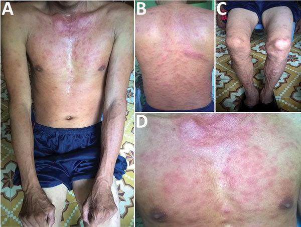
This composite clinical photograph displays a 35-year-old male patient presenting with a generalized maculopapular rash secondary to scrub typhus (Orientia tsutsugamushi infection). The four panels (A-D) illustrate the distribution across different anatomical regions: (A) anterior trunk and arms, (B) back, (C) lower legs, and (D) a close-up of the chest. The rash is characterized by numerous erythematous macules and small papules. The density is highest on the trunk and chest, where lesions show a confluent, intensely red appearance, particularly around the upper chest and neck. On the back and extremities, the rash appears more discrete and sparse, with individual lesions showing clearer margins against unaffected skin. This presentation is a classic dermatological manifestation of rickettsial diseases, typically appearing several days after the onset of fever. The visual findings assist in the clinical diagnosis of scrub typhus in endemic regions, often found in conjunction with a pathognomonic eschar at the mite bite site.
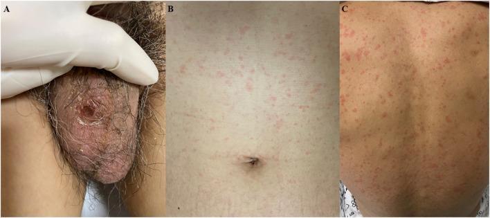
This composite of clinical photographs illustrates the dermatological manifestations of scrub typhus (Orientia tsutsugamushi infection). Panel A displays a characteristic eschar located on the scrotum. The lesion is approximately 1 x 1 cm, presenting as an erythematous to black ulcerative area with a central necrotic crust. Panels B and C show the associated systemic skin involvement, characterized by a diffuse, blanching maculopapular rash distributed across the trunk (B) and the back (C). The rash consists of multiple small, pink-to-red macules and papules that are non-confluent. These findings are diagnostic hallmarks of rickettsial diseases, where the eschar represents the site of the chigger bite and the generalized rash indicates hematogenous dissemination. This clinical presentation, combined with a history of outdoor exposure, is used to identify vector-borne infectious diseases in endemic regions.
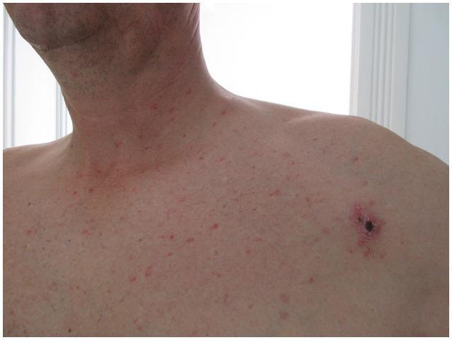
This clinical photograph displays two distinct dermatological manifestations on the upper torso and neck of an adult patient, captured on the 15th day of illness. Primary finding: A prominent 'tache noire' or inoculation eschar located in the left infraclavicular region. This lesion is characterized by a central, dark-black necrotic crust (eschar) surrounded by an erythematous halo indicating localized inflammation. Secondary finding: A generalized, non-pruritic, erythematous macular rash. The rash consists of diffuse, small, reddish macules approximately 2-5mm in diameter, distributed across the chest, shoulders, and lateral neck, creating a speckled appearance. These findings are clinically significant in the diagnosis of rickettsial diseases, such as Mediterranean Spotted Fever or scrub typhus, where an inoculation eschar serves as the primary portal of entry followed by a systemic maculopapular eruption. The image serves as an educational resource for identifying characteristic skin signs of zoonotic infectious diseases.
Scrub Typhus - Treatment
Causative Agent & Transmission
Treatment Regimens
First-Line Therapy
| Drug | Dose | Duration |
|---|---|---|
| Doxycycline | 100 mg orally twice daily | 7-15 days |
| Azithromycin | 500 mg orally once daily | 3 days |
- Doxycycline and tetracyclines are the cornerstone of treatment and typically produce rapid clinical improvement.
- Azithromycin has similar efficacy and is the preferred option when doxycycline is contraindicated.
- Prompt doxycycline treatment leads to rapid improvement including reversal of renal impairment (Brenner & Rector's Kidney).
Alternative / Second-Line
| Drug | Dose | Duration |
|---|---|---|
| Chloramphenicol | 500 mg orally four times daily | 7-15 days |
| Rifampin | Used in resistant cases | Variable |
- Chloramphenicol is less effective than doxycycline and azithromycin and is no longer the preferred alternative.
- Telithromycin is also listed as an alternative in some references.
Severe Scrub Typhus
- Combination therapy: Doxycycline + Azithromycin is recommended over either agent alone - this reduces complications and mortality.
- As per Harrison's 22E (2025): "In severe scrub typhus, the combination of doxycycline and azithromycin is beneficial compared to monotherapy of either agent."
Drug-Resistant Cases (Thailand)
- Some cases in Thailand are poorly responsive to doxycycline and chloramphenicol.
- These cases tend to respond to azithromycin and rifampin.
Special Populations
| Population | Preferred Drug | Rationale |
|---|---|---|
| Pregnant women | Azithromycin | Doxycycline contraindicated in pregnancy |
| Children | Azithromycin | Safer profile; doxycycline avoided in young children |
| Severe disease (any age) | Doxycycline + Azithromycin | Combination reduces mortality |
Clinical Features (for Diagnosis Before Treatment)
- Eschar - painless black necrotic crust at the chigger bite site (pathognomonic but absent in many)
- Maculopapular rash - appears around day 4-6 of illness
- Regional lymphadenopathy
Severe Complications Requiring Aggressive Management
- Encephalitis / meningoencephalitis
- Interstitial pneumonia / ARDS
- Acute kidney injury (AKI in 8-60% of patients; hematuria and proteinuria in up to 80%)
- Myocarditis, pericarditis
- Septic shock, DIC
Diagnosis (to Guide Empiric Treatment)
- Serology: Indirect fluorescent antibody (IFA) is gold standard; ELISA, lateral flow rapid tests
- PCR: Effective from eschar biopsy; less sensitive from blood
- Empiric treatment should be started without waiting for confirmation when clinical suspicion is high, as serologic tests may be negative early in the illness.