
Mrcp
What would you like to know about MRCP?
MRCP magnetic resonance cholangiopancreatography
MRCP magnetic resonance cholangiopancreatography biliary pancreatic duct
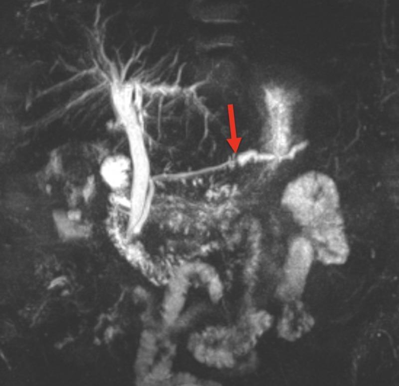
This coronal Magnetic Resonance Cholangiopancreatography (MRCP) image displays the biliary tree and pancreatic duct. The biliary system is visualized as a hyperintense, tree-like branching network in the upper quadrant, featuring the intrahepatic ducts and a prominent common bile duct. The primary focus is the main pancreatic duct, which exhibits significant ductal dilation in its distal portion. A red arrow indicates a point of abrupt transition where the duct diameter suddenly narrows, transitioning from the dilated distal segment to a more constricted proximal segment. This visual finding is characteristic of an obstructive pathology, such as a large pancreatic ductal stone or calculus located within the body of the pancreas. The image provides a clear example of how MRCP can identify obstructive lesions and their secondary effects on ductal morphology in the hepatobiliary system.
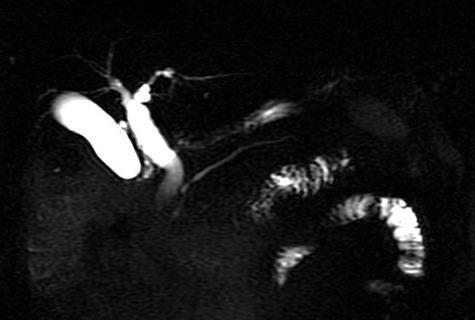
A Magnetic Resonance Cholangiopancreatography (MRCP) image providing a high-contrast visualization of the biliary tree and gallbladder. The image demonstrates high signal intensity (bright) fluid within the biliary system against a dark, suppressed background. Key anatomical features visible include the intrahepatic bile ducts, the gallbladder, and the extrahepatic biliary tree, including the common bile duct. The biliary tree appears patent with no evident filling defects or intraluminal obstructions, suggesting the absence of choledocholithiasis or strictures at the time of imaging. The common bile duct is noted to be approximately 12 mm in diameter, which may represent mild dilation depending on clinical history. The pancreatic duct is also partially visualized. This diagnostic image is used in gastroenterology and radiology to assess for biliary pathologies such as gallstones, bile duct dilation, or masses. The image highlights a post-symptomatic state where previous obstructions may have resolved.
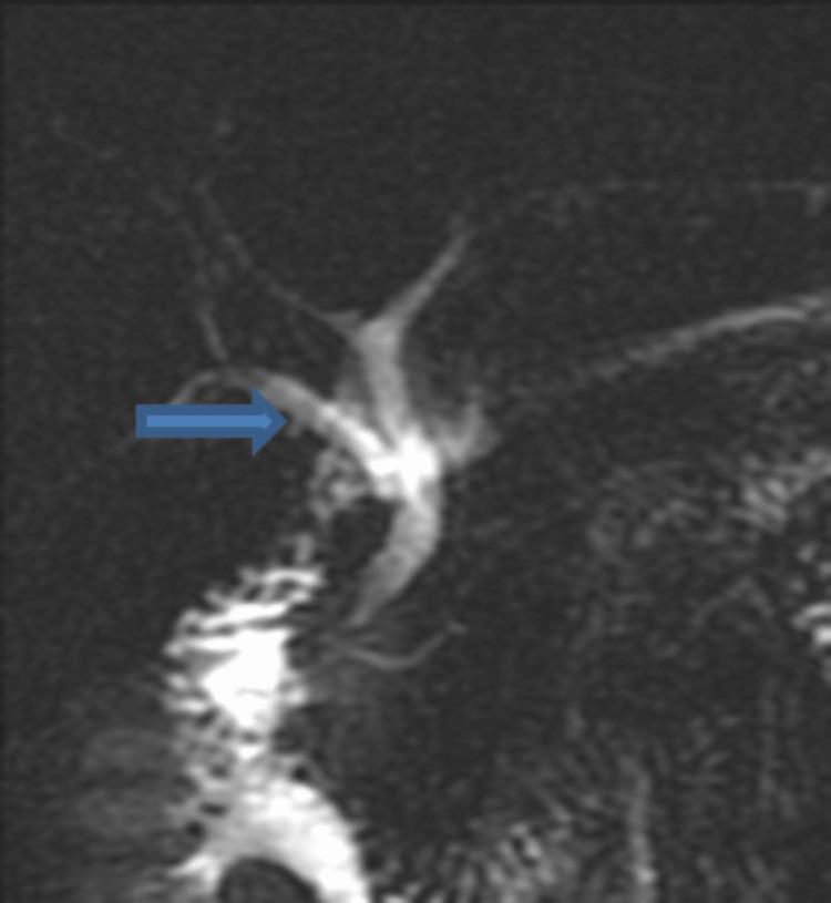
A Magnetic Resonance Cholangiopancreatography (MRCP) or T2-weighted diagnostic image demonstrating an anatomical variation of the biliary tree. The image shows high-signal intensity (fluid-filled) tubular structures representing the bile ducts against a dark background. A blue arrow points to the right posterior sectoral duct, which is clearly visualized merging directly into the common bile duct rather than its standard confluence with the right anterior sectoral duct. The biliary anatomy displays a branching pattern where multiple segmental ducts converge, and the common bile duct is seen descending towards the duodenum. This diagnostic image is used in clinical education to illustrate congenital anatomical variations that are critical for preoperative planning in hepatobiliary and pancreatic surgeries.
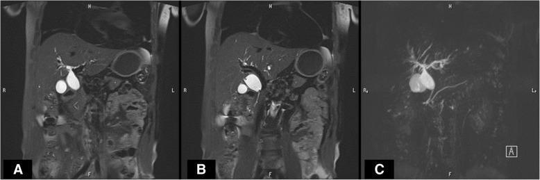
Diagnostic Magnetic Resonance Cholangiopancreatography (MRCP) showing the biliary and pancreatic ductal systems in coronal planes. Panels A and B are T2-weighted source images showing anatomical landmarks, while Panel C is a 3D maximum intensity projection (MIP) reconstruction focusing on fluid-filled structures. The imaging reveals significant fusiform dilatation of the common bile duct (CBD), extending from the common hepatic duct through the middle bile duct, consistent with a choledochal cyst. Panel C clearly demonstrates a pancreaticobiliary maljunction (PBM), characterized by an anomalous union where the pancreatic duct and common bile duct merge into a long common channel outside the duodenal wall. The intrahepatic biliary ducts show mild arborization, and the gallbladder is visible and unremarkable. This case illustrates the diagnostic utility of MRCP in identifying structural biliary anomalies and congenital maljunctions that predispose patients to biliary stasis and malignancy.
MRCP - Magnetic Resonance Cholangiopancreatography
What Is It?
- Schwartz's Principles of Surgery: "MRCP enables rapid, noninvasive depiction of both the biliary tree and the pancreatic duct without the use of ionizing radiation or intravenous contrast media."
- Goldman-Cecil Medicine: MRCP "has replaced ERCP for many diagnostic indications."
How It Works (Technique)
- Uses heavily T2-weighted sequences - fluid-filled structures are maximally bright
- 3D maximum intensity projection (MIP) reconstructions allow visualization of the entire ductal system in one image
- No oral or IV contrast required for standard MRCP
- Can be enhanced with liver-specific MRI contrast agents (e.g., mangafodipir) secreted into bile for special indications
- Secretin-enhanced MRCP (S-MRCP): IV secretin (0.2 µg/kg over 1 min) stimulates pancreatic secretion; T2 images are acquired every 30 seconds for 10 minutes to detect early chronic pancreatitis and assess pancreatic duct compliance
Indications
| Category | Specific Use |
|---|---|
| Choledocholithiasis | Suspected common bile duct stones (sensitivity 81-100%, specificity 96-100%) |
| Biliary obstruction | Level and cause of ductal dilation (malignant vs. benign strictures) |
| Primary Sclerosing Cholangitis (PSC) | Assess multifocal strictures of intra- and extrahepatic ducts |
| Choledochal cysts | Diagnostic procedure of choice; shows cyst morphology and pancreaticobiliary maljunction |
| Chronic pancreatitis | Pancreatic duct dilation, side branch changes, filling defects (stones) |
| Biliary anatomy variants | Pre-surgical planning (e.g., aberrant right posterior sectoral duct) |
| Post-liver transplant | Evaluate biliary anastomotic complications |
| Malignant masses | Relationship of tumors to biliary/pancreatic ducts |
| Low-risk choledocholithiasis | Preferred over ERCP to avoid procedure risk when suspicion is low |
Diagnostic Performance
| Condition | Sensitivity | Specificity |
|---|---|---|
| Choledocholithiasis | 81-100% | 96-100% |
| Biliary strictures | High | High |
| Pancreatic duct (vs ERCP) agreement | 70-80% overall; 83-92% for dilation/narrowing | - |
- MRCP visualizes the main pancreatic duct in the head in 79%, body in 64%, tail in 53% of cases (vs ERCP reference standard)
- Clinical Gastrointestinal Endoscopy: "Correlation with ERCP with respect to main pancreatic duct dilation, narrowing, and filling defects was 83% to 92%, 70% to 92%, and 92% to 100%."
Advantages Over ERCP
| Feature | MRCP | ERCP |
|---|---|---|
| Invasiveness | Non-invasive | Invasive (endoscopic) |
| Radiation | None | Yes (fluoroscopy) |
| Contrast | Not required | Iodinated contrast |
| Complication risk | Minimal | ~5-10% (pancreatitis, perforation, bleeding) |
| Therapeutic capability | Diagnostic only | Diagnostic + therapeutic |
| Spatial resolution | Lower | Higher (sees subtle side branch changes) |
Limitations
- Lower spatial resolution than direct cholangiography - cannot detect subtle early side-branch changes of chronic pancreatitis
- Cannot perform therapeutic interventions (stone extraction, stenting, sphincterotomy) - ERCP is still required for treatment
- False positives: pneumobilia (air in bile ducts) can mimic filling defects
- Motion artifact can degrade image quality
- Limited availability and higher cost compared to ultrasound
- S-MRCP requires specialized centers and expertise
When MRCP vs. ERCP vs. EUS?
- Low/intermediate suspicion of CBD stones → MRCP first (avoid ERCP risk)
- High suspicion + therapeutic intent (stone removal, stenting) → ERCP directly
- Idiopathic acute pancreatitis → MRCP and EUS both acceptable; a 2026 systematic review (PMID 41015654) found EUS and MRCP are comparable for etiological evaluation of idiopathic acute pancreatitis
- Early chronic pancreatitis → S-MRCP or EUS (more sensitive than standard MRCP)
- Post-cholecystectomy biliary leak/stricture → MRCP for anatomy, then ERCP for repair
MRCP Images
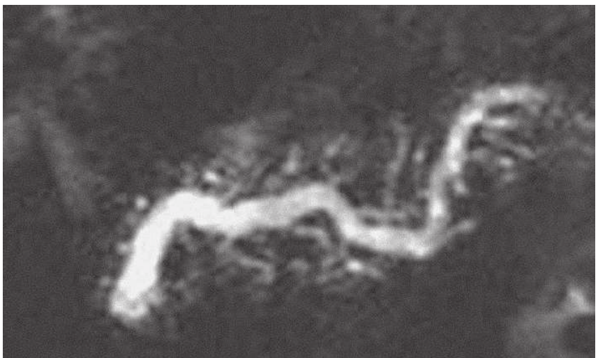

Which is mrcp
- SPINE (MRI-CV Junction, Cervical, Dorsal, Lumbar, etc.)
- ANGIOGRAM (Cerebral, Neck, Aortogram, Pulmonary, Renal, Limb)
- ADVANCED IMAGING (Spectroscopy, Cisternogram, Perfusion, CSF Flow, DTI, Enteroclysis, Arthrogram, Urogram, Sialogram, Functional MRI, Cardiac MRI)
- "/ Contrast"
- "N / MRA/MRV"
- "A + MRV"
- "/ Contrast"
- "BONE/INNER EAR (Plain / Contrast)"