
Can you please answer all the question along witth diagram
Please share the questions you'd like answered. You can:
About head and neck topic specificall ...answer the question basedon marks along with daigram
python3 << 'EOF' import pdfplumber pdf_path = "/tmp/workspace/attachments/d20d9f6a-e08c-40d6-8c6e-ad64f7480eb5/RGUHS-MBBS-5-YEARS-OLD-ANATOMY-Q-PAPERS.pdf" with pdfplumber.open(pdf_path) as pdf: print(f"Total pages: {len(pdf.pages)}") # Extract first 10 pages to understand structure for i, page in enumerate(pdf.pages[:10]): text = page.extract_text() if text: print(f"\n--- PAGE {i+1} ---") print(text[:2000]) EOF
python3 << 'EOF' import pdfplumber pdf_path = "/tmp/workspace/attachments/d20d9f6a-e08c-40d6-8c6e-ad64f7480eb5/RGUHS-MBBS-5-YEARS-OLD-ANATOMY-Q-PAPERS.pdf" # Extract ALL text from all pages all_text = [] with pdfplumber.open(pdf_path) as pdf: for i, page in enumerate(pdf.pages): text = page.extract_text() if text: all_text.append(f"\n=== PAGE {i+1} ===\n{text}") full_text = "\n".join(all_text) # Now filter for Head & Neck related keywords import re head_neck_keywords = [ 'larynx', 'laryngeal', 'tongue', 'parotid', 'thyroid', 'carotid', 'jugular', 'cervical fascia', 'neck', 'nasal', 'nose', 'facial artery', 'facial nerve', 'pharynx', 'palate', 'salivary', 'mandible', 'tonsil', 'orbit', 'eye', 'extraocular', 'pterygoid', 'infratemporal', 'temporomandibular', 'scalp', 'cranial', 'submandibular', 'parapharyngeal', 'branchial', 'brachial plexus cervical', 'thyroid gland', 'parathyroid', 'waldeyer', 'lymph node of tongue', 'external carotid', 'internal jugular', 'deep cervical', 'turbinate', 'prevertebral', 'pretracheal', 'investing layer', 'facial', 'Facial', 'Thyroid', 'Larynx', 'Tongue', 'Parotid', 'Nasal', 'Neck', 'Cervical', 'Salivary', 'Carotid', 'Jugular', 'Pharynx', 'Tonsil', 'Palate', 'Waldeyer', 'Orbit', 'muscles of tongue', 'blood supply of thyroid', 'branches of external carotid', 'lateral wall of nose' ] print("=== HEAD & NECK QUESTIONS EXTRACTED ===\n") print(full_text[0:500]) # Print all pages looking for H&N content with pdfplumber.open(pdf_path) as pdf: for i, page in enumerate(pdf.pages): text = page.extract_text() if text: text_lower = text.lower() hn_found = any(kw.lower() in text_lower for kw in head_neck_keywords) if hn_found: print(f"\n=== PAGE {i+1} (HEAD & NECK CONTENT) ===") print(text) print("---") EOF
larynx interior cavity intrinsic muscles anatomy diagram
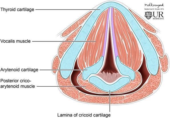
Anatomical diagram of the larynx in axial cross-section at the level of the glottis, illustrating the spatial relationship between cartilaginous structures and intrinsic musculature. The thyroid cartilage forms a prominent, V-shaped anterior boundary. Located posteriorly and inferiorly is the lamina of the cricoid cartilage, which supports the paired arytenoid cartilages. The true vocal folds extend from the anterior midline of the thyroid cartilage to the vocal processes of the arytenoid cartilages, creating the glottic opening. Key intrinsic muscles are labeled, including the vocalis muscle, which runs parallel within the vocal folds, and the posterior cricoarytenoid muscle, situated at the posterior aspect of the cricoid lamina. This illustration is optimized for educational use in otolaryngology and radiology, specifically for correlating axial CT imaging anatomy with laryngeal structure and function, such as vocal fold apposition and glottic patency.
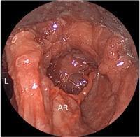
This endoscopic clinical photograph displays the internal anatomy of a human larynx, focusing on the right arytenoid region. The laryngeal lumen (L) is visible as a dark central cavity. The right arytenoid area (AR) is located at the lower portion of the frame, showing characteristic reddish, mucosal tissue. An anatomical variation is highlighted with an asterisk (*), demonstrating an ascending branch of the right superior laryngeal artery (SLA). Unlike the more common descending course, this specific arterial branch exhibits an upward trajectory from the arytenoid region toward the supraglottic space. The image serves as a cadaveric reference for laryngeal microvascular anatomy and surgical planning, illustrating the variability of blood supply to the posterior larynx and intrinsic muscles. This material is relevant for advanced otorhinolaryngology students and surgeons studying laryngeal neurovascular pathways and potential variations encountered during endoscopic or surgical procedures.
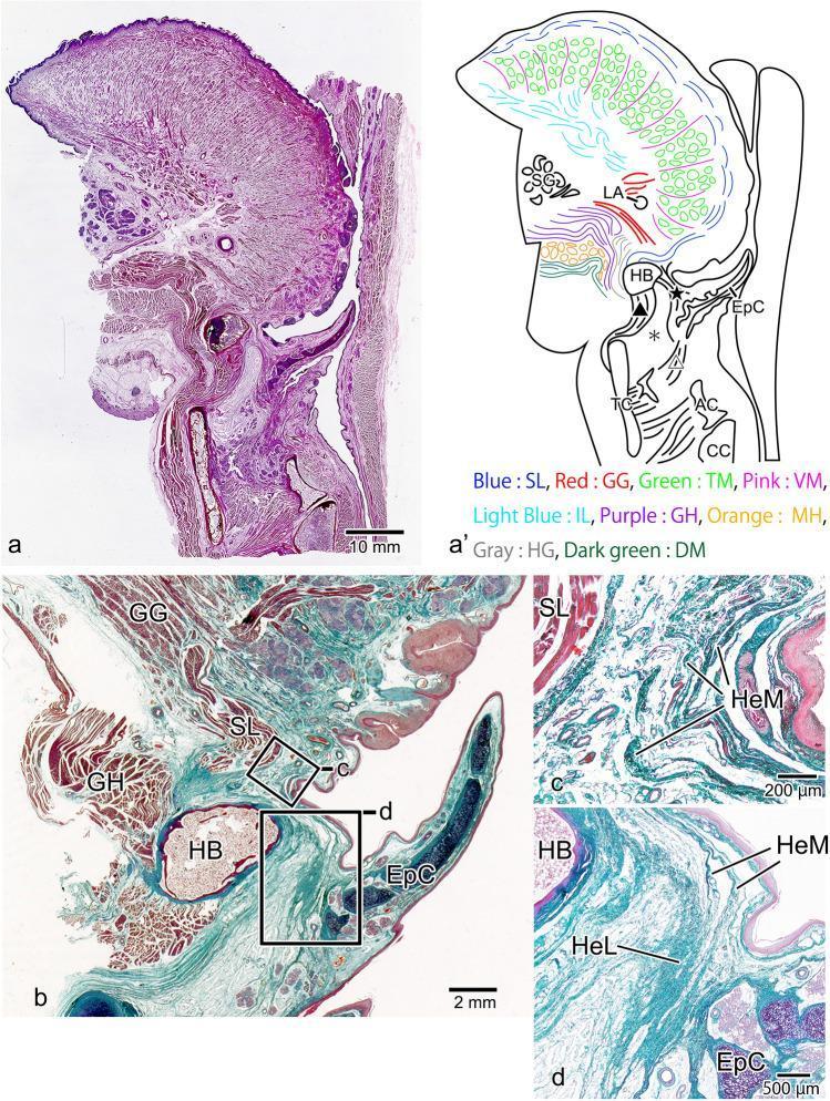
This figure presents a sagittal histological section of the tongue root and larynx, approximately 6-mm lateral to the midline, combining light microscopy with anatomical schematics. Panels (a) and (a') provide a macro-view and corresponding diagram identifying the intrinsic and extrinsic lingual muscles: superior longitudinal (SL), genioglossus (GG), vertical muscle (VM), and transverse muscle (TM). The GG muscle is notably restricted to the posterior region. Panels (b), (c), and (d) provide magnified views of the pre-epiglottic space. The histological images demonstrate the superior longitudinal lingual muscle (SL) exhibiting high continuity, effectively separating the genioglossus (GG) from the epiglottic cartilage (EpC). The hyoepiglottic membrane (HeM) is seen converging before reaching the SL muscle. Furthermore, the HeM merges with the hyoepiglottic ligament (HeL) to attach to the epiglottic cartilage. Key landmarks include the hyoid bone (HB), thyroid cartilage (TC), and cricoid cartilage (CC). This material illustrates the complex fibro-muscular relationships essential for understanding epiglottic movement and swallowing mechanics.
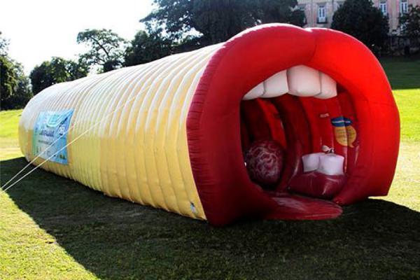
This photograph depicts a large-scale, walk-through educational tool known as a 'giant larynx' model, used in public health campaigns to teach human anatomy and voice production. The structure is an elongated inflatable tunnel with a segmented yellow exterior. One end is designed as a large open mouth featuring oversized red lips and white anatomical representations of upper teeth. The red interior visible through the mouth opening represents the pharynx and larynx, including structural interpretations of the tongue and vocal cords. This interactive anatomical diagram serves as a public education device for the specialty of otolaryngology (ENT), focusing on the upper respiratory tract and phonation. It is designed to simplify complex internal structures for a general audience, demonstrating the pathway from the mouth through the throat.
tongue muscles nerve supply lymphatic drainage anatomy
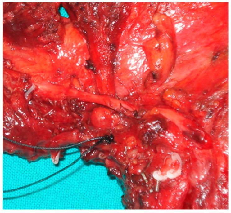
A high-magnification intraoperative clinical photograph of a surgical specimen following a compartment tongue resection for oncology. The image displays the macroscopic anatomy of the T-N (Tumor-Node) tract, focusing on the metastasis of lingual lymph nodes. The surgical field is predominantly characterized by deep red, striated muscle tissue, likely representing the extrinsic tongue muscles (hyoglossus and genioglossus). Central to the image is a discrete, rounded, firm mass representing a metastatic lingual lymph node, positioned along the lymphatic drainage pathway. Linear, pale structures indicative of neurovascular bundles or large-caliber nerves (greater than 1 mm) are visible traversing the field, potentially serving as conduits for perineural neoplastic progression. Surgical artifacts including black silk sutures and metallic hemoclips are present, marking key anatomical landmarks or ligated vessels. The tissue exhibits varied textures from smooth vascular sheaths to irregular, congested areas suspicious for neoplastic infiltration or 'in transit' lymphatic metastasis. This visual serves to educate on the anatomical pathways of oral cancer spread and the principles of compartmental surgery.
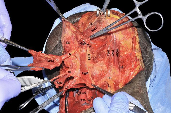
A clinical photograph of a cadaveric dissection illustrating the deep surgical anatomy of the tongue and submandibular region, relevant to compartmental surgery. The image shows several extrinsic tongue muscles and neurovascular structures held with surgical instruments to demonstrate spatial relationships. Key anatomical landmarks labeled include the hyoglossus muscle (HgM), genioglossus muscle (GgM), and mylohyoid muscle (MhM). The hypoglossal nerve (XII) is shown running superficial to the hyoglossus muscle, while the lingual artery (LA) is positioned deep to it. The lingual nerve (LN) is visible in proximity to the sublingual gland (SlG) and retracted mylohyoid muscle. Other identified structures include the common carotid artery (CCA), external carotid artery (ECA), styloglossus muscle (SgM), hyoid bone (HB), and the median lingual septum (MLS). This visual facilitates the understanding of tumor spread pathways (T-N tract) along muscular and neurovascular bundles in head and neck oncology.
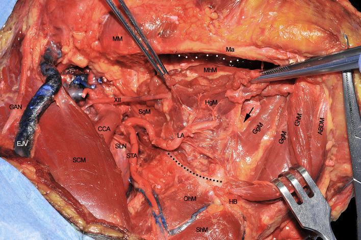
A clinical photograph of a cadaveric dissection illustrating the surgical anatomy of the submandibular and submental regions during a compartmental surgery of the tongue. The image demonstrates the intricate relationships between extrinsic tongue muscles, vascular structures, and nerves. Key anatomical landmarks include the mandible (Ma), hyoid bone (HB), and the mylohyoid (MhM), hyoglossus (HgM), genioglossus (GgM), and geniohyoid (GhM) muscles. Vascular structures shown include the common carotid artery (CCA) branching into the superior thyroid artery (STA) and the lingual artery (LA), the latter of which is seen coursing deep to the hyoglossus muscle. The hypoglossal nerve (XII) and superior laryngeal nerve (SLN) are clearly identified. Superficially, the sternocleidomastoid muscle (SCM), external jugular vein (EJV), and great auricular nerve (GAN) are visible. Dotted lines delineate the mylohyoid line of the mandible and the great hyoid cornu, while a black arrow points to the paramedian septum of the tongue, providing critical orientation for oncological resection pathways.
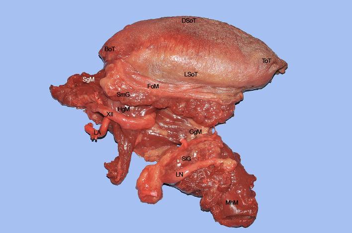
This clinical photograph of a surgical specimen illustrates the complex anatomy of the human tongue and floor of mouth (FoM), relevant for compartmental surgery in head and neck oncology. The specimen includes the dorsal (DSoT) and lateral (LSoT) surfaces of the tongue, the tip (ToT), and the base (BoT). Key neurovascular and muscular structures are annotated to show their spatial relationships. The lingual nerve (LN) is shown in close proximity to the sublingual gland (SlG), running ventrally to the mylohyoid muscle (MhM). The hypoglossal nerve (XII) is visible running superficial to the hyoglossus muscle (HgM), while the lingual artery (LA) is situated deeper. The fan-shaped genioglossus muscle (GgM) constitutes a significant portion of the tongue's volume. Posteriorly, the styloglossus muscle (SgM) is identified as it approaches the lateral aspect of the tongue base. This anatomical demonstration highlights the primary routes for tumor spread along extrinsic muscles and neurovascular bundles, emphasizing the surgical margins required for total hemiglossectomy.
thyroid gland anatomy relations blood supply
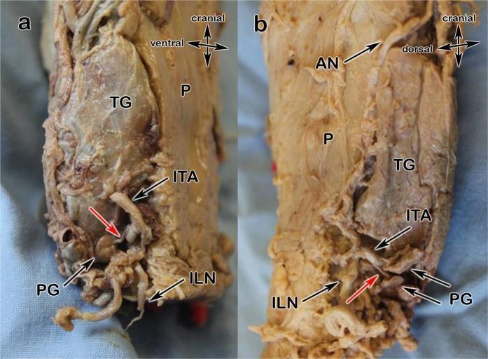
This medical specimen photograph consists of two panels (a and b) illustrating the neurovascular anatomy and arterial supply variations of the human parathyroid glands. Panel (a) shows Type a vascularization where the parathyroid glands (PG) receive their primary blood supply (red arrows) from branches of the inferior thyroid artery (ITA). The thyroid gland (TG) is positioned ventrally to the pharynx (P), with the inferior laryngeal nerve (ILN) serving as a critical surgical landmark. Panel (b) demonstrates Type b vascularization, characterized by the presence of prominent anastomoses (AN) between the superior thyroid artery and the inferior thyroid artery (ITA), enhancing collateral circulation to the parathyroids (PG). Both images provide a lateral or dorsal-lateral view of the larynx/pharynx region, emphasizing the spatial relationships between the thyroid lobe, the recurrent (inferior) laryngeal nerve, and the adjacent parathyroid tissue. These images are intended for surgical education to aid in the preservation of parathyroid function and nerve integrity during thyroidectomy.
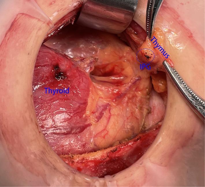
This clinical photograph shows an intraoperative view of a thyroidectomy or neck dissection, highlighting the surgical anatomy of the thyroid gland, thymus, and inferior parathyroid gland (IPG). The thyroid gland is a large, reddish, vascularized structure visible on the left side. Surgical forceps are used to retract the thymus, an elongated, yellowish-tan tissue located superior and lateral to the thyroid. The IPG is visible as a small, distinct ovoid nodule closely associated with the superior aspect of the thymic capsule, circled and labeled in blue. This visual demonstrates the 'in situ reservation' technique, where the IPG and thymus are preserved as an organic unit to maintain parathyroid blood supply during central lymph node dissection. The surgical field displays exposed soft tissue, retractor placement, and visible vasculature, providing educational context for endocrine surgeons and residents regarding the preservation of parathyroid function.
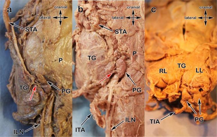
This set of three clinical photographs (a–c) depicts anatomical dissections of the thyroid region, focusing on the arterial blood supply variants to the parathyroid glands (PG). Panel (a) shows Type x supply where the parathyroid gland receives its primary arterial branch (red arrow) from the superior thyroid artery (STA), with the inferior laryngeal nerve (ILN) and pharynx (P) visible as landmarks. Panel (b) illustrates a variant where the blood supply arises directly from the thyroid gland (TG) parenchyma (red arrow); the superior thyroid artery (STA), inferior thyroid artery (ITA), and ILN are identified. Panel (c) demonstrates an anatomical variant where the parathyroid glands located at the inferior aspect of the right lobe (RL) and left lobe (LL) are supplied by a prominent thyroid ima artery (TIA) ascending from the mediastinum. These images provide critical surgical context for thyroidectomy and parathyroidectomy, highlighting the high degree of vascular variability and the necessity of preserving the parathyroid blood supply to prevent postoperative hypocalcemia.
cavernous sinus relations contents tributaries diagram
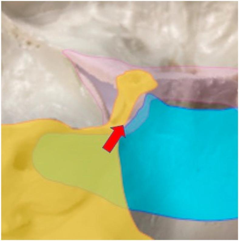
Anatomical Diagram and intraoperative overlay illustrating the arterial blood supply to the cavernous sinus, specifically regarding the feeding vessels of skull base meningiomas. The cavernous sinus is highlighted by a pink outline. The arterial distribution is color-coded: the Tentorial Artery (TA) is represented in yellow, demonstrating its supply to the superolateral aspect of the posterior cavernous sinus. The Dural Meningeal Artery (DMA) is shown in blue, highlighting its coverage of the inferomedial portion of the posterior cavernous sinus. A red arrow identifies the interface where cranial nerves are typically situated between these two arterial territories. The image serves as a clinical reference for neurosurgical planning, helping to differentiate between petrotentorial and petroclival meningioma attachments based on their primary feeding arteries and their spatial relationship to critical neurovascular structures within the skull base.
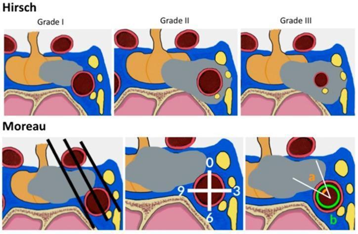
Educational medical diagram illustrating two radiological classification systems for cavernous sinus (CS) invasion by sellar tumors, such as pituitary adenomas, shown in coronal section. The top panel depicts the Hirsch classification (Grades I-III): Grade I shows the tumor (grey) partially touching the internal carotid artery (ICA); Grade II shows total ICA encasement without luminal narrowing; and Grade III shows encasement with significant ICA diameter reduction (stenosis). The bottom panel illustrates the Moreau criteria for assessing CS invasion: the first frame shows three intercarotid lines (medial, median, and lateral) intersecting the ICA and tumor; the second frame demonstrates the subdivision of the cavernous ICA into quadrants (0, 3, 6, 9 o'clock positions); and the third frame uses angular analysis (angle 'a') to quantify the degree of tumor-ICA contact. Key anatomical landmarks include the pituitary gland (orange), sphenoid sinus (pink), and the cavernous sinus venous spaces (blue/yellow).
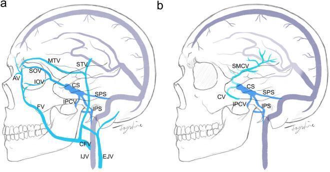
Two-panel anatomical diagram illustrating venous access routes for transvenous cavernous sinus (CS) embolization. Panel (a) highlights superficial venous pathways, showing the angular vein (AV) connecting to the superior ophthalmic vein (SOV), inferior ophthalmic vein (IOV), and facial vein (FV). The facial vein merges into the common facial vein (CFV) before draining into the internal jugular vein (IJV) and external jugular vein (EJV). Other highlighted superficial vessels include the middle temporal vein (MTV) and superficial temporal vein (STV). Panel (b) illustrates intracranial venous access routes, highlighting the cortical veins (CV) and superficial middle cerebral vein (SMCV) as they drain into the cavernous sinus. Both panels depict deep connections through the superior petrosal sinus (SPS), inferior petrosal sinus (IPS), and inferior petroclival vein (IPCV) as primary drainage pathways toward the jugular system. This medical illustration is designed to guide endovascular planning for treating carotid-cavernous fistulas or dural arteriovenous fistulas by identifying surgical and endovascular approach routes.
parotid gland anatomy relations structures within it
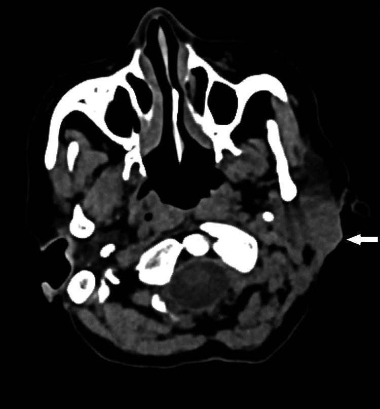
This diagnostic image is an axial computed tomography (CT) scan of the head and neck at the level of the parotid glands. The scan demonstrates the cross-sectional anatomy of the maxillofacial region, including the nasal cavity, maxillary sinuses, and the base of the skull with the foramen magnum and vertebral structures visible posteriorly. The primary focus is the left parotid gland, which is clearly visualized in its relationship to the mandible and the parapharyngeal space. A white arrow points to a distinct bulge and increased prominence within the superficial lobe of the left parotid gland. This finding represents a localized soft tissue mass or lesion that is relatively iso-dense to the surrounding glandular parenchyma. The deep lobe of the parotid gland appears unaffected, maintaining its position medial to the plane of the facial nerve (the fasciovenous plane of Patey). The image is clinically significant for evaluating parotid gland tumors, such as mucoepidermoid carcinoma, and determining their extent relative to the superficial and deep lobes.
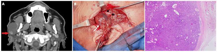
This composite figure illustrates a clinical case of a parotid gland tumor across three modalities: diagnostic imaging, intraoperative findings, and histopathology. Panel A (Axial Contrast-Enhanced CT Scan): Shows a unilateral, well-defined, multilocular cystic lesion within the superficial lobe of the right parotid gland. A small, hyperdense, contrast-enhancing solid nodule (red arrow) is visible in the distal portion of the cyst. Panel B (Intraoperative Clinical Photograph): Displays the surgical field during a superficial parotidectomy. A thin-walled multilocular cyst (white arrow) is identified, positioned laterally to preserved branches of the facial nerve (black arrows), demonstrating the intimate spatial relationship between the pathology and neurovascular structures. Panel C (Histopathology): A Hematoxylin and Eosin (H&E) stained micrograph at 200x magnification reveals the tumor morphology. It shows a dense proliferation of mixed small cells, featuring both epithelioid and spindle-shaped cells without significant cytological atypia, arranged in characteristic trabecular patterns. This sequence demonstrates the diagnostic pathway for salivary gland neoplasms, emphasizing the correlation between radiological cystic components and surgical anatomy.
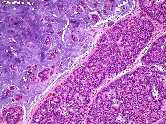
Imaging modality: light microscopy of hematoxylin and eosin stained paraffin-embedded salivary gland tissue from the parotid region. Anatomy: parotid gland tissue showing a classic biphasic neoplasm with epithelial elements intimately admixed with mesenchymal-like stroma. Description: Epithelial components form acinar and tubular structures, composed of duct‑forming cells and myoepithelial-like cells, set within a markedly hypocellular myxoid to chondromyxoid stroma. The epithelial nests are hypercellular in places and juxtaposed to expansive, basophilic stroma; the interface produces a biphasic appearance. The stroma often shows loose, lobulated myxoid material with mild chondroid differentiation, creating the distinctive mixed morphology. Overall, this pattern is typical of pleomorphic adenoma, a benign salivary gland tumor with epithelial–mesenchymal differentiation and typically a well circumscribed capsule. Diagnostic significance: the combination of acinar/tubular epithelial differentiation within a myxoid/chondromyxoid stroma strongly supports pleomorphic adenoma; differentiates from malignant salivary neoplasms. Differential considerations: mucoepidermoid carcinoma, adenoid cystic carcinoma, basal cell adenoma. Clinical correlation: presents as a slow-growing, painless parotid mass in adults; treatment usually involves complete surgical excision with facial nerve preservation and careful follow-up for recurrence. Potential applications: educational histology exemplar, pathology teaching repository, and radiology-pathology correlation studies. This image exemplifies classic pleomorphic adenoma histology suitable for teaching and diagnostic training in pathology education.
facial artery origin course branches face neck
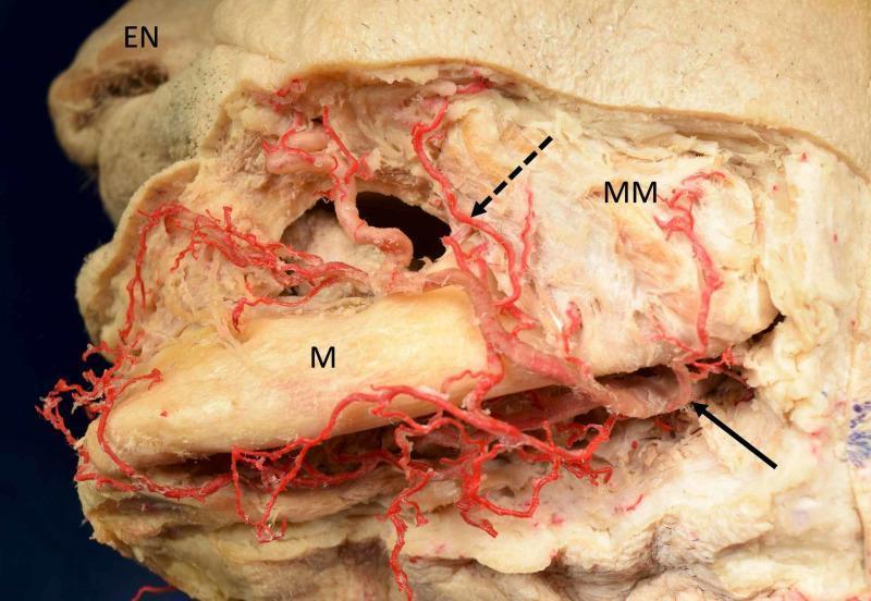
This clinical anatomical photograph shows a lateral view of a dissected Caucasian cadaveric specimen focusing on the vascular supply of the left lower face. The facial artery is highlighted by a solid arrow as it crosses the inferior border of the mandible (M) to enter the face. A prominent premasseteric branch (dotted arrow), also known as the masseteric or posterior branch, is seen ascending superficially over the masseter muscle (MM). The facial artery exhibits a tortuous course as it travels anteriorly toward the external nose (EN), giving off multiple smaller branches. The relationship between the arterial network and underlying musculoskeletal landmarks, specifically the mandible and masseter muscle, is clearly demonstrated. This image serves as an educational resource for plastic surgery, maxillofacial anatomy, and vascular studies, highlighting anatomical variations where the premasseteric branch may be strongly developed, especially in cases with variations in the maxillary artery.
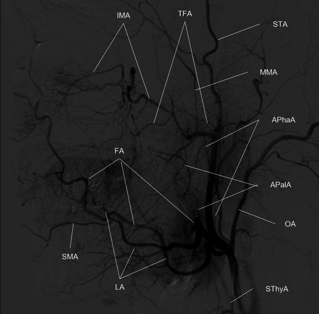
This diagnostic image is a Digital Subtraction Angiogram (DSA) of the human head and neck, specifically showing a lateral view of the external carotid artery (ECA) and its branching patterns. The image serves as a high-resolution anatomical reference for the vascular supply to the face, scalp, and deep facial structures. Key vessels identified via labels include the superior thyroid artery (SThyA), lingual artery (LA), facial artery (FA), occipital artery (OA), and the ascending pharyngeal artery (APhaA). The facial artery (FA) is shown emerging anteriorly, giving rise to the ascending palatine (APalA) and submental (SMA) arteries. Superiorly, the internal maxillary artery (IMA) is visible with its mandibular and pterygoid segments, alongside the middle meningeal artery (MMA). The terminal branches of the ECA, including the superficial temporal artery (STA) and the transverse facial artery (TFA), are also clearly delineated. This visualization is characteristic of interventional radiology and vascular anatomy studies used for planning procedures such as intra-arterial chemotherapy or embolization for head and neck cancers.
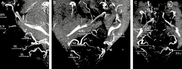
Diagnostic diagnostic imaging showing the arterial anatomy of the human face via 3D Magnetic Resonance Angiography (MRA) with Maximum Intensity Projection (MIP) reconstructions. The image comprises three views: (A) Lateral, (B) Right oblique, and (C) Anteroposterior (AP). Key vascular structures are labeled to demonstrate the course and branching patterns of the facial arterial system. The facial artery (F) is seen giving rise to the inferior labial (IL) and superior labial (SL) arteries before continuing superiorly as the angular artery (Ang/A). Distal branches including the lateral nasal (LN), dorsal nasal (DN), supratrochlear (STr), and supraorbital (SO) arteries are identified, illustrating the complex terminal arborization around the nose and orbits. Additionally, the angular vein (vA/Av) is visible, providing a clinical comparison between arterial and venous calibers in the midface. This imaging is highly relevant for plastic surgery, dermatology, and maxillofacial surgery planning, particularly for understanding vascular danger zones during aesthetic filler injections or reconstructive procedures.
internal jugular vein formation tributaries termination
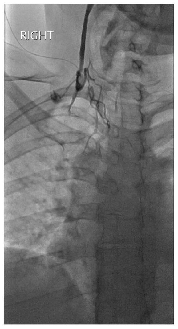
This diagnostic fluoroscopic image displays a right internal jugular venogram obtained via micropuncture access. The radiograph reveals an abrupt termination of contrast medium flow within the proximal right internal jugular vein, signifying a complete vascular occlusion. Distal to the site of obstruction, there is a total absence of contrast filling in the expected anatomical course of the vein toward the superior vena cava. To compensate for the blockage, multiple small, tortuous collateral vessels are visible, branching out to bypass the occluded segment. The underlying skeletal anatomy, including the cervical and upper thoracic vertebrae, clavicle, and ribs, serves as dense radio-opaque landmarks for spatial orientation. This image is clinically significant for demonstrating venous thrombosis or mechanical occlusion in the neck vasculature, illustrating the development of collateral circulation in response to chronic or acute venous obstruction.
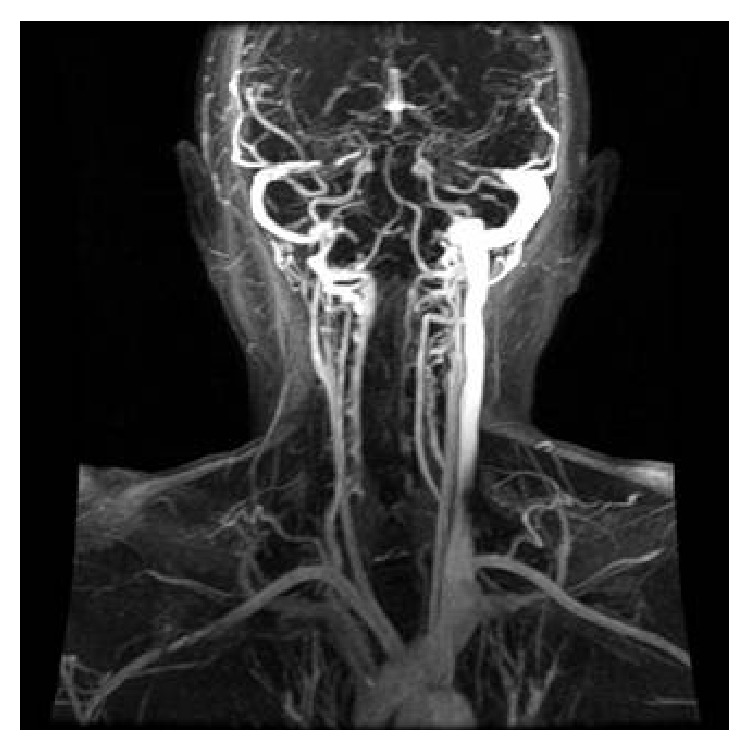
**Imaging Modality:** Coronal maximum intensity projection (MIP) reconstruction of a dynamic contrast-enhanced Magnetic Resonance Venogram (MRV). **Anatomical Region:** Head and neck, focusing on the dural venous sinuses and the internal jugular veins (IJVs). **Observed Pathology:** There is a significant asymmetry in the venous outflow. The right internal jugular vein demonstrates a near-complete lack of contrast opacification distal to the jugular bulb, indicating an occlusion or high-grade stenosis. In contrast, the left internal jugular vein is patent but appears markedly dilated (ectatic) and thickened, likely representing compensatory flow. **Characteristic Visual Features:** * **Filling Defect:** Absence of the normal high-signal column in the right IJV. * **Vascular Morphometry:** Prominent enlargement of the contralateral (left) cervical venous system. * **Collateralization:** Visible smaller venous collaterals in the cervical region. **Differentiating Features:** The sudden termination of the contrast column on the right at the level of the jugular foramen suggests internal jugular vein thrombosis or extrinsic compression, while the compensatory dilatation of the left IJV is a secondary hemodynamic response.
deep cervical fascia layers investing pretracheal prevertebral neck
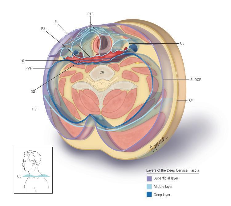
This medical anatomical diagram illustrates a transverse section of the neck at the C6 vertebral level, detailing the layers of the deep cervical fascia and the potential spaces they define. The illustration categorizes the fascia into three layers: superficial (yellow), middle (light blue), and deep (dark blue). Key anatomical structures labeled include the superficial fascia (SF), the superficial layer of deep cervical fascia (SLDCF), and the pretracheal fascia (PTF) enclosing the visceral compartment. Centrally, the diagram highlights the alar fascia (marked with an asterisk), which is situated between the retropharyngeal fascia (RF) anteriorly and the prevertebral fascia (PVF) posteriorly. This arrangement delineates two critical clinical spaces: the retropharyngeal space (RS), located between the RF and the alar fascia, and the 'danger space' (DS), situated between the alar fascia and the PVF. The carotid sheath (CS) is shown laterally. This visualization is essential for understanding the pathways of infection spread between the neck and the mediastinum, as well as providing anatomical guidance for surgical procedures in the prevertebral region.
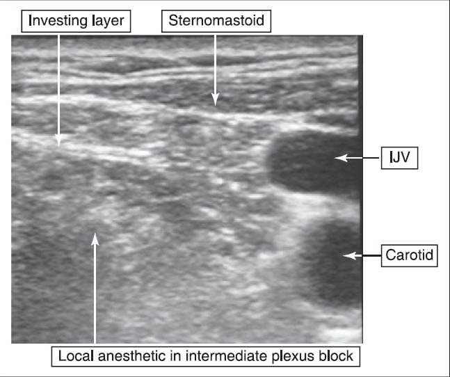
**Imaging Modality:** Ultrasound (B-mode) **Anatomical Region:** Lateral cervical region (neck) **Key Landmarks:** * **Musculature:** Sternocleidomastoid muscle (labeled as sternomastoid) positioned superficially. * **Vasculature:** Internal Jugular Vein (IJV) and Carotid Artery, visualized as anechoic circular structures on the right aspect of the frame. * **Fascial Layers:** The investing layer of the deep cervical fascia is identified. **Procedural Findings:** The image demonstrates an intermediate cervical plexus block. A hypoechoic collection of local anesthetic is visible deep to the investing fascia of the neck and the sternocleidomastoid muscle, but superficial to the prevertebral fascia. The fluid distribution is noted surrounding the cervical plexus nerves, lateral to the vascular bundle (IJV and carotid). **Diagnostic/Visual Features:** The ultrasound captures the hydrodissection of fascial planes. The contrast between the hyperechoic fascial lines and the hypoechoic local anesthetic fluid confirms appropriate needle placement for a regional anesthesia technique targeting the intermediate cervical plexus.
lateral wall nasal cavity turbinates features blood supply nerve supply
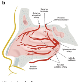
Summary : This is a labeled anatomical illustration of the arterial blood supply to the lateral wall of the human nasal cavity, showing the main arteries and their branches in relation to nasal structures. illustration: # Scene Overview : • Main subject is the lateral wall of the nasal cavity, depicted in a sagittal section. • Shows the distribution of arteries supplying the nasal mucosa. • Key anatomical landmarks labeled: superior turbinate, middle turbinate, inferior turbinate. # Technical Details : • No scale bar or magnification indicated. • Arteries are shown in red, with branching patterns clearly illustrated. • Labeled arteries: anterior ethmoidal artery, posterior ethmoidal artery, sphenopalatine artery, greater palatine artery. # Spatial Relationships : • The anterior ethmoidal artery and posterior ethmoidal artery are shown entering the nasal cavity from the upper region. • The sphenopalatine artery and greater palatine artery supply the lower and posterior regions. • The turbinates (superior, middle, inferior) are used as reference points for arterial distribution. # Analysis : • The illustration highlights the rich vascular network of the nasal cavity, with multiple arteries converging in the region of the turbinates. • The sphenopalatine artery appears to be a major supplier to the posterior and inferior regions. • The ethmoidal arteries supply the upper and anterior regions. • This figure is useful for understanding the sources of nasal blood supply, relevant for clinical contexts such as epistaxis (nosebleeds).
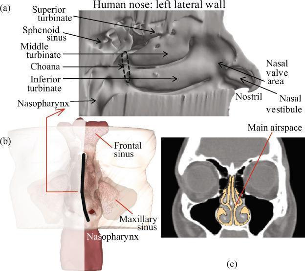
This medical illustration presents three anatomical views of the human sinonasal cavity. Panel (a) shows a detailed sagittal cut-away of the left lateral nasal wall, labeling the superior, middle, and inferior turbinates, the sphenoid sinus, the posterior choana, and the nasopharynx. Anterior features include the nasal valve area, nostril, and nasal vestibule. Panel (b) provides a 3D volumetric rendering of the sinonasal complex, highlighting the frontal and maxillary sinuses and the nasopharynx, with a black line indicating the sagittal plane used in the first view. Panel (c) is a representative coronal computed tomography (CT) scan through the mid-nasal cavity, where the main nasal passage and airway are highlighted in a contrasting light yellow color to demonstrate the cross-sectional patency. This figure is designed for educational instruction on nasal anatomy, specifically focusing on the complex geometry relevant to intranasal drug delivery and airflow dynamics within the respiratory tract.
RGUHS ANATOMY - HEAD & NECK: COMPLETE QUESTION BANK WITH ANSWERS
LONG ESSAYS (10 Marks)
Q1. LARYNX - Cavity / Intrinsic Muscles / Nerve Supply + Applied Anatomy
| Compartment | Boundaries | Features |
|---|---|---|
| Vestibule (supraglottic) | From aditus to vestibular folds | Aryepiglottic folds laterally |
| Ventricle (Sinus) | Between vestibular & vocal folds | Laryngeal appendix extends upward |
| Infraglottic cavity (subglottis) | Below vocal folds to trachea | Widens to become trachea |
- Glottis = the vocal folds + rima glottidis (the narrowest part, most important for phonation)
- Rima glottidis = narrowest part in adults; in children the subglottis is narrowest (site of croup)
- 3 unpaired cartilages: Thyroid, Cricoid, Epiglottis
- 3 paired cartilages: Arytenoids, Corniculate, Cuneiform
| Muscle | Action |
|---|---|
| Posterior cricoarytenoid | ONLY abductor of vocal cords (opens rima glottidis) |
| Lateral cricoarytenoid | Adductor - closes rima glottidis |
| Transverse arytenoid | Adductor |
| Oblique arytenoid | Adductor + closes laryngeal inlet |
| Cricothyroid | Tenses (elongates) vocal cords - increases pitch |
| Thyroarytenoid (vocalis) | Relaxes vocal cords - decreases pitch |
| Aryepiglottic | Closes laryngeal inlet during swallowing |
| Thyroepiglottic | Opens laryngeal inlet |
| Nerve | Origin | Supplies |
|---|---|---|
| Superior laryngeal nerve | Vagus at nodose ganglion | Internal branch: sensory above vocal cords; External branch: motor to cricothyroid only |
| Recurrent laryngeal nerve | Loops under aortic arch (left) / subclavian (right) | Motor to ALL intrinsic muscles EXCEPT cricothyroid; Sensory below vocal cords |
- Recurrent laryngeal nerve (RLN) runs in the groove between trachea and esophagus, close to the inferior thyroid artery. It is at risk during thyroidectomy.
- Unilateral RLN injury: hoarseness (the cord lies in paramedian position, the other cord compensates)
- Bilateral RLN injury: respiratory distress + aphonia - emergency tracheostomy needed
- External branch of superior laryngeal nerve runs with the superior thyroid artery close to the superior pole. Injury → loss of high-pitched phonation (cricothyroid paralysis) - "singer's nerve"
- Why the thyroid gland moves with swallowing: It is enclosed in the pretracheal fascia (which is attached to the larynx and trachea), so it rises with the larynx during deglutition.
Q2. THYROID GLAND - Parts / Capsule / Relations / Blood Supply + Applied Anatomy
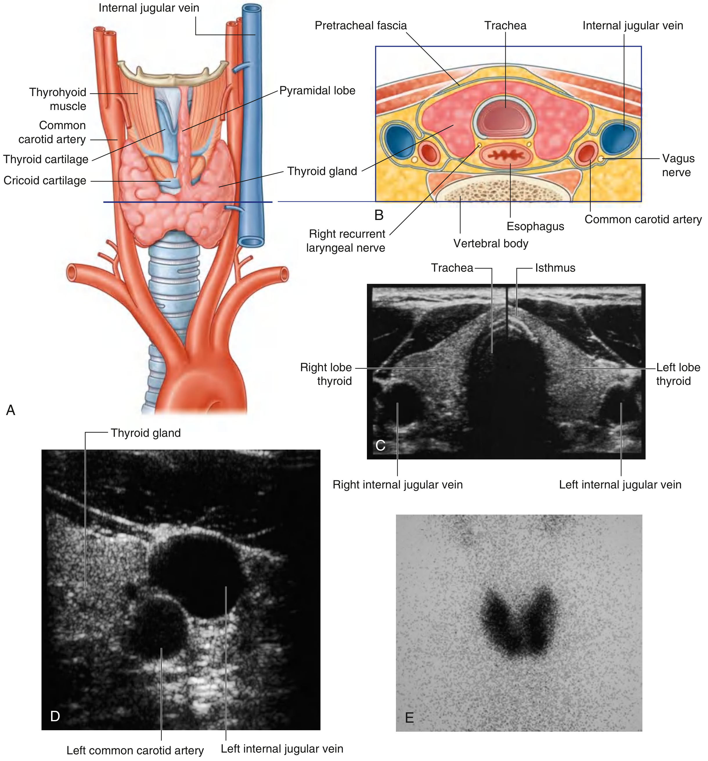
- Two lateral lobes (right & left) - cover anterolateral surfaces of C5-T1 vertebrae
- Isthmus - crosses 2nd and 3rd tracheal rings anteriorly
- Pyramidal lobe (developmental remnant of thyroglossal duct) - present in ~50% of people, ascending from the isthmus
- True capsule (inner): thin, dense fibrous capsule from which septa pass into the gland dividing it into lobules
- False capsule (outer): derived from the pretracheal layer of deep cervical fascia. The space between true and false capsule contains blood vessels and parathyroid glands. The false capsule condenses posteriorly to form the suspensory ligament of Berry (attaches gland to trachea and cricoid).
| Artery | Origin | Supplies |
|---|---|---|
| Superior thyroid artery | First branch of external carotid artery | Superior pole of each lobe |
| Inferior thyroid artery | Thyrocervical trunk (from 1st part of subclavian) | Lower and posterior parts |
| Thyroid ima artery | Brachiocephalic trunk or aortic arch | Isthmus (present in ~10%) |
- Thyroid moves with swallowing: attached to larynx via pretracheal fascia - distinguishes thyroid swelling from other neck swellings
- Thyroglossal cyst: at foramen cecum to isthmus; moves with protrusion of tongue
- Recurrent laryngeal nerve injury: hoarseness post-thyroidectomy
- Hypoparathyroidism: if parathyroids inadvertently removed
- Thyroid ima artery: risk during emergency tracheostomy
Q3. CAVERNOUS SINUS - Relations / Contents / Tributaries / Applied Anatomy
- Superiorly: Optic chiasma, pituitary gland
- Inferiorly: Foramen lacerum, greater wing of sphenoid
- Medially: Pituitary fossa, sphenoidal air sinus
- Laterally: Temporal lobe of brain
- Anteriorly: Superior orbital fissure (communicates with orbit)
- Posteriorly: Dorsum sellae, basilar plexus
- Internal carotid artery (with its sympathetic plexus) - passes through the sinus in an S-shaped curve (carotid siphon)
- Abducent nerve (CN VI) - lies freely within the sinus (most vulnerable to pressure)
- Oculomotor nerve (CN III)
- Trochlear nerve (CN IV)
- Ophthalmic branch of trigeminal (V1)
- Maxillary branch of trigeminal (V2) - in the lower part of the lateral wall
| Tributary | From |
|---|---|
| Superior ophthalmic vein | Orbit (connects face to sinus - spread of infection) |
| Inferior ophthalmic vein | Orbit |
| Sphenoparietal sinus | Anterior |
| Superficial middle cerebral vein | Brain surface |
| Central vein of retina | Through optic canal |
| Basilar plexus | Connects the two cavernous sinuses |
- Cavernous sinus thrombosis: Infection from the "dangerous area of face" spreads via facial vein → angular vein → superior ophthalmic vein → cavernous sinus. Features: proptosis, chemosis, ophthalmoplegia, fever, meningism.
- Pituitary tumor compression: CN III palsy first (ptosis, dilated pupil, eye turned down and out), then CN IV, V1, VI
- Carotid-cavernous fistula (after trauma): pulsating exophthalmos, bruit over eye
- CN VI (Abducent) is most vulnerable: runs freely in sinus, so lateral rectus palsy (convergent squint) occurs early in any sinus pathology
Q4. TONGUE - Muscles / Nerve Supply / Lymphatic Drainage
| Muscle | Action |
|---|---|
| Superior longitudinal | Curls tip upward, shortens tongue |
| Inferior longitudinal | Curls tip downward, shortens tongue |
| Transverse | Narrows and elongates tongue |
| Vertical | Flattens and broadens tongue |
| Muscle | Origin | Insertion | Action |
|---|---|---|---|
| Genioglossus | Genial tubercle of mandible | Dorsum of tongue + hyoid | Protrudes tongue (main protruder) |
| Hyoglossus | Body & greater horn of hyoid | Side of tongue | Depresses and retracts |
| Styloglossus | Styloid process | Side and undersurface | Retracts and elevates |
| Palatoglossus | Palatine aponeurosis | Side of tongue | Elevates tongue base, closes oropharyngeal isthmus |
| Region | Sensation (General) | Taste (Special) | Motor |
|---|---|---|---|
| Anterior 2/3 | Lingual nerve (V3 branch of trigeminal) | Chorda tympani (VII) via lingual nerve | CN XII |
| Posterior 1/3 | Glossopharyngeal (IX) | Glossopharyngeal (IX) | CN XII |
| Extreme base (vallecule) | Internal laryngeal nerve (X) | Vagus (X) | CN XII |
| Palatoglossus | - | - | Vagus (pharyngeal plexus) |
- Tip → Submental nodes (then to deep cervical nodes)
- Anterior 2/3 (lateral margins) → Submandibular nodes → Jugulodigastric → Deep cervical chain
- Posterior 1/3 → Jugulodigastric nodes directly (most important lymph node of tongue)
- Central tongue (bilateral drainage) → Both right and left deep cervical nodes
Q5. LATERAL WALL OF NOSE - Features / Blood Supply / Nerve Supply / Applied
- Superior turbinate - part of ethmoid
- Middle turbinate - part of ethmoid
- Inferior turbinate - separate bone
| Meatus | Opening(s) Into It |
|---|---|
| Superior meatus | Posterior ethmoidal air sinuses, Sphenoethmoidal recess (for sphenoid sinus) |
| Middle meatus | Frontal sinus (via frontonasal duct), anterior & middle ethmoidal sinuses, maxillary sinus (via hiatus semilunaris) |
| Inferior meatus | Nasolacrimal duct (anteromedial part) |
- Sphenopalatine artery (from maxillary artery) - main supply, posterior and inferior region
- Anterior ethmoidal artery (from ophthalmic artery) - anterosuperior region
- Posterior ethmoidal artery (from ophthalmic artery) - posterosuperior region
- Greater palatine artery - floor of nose
- Septal branches of superior labial artery - vestibule and anterior septum
| Region | Nerve |
|---|---|
| Anterosuperior | Anterior ethmoidal nerve (V1) |
| Posterosuperior | Posterior superior nasal nerves (pterygopalatine ganglion, V2) |
| Posteroinferior | Greater palatine nerve (V2) |
| Olfactory mucosa (upper 1/3) | Olfactory nerve (CN I) |
| Septal branches | Nasopalatine nerve (V2) |
- Epistaxis: Most common at Little's area (anterior bleeding, from ethmoidal and superior labial branches)
- Posterior bleeds from sphenopalatine artery - more severe, may require endoscopic cauterization
- Rhinitis/sinusitis: Obstruction at middle meatus → frontal/ethmoidal/maxillary sinusitis
- Turbinate hypertrophy: Causes nasal obstruction - treated by turbinoplasty
- Nasolacrimal duct blockage: Epiphora - opens into inferior meatus
SHORT ESSAYS (5 Marks)
Q6. FACIAL ARTERY - Origin, Course, Branches
- Arises from external carotid artery in the carotid triangle
- Passes upward and forward, deep to digastric and stylohyoid muscles
- Grooves the posterior surface of the submandibular gland (then passes through/over it)
- Winds around the inferior border of the mandible at the anterior border of masseter (where it can be palpated)
- Ascending palatine artery
- Tonsillar artery (main supply to palatine tonsil)
- Glandular branches to submandibular gland
- Submental artery
- Inferior labial artery
- Superior labial artery (supplies upper lip and gives septal branch)
- Lateral nasal artery (to ala nasi)
- Angular artery (terminal branch - anastomoses with dorsal nasal branch of ophthalmic artery)
Q7. INTERNAL JUGULAR VEIN - Formation, Relations, Tributaries, Termination
- Descends in the neck within the carotid sheath
- Lies lateral to the internal carotid artery (upper part) and lateral to the common carotid artery (lower part)
- Vagus nerve (CN X) runs posteriorly between artery and vein
- At its lower end, there is another dilatation - the inferior bulb - which contains a bicuspid valve
- Inferior petrosal sinus
- Facial vein (common facial vein)
- Lingual veins
- Pharyngeal veins
- Superior thyroid vein
- Middle thyroid vein
- Sometimes occipital vein
Q8. DEEP CERVICAL FASCIA (Investing Layer) - Vertical Disposition and Modifications
- External occipital protuberance → superior nuchal lines → mastoid process → zygomatic arch → lower border of mandible → hyoid bone
- Manubrium sterni (splits to enclose the suprasternal space of Burns), clavicle, acromion, spine of scapula
- Trapezius muscle (posterior)
- Sternocleidomastoid muscle (anterolateral)
- Parotid gland (parotid capsule)
- Submandibular gland (floor of the mouth)
- Roof of the posterior triangle
- Roof of the anterior triangle
- Parotid capsule - tightly adherent (limits expansion in parotitis → severe pain)
- Masseteric fascia
- Pretracheal fascia fuses with it anteriorly (visceral compartment)
Q9. POSTERIOR TRIANGLE OF NECK - Boundaries and Contents
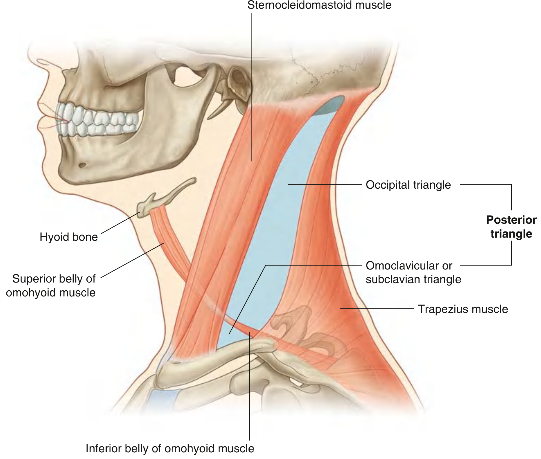
- Anterior wall: Posterior border of sternocleidomastoid
- Posterior wall: Anterior border of trapezius
- Base (inferior): Middle 1/3 of clavicle
- Apex: Occipital bone (where SCM and trapezius meet behind mastoid)
- Roof: Investing layer of deep cervical fascia
- Floor: Prevertebral fascia covering muscles (from above: splenius capitis, levator scapulae, posterior/middle/anterior scalene)
- Occipital triangle (larger, superior)
- Omoclavicular (subclavian) triangle (smaller, inferior)
- Accessory nerve (CN XI) - crosses it obliquely from SCM to trapezius (most important content - vulnerable to injury)
- Cutaneous branches of cervical plexus (at Erb's point - posterior border of SCM at its midpoint):
- Lesser occipital (C2)
- Great auricular (C2, C3)
- Transverse cervical (C2, C3)
- Supraclavicular (C3, C4)
- Branches of brachial plexus (trunks)
- Phrenic nerve (C3, C4, C5) - crosses anterior scalene on its floor
- External jugular vein (most superficial)
- Subclavian artery (3rd part) + Transverse cervical artery
- Suprascapular artery
- Occipital artery (at apex)
- Cervical lymph nodes
- Part of the brachial plexus
Q10. SUBMANDIBULAR GANGLION - Pathway (Sensory, Sympathetic, Parasympathetic)
| Component | Pre-ganglionic Origin | Relay | Post-ganglionic Distribution |
|---|---|---|---|
| Parasympathetic (secretomotor) | Superior salivatory nucleus → chorda tympani (VII) → joins lingual nerve (V3) | Synapses in submandibular ganglion | To submandibular gland (secretomotor) and sublingual gland |
| Sympathetic | T1 sympathetic trunk → superior cervical ganglion | NO relay in ganglion (passes through) | Vasoconstriction to salivary glands |
| Sensory | Cell bodies in trigeminal ganglion → lingual nerve (V3) | NO relay in ganglion (passes through) | Sensory to anterior 2/3 of tongue (lingual nerve) |
Q11. PAROTID GLAND - Location, Relations, Blood Supply, Nerve Supply, Applied
- Anterior: Masseter muscle, ramus of mandible, medial pterygoid
- Posterior: Mastoid process, posterior belly of digastric, SCM
- Superior: External acoustic meatus, temporomandibular joint
- Medial (deep part): Styloid apparatus, carotid sheath
- Facial nerve (CN VII) - divides into 5 branches within gland (temporal, zygomatic, buccal, marginal mandibular, cervical)
- Retromandibular vein (formed by superficial temporal vein + maxillary vein)
- External carotid artery - divides into superficial temporal and maxillary arteries within gland
- Emerges from anterior border
- Crosses masseter
- Pierces buccinator
- Opens into vestibule of mouth opposite upper 2nd molar tooth
- Parasympathetic (secretomotor): Inferior salivatory nucleus → CN IX (glossopharyngeal) → tympanic nerve → tympanic plexus → lesser petrosal nerve → relays in otic ganglion → auriculotemporal nerve → parotid
- Sympathetic: superior cervical ganglion → via middle meningeal artery
- Parotitis (mumps): Severe pain because the investing fascia is tightly adherent (does not yield to swelling)
- Frey's syndrome (auriculotemporal syndrome): After parotidectomy, parasympathetic secretomotor fibers regenerate and misdirect to sweat glands → sweating and flushing during eating (gustatory sweating)
- Parotid tumor: Pleomorphic adenoma (benign) most common - painless, slow-growing. Facial nerve preservation essential in surgery.
- Parotid abscess: Cannot feel fluctuation due to tight capsule
Q12. MUSCLES OF MASTICATION - Attachments, Nerve Supply, Action
| Muscle | Origin | Insertion | Action | Nerve |
|---|---|---|---|---|
| Masseter | Lower border + medial surface of zygomatic arch | Lateral surface of ramus & angle of mandible | Elevation (jaw closure), some protrusion | Masseteric nerve (V3) |
| Temporalis | Temporal fossa (floor) | Coronoid process + anterior border of ramus | Elevation + retraction (posterior fibers) | Deep temporal nerves (V3) |
| Medial pterygoid | Medial surface of lateral pterygoid plate + pyramidal process of palatine | Medial surface of ramus, below mandibular foramen | Elevation + protrusion + lateral excursion | Medial pterygoid nerve (V3) |
| Lateral pterygoid | Upper head: Infratemporal crest of greater wing of sphenoid; Lower head: Lateral surface of lateral pterygoid plate | Upper head: Articular disc + capsule of TMJ; Lower head: Pterygoid fovea of mandibular condyle | Protrusion (both heads), lateral excursion, depression (lower head), opens mouth | Lateral pterygoid nerve (V3) |
- Lateral pterygoid is the key muscle in jaw opening (with gravity + digastric/mylohyoid)
- Trismus (jaw locking): spasm of masseter + medial pterygoid; seen in peritonsillar abscess, tetanus
- TMJ dysfunction often involves lateral pterygoid pathology
SHORT ANSWERS (3 Marks)
Q13. BLOOD SUPPLY OF THYROID GLAND (3 marks)
- Superior thyroid artery - 1st branch of external carotid artery → superior pole
- Inferior thyroid artery - from thyrocervical trunk (1st part of subclavian) → inferior pole
- Thyroid ima artery - from brachiocephalic trunk or arch of aorta (10% people) → isthmus
- Veins: Superior + middle thyroid veins → Internal jugular vein; Inferior thyroid veins → Left brachiocephalic vein
- Lymphatics: Prelaryngeal, pretracheal, paratracheal nodes
Q14. COMPONENTS OF WALDEYER'S RING (3 marks)
| Component | Location |
|---|---|
| Pharyngeal tonsil (adenoids) | Roof and posterior wall of nasopharynx |
| Tubal tonsils | Near opening of Eustachian tube (×2) |
| Palatine tonsils | Tonsillar fossae between anterior and posterior pillars (×2) |
| Lingual tonsil | Posterior 1/3 of tongue dorsum |
| Lateral pharyngeal bands | Posterior to palatopharyngeal arches |
Q15. BRANCHES OF EXTERNAL CAROTID ARTERY (3 marks)
| Branch | Mnemonic | Region |
|---|---|---|
| Superior thyroid | S | First branch, anteriorly |
| Ascending pharyngeal | A | Medially |
| Lingual | L | Anteriorly |
| Facial | F | Anteriorly |
| Occipital | O | Posteriorly |
| Posterior auricular | P | Posteriorly |
| Superficial temporal | S | Terminal branch |
| Maxillary | M | Terminal branch |
Q16. DANGEROUS LAYER OF SCALP (3 marks)
- Subaponeurotic space = the 4th layer of the scalp (loose areolar tissue between epicranial aponeurosis and pericranium)
- Also called the dangerous area or dangerous layer of the scalp
- Why dangerous?:
- Infection can spread freely across the entire scalp within this loose areolar space
- Emissary veins in this layer connect scalp veins (outside) to intracranial dural venous sinuses (inside) - allowing spread of infection intracranially (meningitis, cavernous sinus thrombosis)
- Hematoma in this layer is extensive ("black eye" can result from spread)
- Emissary veins involved: Parietal, mastoid, frontal emissary veins
Q17. MUSCLES OF SOFT PALATE (3 marks)
| Muscle | Origin | Insertion | Nerve Supply | Action |
|---|---|---|---|---|
| Tensor veli palatini | Scaphoid fossa, spine of sphenoid, cartilage of Eustachian tube | Palatine aponeurosis | Medial pterygoid nerve (V3) - ONLY muscle NOT by pharyngeal plexus | Tenses soft palate, opens Eustachian tube |
| Levator veli palatini | Petrous temporal, cartilage of Eustachian tube | Palatine aponeurosis | CN X (pharyngeal plexus) | Elevates soft palate during swallowing (main action) |
| Palatoglossus | Palatine aponeurosis | Side of tongue | CN X (pharyngeal plexus) | Elevates tongue, closes oropharyngeal isthmus |
| Palatopharyngeus | Palatine aponeurosis | Posterior border of thyroid cartilage | CN X (pharyngeal plexus) | Elevates pharynx and larynx during swallowing |
| Musculus uvulae | Posterior nasal spine | Mucosa of uvula | CN X (pharyngeal plexus) | Shortens and thickens uvula |
Q18. NERVE SUPPLY OF ANTERIOR 2/3 TONGUE (3 marks)
- General sensation (touch, pain, temperature): Lingual nerve (branch of V3, mandibular division of trigeminal nerve) - cell bodies in trigeminal ganglion
- Special sense (taste): Chorda tympani nerve (branch of facial nerve/CN VII) - joins the lingual nerve in the infratemporal fossa; cell bodies in geniculate ganglion
- Motor: Hypoglossal nerve (CN XII) to all intrinsic and extrinsic muscles (except palatoglossus)
Q19. LITTLE'S AREA / KIESSELBACH'S PLEXUS (3 marks)
- Location: Anteroinferior part of the nasal septum, in the lower anterior part of Little's area
- Formation: Anastomosis of 5 arteries:
- Anterior ethmoidal artery (from ophthalmic/ICA territory)
- Posterior ethmoidal artery (from ophthalmic/ICA territory)
- Sphenopalatine artery (from maxillary artery/ECA territory)
- Greater palatine artery (from maxillary artery/ECA territory)
- Superior labial artery (from facial artery/ECA territory)
- Clinical importance: Most common site of epistaxis (nosebleeds) - 90% of nosebleeds originate here. Treatment: pressure, vasoconstrictor nasal drops, or silver nitrate cauterization.
Q20. CARTILAGES OF LARYNX (3 marks)
- Thyroid cartilage - largest, hyaline, V-shaped; has a notch superiorly (laryngeal prominence = Adam's apple), inferior horns articulate with cricoid
- Cricoid cartilage - hyaline, signet-ring shaped, only complete ring encircling the airway; base posteriorly, narrow arch anteriorly
- Epiglottis - elastic fibrocartilage (not hyaline), leaf-shaped, attached to back of thyroid cartilage angle
Q21. BLOOD SUPPLY OF SCALP (3 marks)
| Vessel | Origin | Region |
|---|---|---|
| Supratrochlear artery | Ophthalmic (ICA) | Frontal, medial |
| Supraorbital artery | Ophthalmic (ICA) | Frontal, lateral |
| Superficial temporal artery | External carotid | Temporal region |
| Posterior auricular artery | External carotid | Behind ear |
| Occipital artery | External carotid | Occipital region |
Q22. TYMPANIC MEMBRANE - Features (3 marks)
- Pearl-grey, semi-transparent, obliquely placed membrane
- Shape: Funnel-shaped (umbo at the deepest point = tip of malleus handle)
- Layers: 3 layers - outer squamous epithelium, fibrous middle layer (radial + circular), inner mucous membrane
- Parts: Pars tensa (larger, taut, has all 3 layers) + Pars flaccida/Shrapnell's membrane (upper portion, lacks fibrous layer, most vulnerable to perforations)
- Handle of malleus: Runs downward and backward from flaccid part to umbo
- Light reflex (cone of light): Anteroinferior quadrant (5 o'clock position in right ear)
- Nerve supply: Auriculo-temporal nerve (V3) - lateral surface; Glossopharyngeal (IX) via Jacobson's nerve + Vagus (X) - medial surface
Q23. EXTERNAL JUGULAR VEIN (3 marks)
- Formation: Behind the angle of mandible, by union of posterior division of retromandibular vein + posterior auricular vein
- Course: Descends vertically in the superficial fascia of the neck, crossing obliquely over the sternocleidomastoid muscle; pierces the investing layer of deep cervical fascia above the middle of the clavicle
- Termination: Drains into the subclavian vein (variable - sometimes into IJV)
- Tributaries: Posterior external jugular vein, transverse cervical vein, suprascapular vein, anterior jugular vein (via jugular venous arch)
- Applied: Visible at the neck surface in right heart failure (raised JVP). Used for emergency venous access or measurement of JVP clinically.
Q24. SUPERIOR ORBITAL FISSURE - Structures Passing Through (3 marks)
- Oculomotor nerve CN III (upper and lower divisions)
- Abducent nerve CN VI
- Nasociliary nerve (branch of V1)
- Sympathetic root of ciliary ganglion
- Trochlear nerve CN IV
- Frontal nerve (branch of V1)
- Lacrimal nerve (branch of V1)
- Superior ophthalmic vein
Q25. SUBOCCIPITAL TRIANGLE (3 marks)
- Formed by:
- Medial boundary: Rectus capitis posterior major (from spinous process of axis to inferior nuchal line)
- Lateral boundary: Obliquus capitis superior (from transverse process of atlas to inferior nuchal line)
- Inferior boundary: Obliquus capitis inferior (from spinous process of axis to transverse process of atlas)
- Floor: Posterior atlantooccipital membrane + posterior arch of atlas
- Roof: Semispinalis capitis
- Contents:
- Vertebral artery (3rd part - curves medially on posterior arch of atlas)
- Suboccipital nerve (dorsal ramus of C1) - motor only (no cutaneous branch)
- Suboccipital venous plexus
- Actions: Extension and rotation of head at atlantoaxial and atlantooccipital joints
MCQ ANSWERS (Head & Neck Related)
| Question | Answer | Reason |
|---|---|---|
| Lymph node of tongue | Jugulodigastric (C) | Also called the tonsillar node; primary node for tongue drainage |
| Parasympathetic ganglion without secretomotor root | Ciliary (A) | It only has motor (to ciliary + pupillary constrictor) - no secretomotor |
| Muscle pierced by parotid duct | Buccinator (D) | Parotid duct crosses masseter then pierces buccinator to enter the mouth |
| Nerve supply to posterior 1/3 of tongue | Glossopharyngeal (C) | Both general and taste for posterior 1/3 |
| Inferior thyroid vein drains into | Brachiocephalic vein (C) | Drains into left brachiocephalic vein |
| Carotid tubercle vertebral level | C6 (C) | Chassaignac's tubercle - where carotid pulse is compressed |
| General sensory supply to anterior 2/3 of tongue | Lingual nerve (A) | Branch of V3 |
| Motor to pharynx and larynx | Nucleus ambiguus (C) | Contains motor neurons for CN IX, X, XI |
| Left recurrent laryngeal nerve hooks around | Arch of aorta (D) | Right RLN hooks around subclavian artery |
| All cartilages unpaired EXCEPT | Corniculate (C) | Corniculate and cuneiform are paired; Thyroid, cricoid, epiglottis are unpaired |
| Muscles elevating jaw - except | Lateral pterygoid (B) | Lateral pterygoid is a depressor (opener) of jaw |
| Topic | Frequency | Marks |
|---|---|---|
| Thyroid gland | 5+ times | 10 marks |
| Tongue (muscles, nerve supply) | 4+ times | 10 marks |
| Cavernous sinus | 4+ times | 10 marks |
| Larynx | 3+ times | 10 marks |
| Lateral wall of nose | 3+ times | 10 marks |
| Parotid gland | 3+ times | 10 marks |
| Facial artery | 2+ times | 5 marks |
| Deep cervical fascia | 2+ times | 5 marks |
| Posterior triangle | 2+ times | 5 marks |
| Muscles of mastication | 2+ times | 5-10 marks |
| Submandibular ganglion | 2+ times | 5 marks |
All answers sourced from Gray's Anatomy for Students and Cummings Otolaryngology Head and Neck Surgery, cross-referenced with RGUHS question patterns.