What is purpura in easy way

Reading File
This is a simple definitional question, so I'll do a quick targeted lookup.
Finding Sources
Searching Images

purpura skin rash petechiae ecchymosis
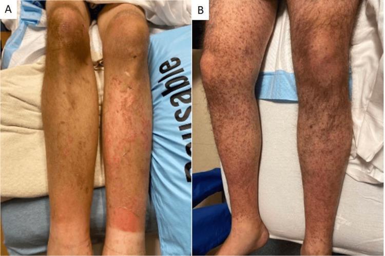
Clinical photograph of bilateral lower extremities from two different patients demonstrating dermatologic manifestations of Vitamin C deficiency (scurvy). Panel A shows extensive non-blanching perifollicular purpura and petechiae across the pretibial areas and calves. There is a large, confluent erythematous rash on the lower right calf, suggestive of ecchymosis or secondary inflammation. Panel B displays diffuse, folliculocentric hyperkeratotic papules and petechiae extending from the shins to the thighs. Characteristic 'corkscrew' hairs (coiled, malformed hair shafts) are visible within the hyperkeratotic follicles. These findings are classic pathognomonic signs of scurvy, resulting from impaired collagen synthesis and capillary fragility. The images serve as an educational resource for identifying nutritional deficiency-related skin disorders in vulnerable populations.
Clinical photograph of bilateral lower extremities from two different patients demonstrating dermatologic manifestations of Vitamin C deficiency (scurvy). Panel A shows extensive non-blanching perifollicular purpura and petechiae across the pretibial areas and calves. There is a large, confluent erythematous rash on the lower right calf, suggestive of ecchymosis or secondary inflammation. Panel B displays diffuse, folliculocentric hyperkeratotic papules and petechiae extending from the shins to the thighs. Characteristic 'corkscrew' hairs (coiled, malformed hair shafts) are visible within the hyperkeratotic follicles. These findings are classic pathognomonic signs of scurvy, resulting from impaired collagen synthesis and capillary fragility. The images serve as an educational resource for identifying nutritional deficiency-related skin disorders in vulnerable populations.

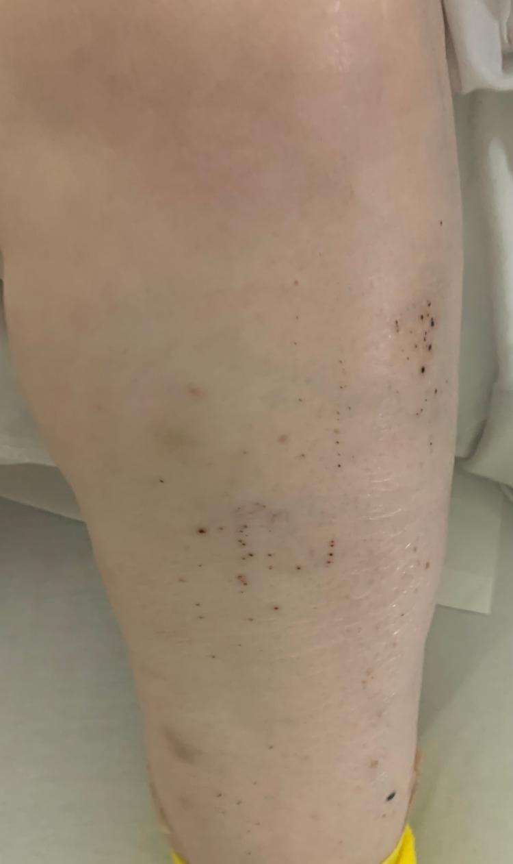
Clinical photograph of the left lower extremity demonstrating a localized rash characterized by numerous small, non-blanching, dark red to purplish-black spots consistent with petechiae. The lesions vary from pinpoint size to approximately 2-3 mm in diameter and are distributed in a scattered, patchy pattern across the anterior and lateral leg surface. Centrally, a larger, faint purplish area suggests underlying ecchymosis. The surrounding skin appears otherwise intact without evidence of ulceration or significant scaling. This presentation is highly characteristic of profound thrombocytopenia, such as Immune Thrombocytopenic Purpura (ITP), where low platelet counts lead to spontaneous microvascular hemorrhages within the dermis.
Clinical photograph of the left lower extremity demonstrating a localized rash characterized by numerous small, non-blanching, dark red to purplish-black spots consistent with petechiae. The lesions vary from pinpoint size to approximately 2-3 mm in diameter and are distributed in a scattered, patchy pattern across the anterior and lateral leg surface. Centrally, a larger, faint purplish area suggests underlying ecchymosis. The surrounding skin appears otherwise intact without evidence of ulceration or significant scaling. This presentation is highly characteristic of profound thrombocytopenia, such as Immune Thrombocytopenic Purpura (ITP), where low platelet counts lead to spontaneous microvascular hemorrhages within the dermis.

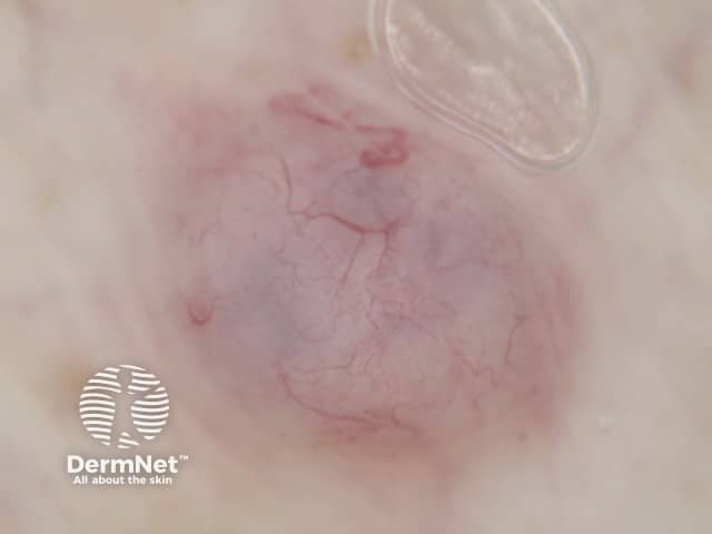
This is a macroscopic, frontal-view clinical photograph of a solitary cutaneous lesion. The exact anatomic site is not specified. The lesion appears as a violaceous-red patch with irregular, ill-defined borders, measuring several centimeters in diameter. The surface is relatively smooth with subtle translucency; underlying vasculature is visible as serpentine superficial vessels within the patch. Mild surrounding erythema and slight edema may be present. The combination of color and vascular pattern raises consideration for superficial bleeding (purpura/ecchymosis) or a vascular lesion such as a capillary malformation, telangiectatic patch, or small-vessel vasculitis; however, a definitive diagnosis cannot be made from a single photograph alone. Observations are suggestive of extravasated blood or mucocutaneous vascular dilation rather than epidermal disruption. Differential diagnoses include ecchymosis, petechiae, palpable purpura, vasculitic rash, pigmented vascular lesions, and inflammatory dermatitis. Clinical significance centers on potential associations with coagulopathy, trauma, infection, or systemic vasculopathy; correlation with history and examination is essential, and if indicated, laboratory testing or skin biopsy may be pursued. This image is educational for recognizing cutaneous vascular phenomena and distinguishing purpuric patches from inflammatory or pigmented lesions, suitable for dermatology atlases and residency training. Additional cases and imaging can improve diagnostic confidence and educational value.

This is a macroscopic, frontal-view clinical photograph of a solitary cutaneous lesion. The exact anatomic site is not specified. The lesion appears as a violaceous-red patch with irregular, ill-defined borders, measuring several centimeters in diameter. The surface is relatively smooth with subtle translucency; underlying vasculature is visible as serpentine superficial vessels within the patch. Mild surrounding erythema and slight edema may be present. The combination of color and vascular pattern raises consideration for superficial bleeding (purpura/ecchymosis) or a vascular lesion such as a capillary malformation, telangiectatic patch, or small-vessel vasculitis; however, a definitive diagnosis cannot be made from a single photograph alone. Observations are suggestive of extravasated blood or mucocutaneous vascular dilation rather than epidermal disruption. Differential diagnoses include ecchymosis, petechiae, palpable purpura, vasculitic rash, pigmented vascular lesions, and inflammatory dermatitis. Clinical significance centers on potential associations with coagulopathy, trauma, infection, or systemic vasculopathy; correlation with history and examination is essential, and if indicated, laboratory testing or skin biopsy may be pursued. This image is educational for recognizing cutaneous vascular phenomena and distinguishing purpuric patches from inflammatory or pigmented lesions, suitable for dermatology atlases and residency training. Additional cases and imaging can improve diagnostic confidence and educational value.

Purpura, explained simply

Purpura means small purple/reddish-brown spots or patches that appear on the skin (or sometimes inside the mouth) because tiny blood vessels have leaked blood under the skin. Since the blood is sitting under the skin rather than flowing through vessels, the spots don't turn white (blanch) when you press on them - that's the key sign that separates purpura from an ordinary red rash.
Why it happens - basically two reasons:
  1. Blood isn't clotting properly - e.g. low platelet count (as in immune thrombocytopenic purpura/ITP), blood thinners, or clotting disorders.
  2. The blood vessels themselves are fragile or inflamed - e.g. vasculitis, old age (senile purpura), steroid use, vitamin C deficiency (scurvy), or infections like meningococcemia.
Size matters for naming:
  • Tiny pinpoint spots (< 3 mm) = petechiae
  • Larger patches (> 1 cm) = purpura/ecchymosis (bruise-like)
  • If you can feel a bump when you touch the spot ("palpable purpura"), it usually points to vessel inflammation (vasculitis) rather than just a clotting problem.
When it's a red flag: purpura that spreads fast, comes with fever, or occurs in a sick child can signal serious infection (like meningococcemia) or a bleeding/clotting emergency ("purpura fulminans") and needs urgent medical attention.
Here's an example clinical photo showing petechiae/purpura from a low platelet count:
Petechiae and purpura on the leg from immune thrombocytopenia
Sources: Rosen's Emergency Medicine; Dermatology, 2-Volume Set 5e; Andrews' Diseases of the Skin.
If you want, I can go deeper into a specific type (e.g. Henoch-Schönlein purpura, ITP, or purpura fulminans) or explain what tests are done to find the cause.
This is a shared conversation. Sign in to Orris to start your own chat.