


IMG_9717.jpg
af2346f3-a1b9-4cb5-9d51-eb059fb5d9bb.jpg
40 year male presents with fever weight loss loss of appetite since 1 month Usg abdomen- Mild ascites Mild right pleural effusion 2 d echo - Normal How to approach
analyse_media
analyse_media
Reading File
Reading File
Reading File
Reading File
Finding Sources
Finding Sources
Searching the Web
approach fever weight loss ascites pleural effusion miliary pattern CT chest 40 year male
Reading File
Finding Sources
Finding Sources
Searching PubMed
"miliary tuberculosis" diagnosis
Clinical Picture
A 40-year-old man with a 1-month history of fever, weight loss, and anorexia, with ascites, right pleural effusion, a normal echocardiogram (ruling out a cardiac/pericardial cause for the effusions), and a CT chest showing innumerable, diffusely and randomly distributed fine nodules throughout both lungs ("miliary" pattern) - this combination is a classic presentation of disseminated (miliary) tuberculosis with tuberculous serositis (peritonitis + pleuritis), especially in a TB-endemic setting like India. That should be your leading diagnosis, but it needs to be confirmed microbiologically/histologically, not treated on pattern-recognition alone, because the same triad (fever + weight loss + serosal effusions + miliary nodules) can also be produced by disseminated malignancy (lymphoma, miliary metastases, peritoneal carcinomatosis), fungal infection, or sarcoidosis.
Step-by-step approach
1. History and exam clues to actively look for
- Night sweats, cough, hemoptysis, contact with a TB patient, past incompletely treated TB
- Risk factors for immunosuppression: HIV status (mandatory test), diabetes, chronic steroid/immunosuppressant use, malnutrition, alcohol use
- Lymphadenopathy, hepatosplenomegaly, skin lesions
- Fundoscopy for choroidal tubercles - quick, often overlooked, near-pathognomonic bedside sign of miliary TB
2. Basic labs
- CBC (miliary TB can cause pancytopenia from marrow involvement), LFTs (TB hepatitis is common), RFT, ESR/CRP, HIV ELISA, HbA1c/blood sugar, LDH
3. Fluid analysis - this is where the diagnosis is usually made
- Ascitic fluid: SAAG, total protein, cell count, cytology, AFB smear/culture, Xpert MTB/RIF (CBNAAT), and adenosine deaminase (ADA) - ascitic ADA has high sensitivity/specificity for tuberculous peritonitis (Sleisenger & Fordtran's Gastrointestinal and Liver Disease; Murray & Nadel's Textbook of Respiratory Medicine). CA-125 may be raised in TB peritonitis and can mimic ovarian/peritoneal malignancy - don't let a raised CA-125 alone push you toward malignancy.
- Pleural fluid: Light's criteria (expect exudate, lymphocyte-predominant), glucose, cytology, AFB smear/culture, Xpert MTB/RIF, and pleural ADA (>40 U/L strongly supports TB pleuritis) - Goldman-Cecil Medicine; Henry's Clinical Diagnosis and Management by Laboratory Methods.
4. Microbiological confirmation from the chest
- Sputum (even if non-productive, use induced sputum) for AFB smear, culture, and Xpert MTB/RIF
- If sputum is negative/unobtainable: bronchoscopy with BAL and transbronchial lung biopsy - given the diffuse miliary pattern, TBLB has a good yield for granulomas and AFB culture
5. Tissue diagnosis if fluid/sputum are inconclusive
- Pleural biopsy (Abrams needle or CT/USG-guided) for histopathology (caseating granulomas) and culture - higher yield than fluid cytology/AFB alone
- Laparoscopic peritoneal/omental biopsy if ascites persists undiagnosed - considered near gold-standard for TB peritonitis (shows "violin-string" adhesions, whitish tubercles studding the peritoneum) and simultaneously excludes peritoneal carcinomatosis
- Bone marrow aspirate/biopsy with AFB culture - especially useful if there is cytopenia; high yield in disseminated TB
- Liver biopsy if hepatomegaly/deranged LFTs
6. Imaging follow-up
- Contrast CT abdomen: look for necrotic mesenteric/para-aortic lymph nodes, hepatosplenic micro-abscesses, peritoneal thickening/enhancement (favors TB) versus omental caking/nodularity (favors carcinomatosis)
- USG-guided aspiration of any accessible node
7. Adjuncts (supportive, not diagnostic)
- Tuberculin skin test / IGRA - low sensitivity in disseminated TB due to anergy; a negative result does not rule out miliary TB
8. Empirical therapy
- If the patient deteriorates before a tissue/microbiological diagnosis is secured, and pre-test probability is high (endemic region, compatible imaging, no clear alternative), collect all possible samples for AFB smear/culture/PCR first, then start empirical anti-tubercular therapy. A clinical and radiological response within 4-6 weeks supports the diagnosis; failure to respond should prompt reconsideration of the diagnosis (per Harrison's Principles of Internal Medicine, FUO chapter).
Differentials to keep on the list until excluded
- Disseminated fungal infection (histoplasmosis, cryptococcosis) - especially if immunocompromised
- Sarcoidosis (less commonly causes this degree of ascites)
- Lymphoma or miliary metastatic malignancy with serosal spread
- Peritoneal carcinomatosis (send fluid cytology in every case)
Bottom line
Work this up as presumed disseminated TB with TB pleuritis/peritonitis but confirm it - send ascitic and pleural fluid for ADA, cytology, AFB smear/culture and Xpert MTB/RIF, check HIV status, and proceed to bronchoscopy/TBLB or laparoscopic peritoneal biopsy if fluid studies are inconclusive, before or in parallel with starting anti-tubercular therapy.
A quick literature check found no recent systematic reviews that would change this diagnostic pathway for miliary TB (searched PubMed, last 3 years, systematic reviews/meta-analyses - top hits were about miliary TB of the brain/meningitis and stool-based pulmonary TB diagnosis, not directly contradicting this approach). If you want, I can pull up illustrative images of the classic miliary CT pattern or the ADA cutoff data from the primary literature.


+4 more
Interpret this ct scan Patient is kyphoscoliotic and has history of sliding hiatus hernia
Reading File
Reading File
Reading File
Reading File
Reading File
Reading File
Finding Sources
Reading File
Loading Image
CT / CXR Interpretation - Rukmani Bai Khatik, 76F, Kyphoscoliotic, Known Sliding Hiatus Hernia
Technique: AP chest radiograph (rotated projection due to kyphoscoliosis) followed by contrast-enhanced CT chest/upper abdomen, reviewed in mediastinal and lung windows.
Findings
Chest X-ray (AP)
- Markedly rotated, kyphoscoliotic bony thorax with asymmetric rib cage.
- A large, rounded, mottled/bubbly opacity is seen overlying the right cardiophrenic-retrocardiac region, obscuring the right heart border - the lucent, bubbly internal pattern represents air within herniated gastric rugal folds rather than a solid mass.
CT chest (mediastinal window, cranial to caudal)
- A large, well-defined, thin-walled structure with mixed fluid/soft-tissue and gastric-fold-lined gas content lies immediately posterior/adjacent to the heart in the lower posterior mediastinum.
- On sequential caudal slices this structure is seen in direct continuity across the esophageal hiatus with the intra-abdominal stomach - confirming it is herniated stomach, not a mediastinal mass.
- Severe kyphoscoliotic distortion of the thoracic cage (rotated vertebral bodies, crowded/asymmetric ribs) is present throughout.
CT (lung window)
- The adjacent lung (right lower/middle lobe territory) shows compressive atelectasis/crowding from mass effect of the hernia sac; no discrete intrapulmonary nodule or infiltrate is seen - the apparent "opacity" on the CXR is entirely accounted for by the herniated stomach, not primary lung pathology.
Impression
Large paraesophageal / mixed-type (Type III) hiatal hernia with a substantial intrathoracic gastric component, sitting alongside and displacing the heart, in a patient with severe kyphoscoliosis. This appears to represent progression from the previously documented simple sliding (Type I) hernia to a larger mixed hernia - a recognized evolution, particularly in elderly patients where kyphoscoliosis alters thoraco-abdominal pressure gradients and crural diaphragmatic support, favoring hiatal widening and stomach migration into the chest.
For comparison, here is the classic CT appearance of a mixed (Type III) hiatal hernia from Grainger & Allison's Diagnostic Radiology - a large gastric structure lying beside the heart, matching the pattern in this patient's scan:
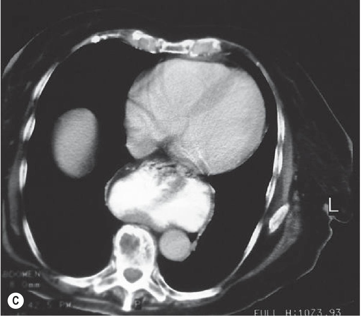
- Grainger & Allison's Diagnostic Radiology, Fig. 20.23C
Points to actively check on the images / clinically
- Rule out gastric volvulus - look for organoaxial/mesenteroaxial rotation of the herniated stomach, a "double air-fluid level," or an abrupt transition point at the hiatus. This is a recognized complication of large intrathoracic stomach and can progress to a surgical emergency (violent retching with minimal vomitus, epigastric pain, inability to pass an NG tube).
- Wall integrity/perfusion of the herniated gastric segment - wall thickening, lack of contrast enhancement, or pneumatosis would suggest ischemia/strangulation.
- Degree of lung compression - relevant given this patient already has restrictive physiology from kyphoscoliosis; the added extrinsic compression from a large hernia can meaningfully worsen respiratory reserve.
- Other herniated contents - confirm whether only stomach, or also colon/omentum/spleen, has migrated through the hiatus (common in large paraesophageal hernias).
- Clinical correlation for reflux, dysphagia, early satiety, postprandial breathlessness or chest discomfort, and check hemoglobin (chronic mucosal ulceration at the hernia neck - "Cameron lesions" - can cause iron-deficiency anemia).
Practical implication
Given the size (now paraesophageal/mixed rather than simple sliding) and this patient's age plus kyphoscoliosis, a surgical opinion for elective repair should be considered because of the risk of incarceration, volvulus, or strangulation with large paraesophageal hernias - but the kyphoscoliosis and likely reduced pulmonary reserve raise the perioperative risk, so this needs a risk-benefit discussion (surgery vs. conservative/symptomatic management) individualized to her overall fitness.
This is a shared conversation. Sign in to Orris to start your own chat.