
Write a short note on triple arthrodesis for Ms orthopedics theory examination gold medal level
triple arthrodesis foot surgery indications technique complications orthopedics
triple arthrodesis foot subtalar talonavicular calcaneocuboid joints fusion diagram
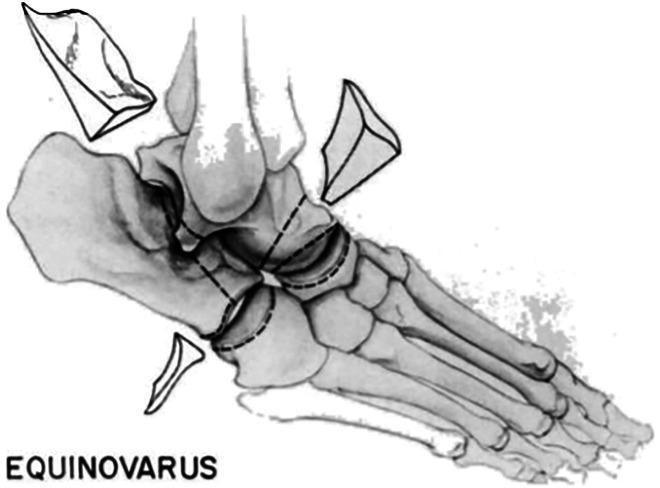
Anatomical diagram illustrating the Hoke technique for triple arthrodesis in the context of equinovarus deformity correction. The illustration depicts a lateral-oblique view of the human foot and ankle skeletal structure. Three distinct wedge-shaped bone resections are shown as detached segments to indicate their removal: one from the talar neck/head, one from the posterior facet of the subtalar joint (calcaneus), and one from the calcaneocuboid joint area. Dashed lines on the main skeletal body mark the precise sites of the osteotomies across the subtalar, talonavicular, and calcaneocuboid joints. The educational focus is on the osseous reshaping required to achieve a plantigrade foot position by reducing the equinus and varus components of the deformity. The diagram labels the condition as 'EQUINOVARUS' to provide clinical context for the surgical intervention, which targets the stabilization and alignment of the hindfoot and midfoot complexes through joint fusion.
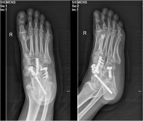
This diagnostic image consists of two postoperative radiographs (anteroposterior and lateral oblique views) of the right foot following a triple arthrodesis procedure, frequently used for managing advanced Mueller-Weiss disease. The images demonstrate extensive metallic internal fixation hardware. A long compression screw is seen traversing from the posterior calcaneus through the subtalar joint into the talar body. Additionally, a combination of orthopedic plates and screws is utilized to stabilize the talonavicular and calcaneocuboid joints, creating a rigid fusion across the hindfoot and midfoot. Another plate and screw construct appears to bridge the naviculocuneiform joint space. The radiograph shows evidence of bone grafting at the site of the navicular, which typically exhibits loss of volume and increased radiodensity in Mueller-Weiss disease. The primary educational focus is the visual demonstration of surgical stabilization and joint fusion (arthrodesis) techniques used to correct midfoot deformity and address chronic pain associated with idiopathic tarsal navicular osteonecrosis.
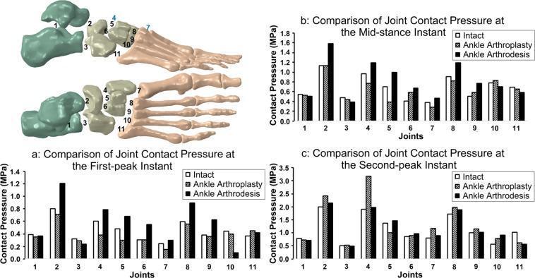
This medical graphic presents a biomechanical comparison of joint contact pressure across three foot models: intact, total ankle arthroplasty (TAA), and ankle arthrodesis. The visual includes an anatomical diagram of the foot bones with 11 numbered joints and three bar graphs corresponding to specific phases of the gait cycle: (a) first-peak, (b) mid-stance, and (c) second-peak instants. The 11 joints analyzed include the subtalar (1), talonavicular (2), calcaneocuboid (3), medial cuneonavicular (4), intermediate cuneonavicular (5), lateral cuneonavicular (6), and the five tarsometatarsal joints (7–11). The data shows that ankle arthrodesis generally results in significantly higher contact pressures, particularly at the talonavicular and medial cuneonavicular joints, compared to the intact foot. TAA generally shows a closer approximation to intact pressure levels, though it exhibits a distinct pressure spike at the medial cuneonavicular joint during the second-peak instant. This educational content illustrates how surgical interventions like fusion or replacement alter load distribution across the midfoot and forefoot during walking.
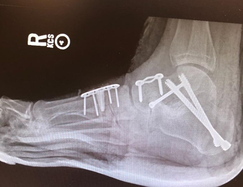
This lateral radiographic image of a right foot demonstrates extensive orthopedic internal fixation hardware following reconstructive surgery, likely a triple arthrodesis and medial column stabilization. In the hindfoot, three large, fully threaded compression screws are visible, inserted from the posterior-inferior calcaneus and extending superior-anteriorly across the subtalar joint into the talus. The midfoot shows a dorsally placed, rectangular multi-hole neutralization plate secured with four screws. Centrally, a two-hole compression staple or plate is visible across the talonavicular or calcaneocuboid region. Notably, a radiopaque titanium wedge is positioned at the first tarsometatarsal joint (Cotton osteotomy or similar) to achieve plantarflexion of the first ray. The image illustrates complex surgical management of rigid foot deformities, such as pes planovalgus or neuromuscular collapse, highlighting the use of diverse metallic implants to maintain correction and facilitate osseous fusion while preventing medial column shortening.
Triple Arthrodesis
Definition
- Subtalar (talocalcaneal) joint
- Talonavicular joint
- Calcaneocuboid joint
Anatomy and Biomechanical Basis
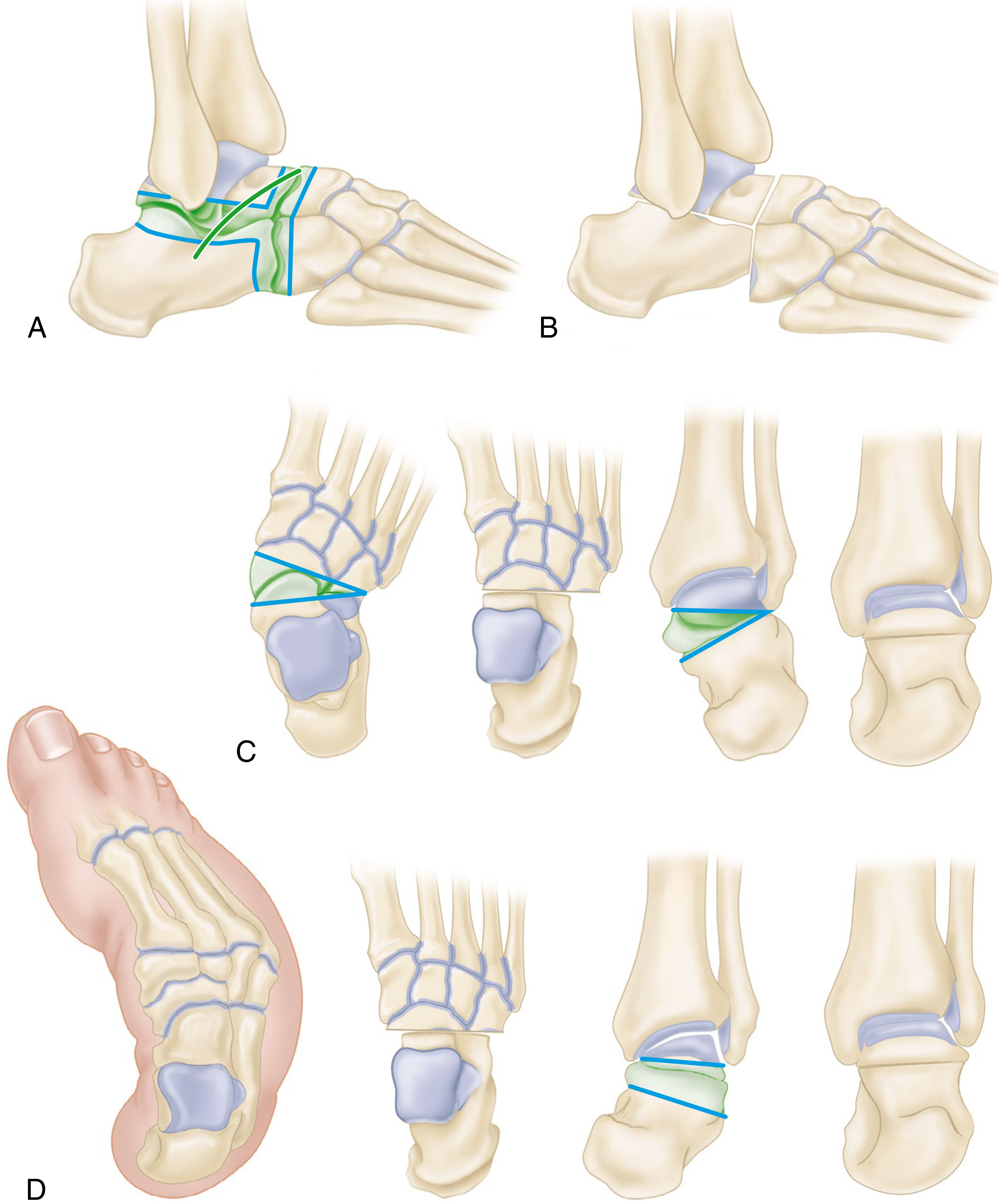
Goals of Triple Arthrodesis
- Obtain stable, static realignment of the foot into a plantigrade position
- Remove deforming forces
- Arrest progression of deformity
- Eliminate pain
- Eliminate the use of a short leg brace, or provide sufficient correction to allow fitting of a long-leg brace to control the knee
- Obtain a more normal-appearing foot
Indications
- Spastic/flaccid paralysis - poliomyelitis, cerebral palsy, spina bifida
- Talipes equinovarus (rigid, uncorrected or recurrent clubfoot in older children/adolescents)
- Talipes equinovalgus
- Cavovarus foot (Charcot-Marie-Tooth disease, Friedreich's ataxia)
- Rigid flatfoot due to posterior tibial tendon dysfunction (PTTD) Stage III/IV
- Posttraumatic arthritis of subtalar or midtarsal joints
- Neglected calcaneal fractures with subtalar arthritis
- Tarsal coalition (late, failed conservative/lesser surgical management)
- Rheumatoid arthritis with hindfoot collapse
- Mueller-Weiss disease (idiopathic navicular osteonecrosis)
Preoperative Planning
- Weight-bearing AP, lateral, and oblique radiographs of the foot and ankle
- A paper tracing technique is used: lateral radiograph traced and cut into three segments (tibiotalar, calcaneal, and foot distal to midtarsal joint); segments are reassembled in corrected position to measure the size and shape of bone wedges required
- CT scan useful for tarsal coalition and complex deformities
- Assessment of ankle joint for coexisting tibiotalar arthritis or valgus/varus tilt (which would negate correction)
- Vascular status assessment, especially in rheumatoid or diabetic patients
Surgical Technique (Hoke/Standard Two-Incision Technique)
Patient Position
Incision
Steps (eTechnique 36.4, Campbell's):
- Identify and protect peroneal tendons and sural nerve
- Reflect the extensor digitorum brevis (EDB) origin distally, clearing the sinus tarsi fat pad
- Incise capsules of all three joints circumferentially to gain maximum mobility
- Excise the anterior articular process of the calcaneus at the sinus tarsi floor (bone preserved for grafting)
- Remove articular cartilage and subchondral bone from the calcaneocuboid joint with osteotomes
- Remove the distal portion of the talar head (talonavicular joint) - only enough to expose cancellous bone unless medial wedge correction is required
- Remove the proximal articular surface of the navicular
- Excise the sustentaculum tali facet and anterior facet of the subtalar joint
- Completely remove articular surfaces of the posterior subtalar joint; use a lamina spreader for exposure
Wedge Resections for Deformity Correction:
| Deformity | Wedge base direction |
|---|---|
| Equinovalgus (flat foot) | Medially-based wedge from talar head/neck |
| Equinovarus (clubfoot) | Laterally-based wedge from midtarsal + subtalar joints |
| Pes cavus | Anteriorly-based wedge from midtarsal + posteriorly-based from subtalar |
- Cut excised bone into small pieces for bone graft; pack around talonavicular joint and sinus tarsi
- Correct foot position: neutral dorsiflexion, 5° hindfoot valgus, neutral forefoot
- Internal fixation: smooth Steinmann pins or Kirschner wires (classically); modern practice uses cannulated screws for compression
- Close EDB muscle belly over sinus tarsi to obliterate dead space
- Close wound over suction drain; apply well-padded short leg cast
Correct Position of Fusion
- Ankle at 90° (neutral dorsiflexion) - critical to enable normal heel-toe gait
- Hindfoot in 5° of valgus (not neutral, not varus) - slight valgus distributes weight along the lateral border and prevents painful lateral column overload
- Forefoot in neutral without supination
Special Techniques
Lambrinudi Triple Arthrodesis
- Designed specifically for drop foot (fixed equinus) with no active dorsiflexion
- A beak-shaped wedge is removed from the inferior talar head and neck; the navicular is locked beneath the talar beak, keeping the ankle in neutral despite absent dorsiflexor power
- Indication: poliomyelitis with paralysis of dorsiflexors but intact plantar flexors
Siffert-Forster-Nachamie Technique
- Used for severe cavus deformity
- Wedge osteotomy of anterior calcaneus, posterior navicular, and inferior talar head/neck; forefoot displaced plantarward and navicular locked beneath the talar remnant
Postoperative Care
- Suction drain removed at 24-48 hours
- Foot elevated to minimize swelling
- Non-weight bearing with crutches/walker (touch-down weight bearing) for 6-8 weeks
- Cast and pins/wires removed at 6-8 weeks; short leg walking cast applied for 4 more weeks until union complete (total ~10-12 weeks immobilization)
- Full weight bearing usually by 3 months
- Return to full activity: 6-12 months; up to 10 months to become completely pain-free
Complications
Early:
- Wound dehiscence / skin necrosis - especially with overcorrection of severe valgus (lateral skin stretching); medial incision approach preferred in high-risk patients
- Infection (superficial / deep)
- Sural nerve injury / neuralgia
- Deep vein thrombosis
Late:
| Complication | Details |
|---|---|
| Pseudarthrosis (non-union) | Most common complication; most frequent at the talonavicular joint due to its small surface area and poor vascularity |
| Adjacent joint arthritis | Especially ankle joint arthritis (from increased stress transfer); also tarsometatarsal joints distally |
| Osteonecrosis of the talus | From excessive bone resection; evident on X-ray at 8-12 weeks post-op |
| Residual/recurrent deformity | Due to insufficient correction, inadequate immobilization, pseudarthrosis, or muscle imbalance |
| Forefoot deformity | Unopposed anterior tibial or peroneal muscle action after hindfoot stabilization; treat by tendon transfer |
| Ankle ligamentous laxity | From altered biomechanics; may require ankle arthrodesis |
| Hardware irritation | Prominent screws/pins requiring removal |
| Malunion | Foot fused in varus - worst outcome |
Outcomes
- When performed for appropriate indications with good alignment, results are generally satisfactory for pain relief and walking
- Lower-impact activities (walking, cycling, swimming) are achievable goals
- Return to high-impact activity is not reliably expected
- Long-term follow-up shows progressive ankle arthritis in many patients due to altered hindfoot kinematics
Key Points for Examination
| Aspect | Detail |
|---|---|
| Three joints fused | Subtalar + Talonavicular + Calcaneocuboid |
| Also called | Hindfoot arthrodesis |
| Popularized by | Ryerson (1923) |
| Most effective stabilizing procedure | In the foot (Campbell's) |
| Motion retained post-op | Plantarflexion and dorsiflexion only |
| Age of preference | ≥12 years (occasionally 8-12 years) |
| Most common complication | Pseudarthrosis (esp. talonavicular joint) |
| Fusion position | Plantigrade: 90°, 5° hindfoot valgus |
| Drop foot variant | Lambrinudi arthrodesis |
| Classic incision | Oblique lateral over sinus tarsi ± medial incision |
Write a short note on spina ventosa for Ms orthopedics theory examination gold medal level
spina ventosa tuberculous dactylitis pathology clinical features treatment orthopedics
spina ventosa tuberculous dactylitis phalanges expansion x-ray
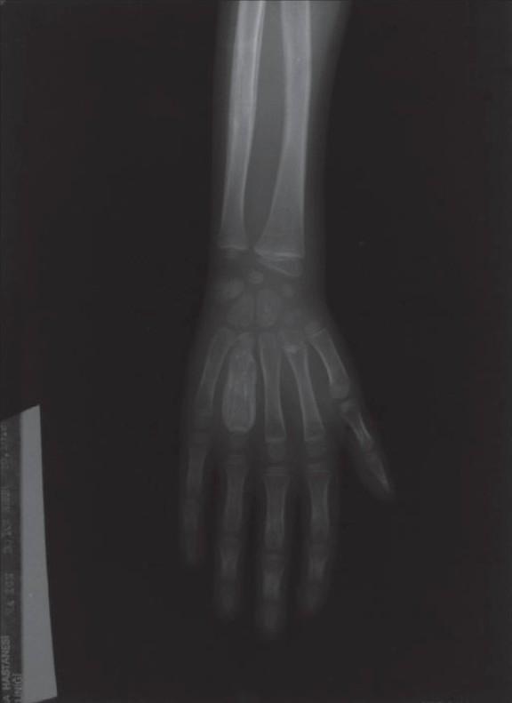
This diagnostic image is a posterior-anterior (PA) X-ray of a pediatric hand and distal forearm demonstrating a classic case of tuberculous dactylitis, also known as spina ventosa. The primary finding is significant fusiform expansion and cortical thinning of the fourth metacarpal bone. The diaphysis appears markedly enlarged and contains heterogeneous, cystic-like material with varying density, suggesting chronic granulomatous inflammation and osteomyelitis. In contrast, the adjacent metacarpals and phalanges maintain a more typical morphology and bone density, though some periosteal reaction may be present in other segments. The carpal bones and distal radius/ulna are visible but appear largely unaffected by the focal metacarpal expansion. This radiological presentation is highly characteristic of skeletal tuberculosis in children, reflecting the cyst-like expansion and internal 'wind-filled' (spina ventosa) appearance of the short tubular bones.
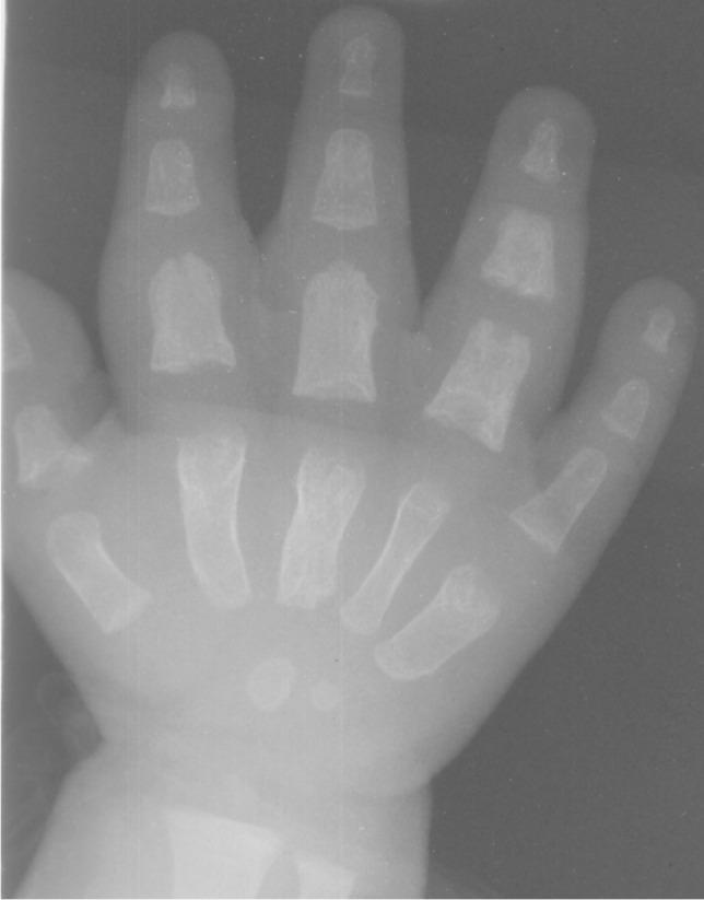
This dorsopalmar radiograph of a pediatric hand (infant) demonstrates classic radiographic findings of tuberculous dactylitis, also known as spina ventosa. The imaging reveals characteristic fusiform expansion of multiple short tubular bones. Specifically, there is prominent spindle-like widening of the second, third, and fifth metacarpals. Similar fusiform swelling and cortical erosion are evident in the first, second, third, and fourth proximal phalanges, as well as the fourth middle phalanx. The bones show rarefaction of the medulla with a thin, expanded cortex and overlying soft tissue swelling, particularly notable in the second through fourth digits. These findings represent the rapid destruction of the marrow space by granulomatous tissue, resulting in internal cyst-like cavities and bone expansion. The lack of significant periosteal reaction and the presence of both expansion and erosion in a young child are highly suggestive of a tuberculous etiology rather than pyogenic osteomyelitis.
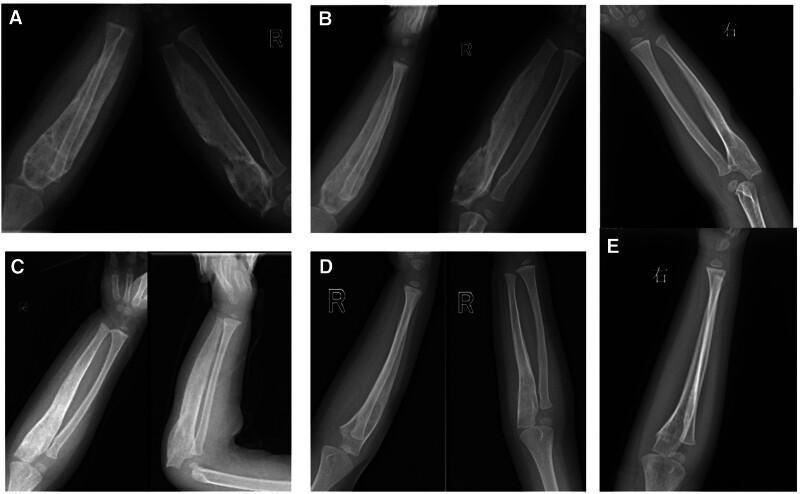
A series of five pediatric X-ray panels (A-E) illustrating the longitudinal progression and resolution of spina ventosa (tuberculous dactylitis) in the right ulna. Panel A (initial presentation) shows dramatic expansile remodeling of the ulnar shaft, characterized by significant bone expansion, thinned cortical bone, and a disorganized, cystic-like trabecular pattern. Panel B shows persistent expansion with an initial increase in radiodensity following intervention. Panel C demonstrates a marked reduction in bone diameter, ulnar remodeling, and cortical thickening. Panel D shows nearly complete resolution of the expansile lesion with restoration of normal bone mineral density. Panel E (final follow-up at 3.5 years old) confirms complete cortical and medullary restoration, exhibiting normal ulnar morphology and a smooth periosteal surface. The series demonstrates the typical radiological evolution of bone tuberculosis under successful antituberculous therapy, moving from extensive bone destruction and expansion to complete structural normalization.
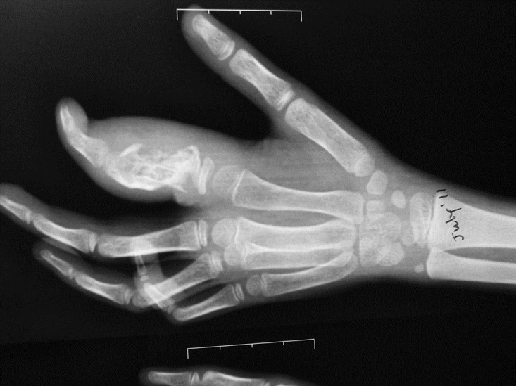
**Imaging Modality:** Plainfilm Radiograph (X-ray). **Anatomical Region:** Oblique view of a pediatric hand and wrist. **Observed Pathology:** Significant lytic destruction and expansile remodeling of the proximal phalanx of the second digit (index finger). The lesion is located distal to the proximal physis. There is associated fusiform soft tissue swelling surrounding the affected phalanx. **Characteristic Visual Features:** * **Osseous Findings:** The affected proximal phalanx demonstrates extensive cortical thinning, trabecular distortion, and a lucent, expansile appearance (dactylitis pattern). The distal aspect of the phalanx appears fragmented or severely eroded. * **Pediatric Context:** Presence of open physes at the base of the metacarpals and phalanges indicates a skeletally immature patient. * **Distribution:** Isolated involvement of the second proximal phalanx, with the remaining visualized metacarpals and phalanges appearing relatively spared of acute destructive changes. **Key Diagnostic Features:** The combination of localized diaphyseal/metaphyseal bone destruction and significant soft tissue expansion in a pediatric patient is characteristic of tuberculous dactylitis (spina ventosa) or chronic fungal/pyogenic osteomyelitis. The preservation of the proximal physis helps localize the primary site of destruction to the metadiaphysis.
Spina Ventosa (Tuberculous Dactylitis)
Etymology and Definition
- Spina - spine/short bones (referring to the short tubular bones of the hands and feet)
- Ventosa - inflated or expanded with air (describing the characteristic balloon-like expansion of the affected bone)
Historical Notes
- First described by the Swedish botanist-physician Carl von Linné (Linnaeus), who coined the term "spina ventosa"
- First radiological description: credited to Feilchenfeld (1896)
- First histological description: given by Rankin (1886)
- Also described by Boyer and Nélaton in early surgical literature
Epidemiology
- Predominantly a disease of children under 6 years (occasionally up to early adolescence)
- Rare in adults; when it does occur in adults, it preferentially involves flexor tendon sheaths, sparing joint synovium
- More common in developing countries with high TB burden; increasing in developed countries due to immigration and HIV/AIDS
- Hands are affected more commonly than feet
- Multiple bones may be involved simultaneously (polyostotic in 20-30% of cases)
Aetiology and Route of Infection
- Causative organism: Mycobacterium tuberculosis (rarely non-tuberculous mycobacteria)
- Route: haematogenous spread from a primary focus (usually pulmonary or lymph nodal TB)
- In some cases, direct extension from adjacent soft tissue TB
Pathogenesis
-
In children, the phalangeal and metacarpal medullary canal contains active red marrow - a nutrient-rich environment favorable for tuberculous bacilli to seed and multiply after haematogenous dissemination
-
TB bacilli lodge in the diaphyseal marrow and set up a tuberculous granuloma - characterized by epithelioid cells, Langhans giant cells, lymphocytes, and central caseating necrosis
-
The granulomatous process slowly involves the entire marrow space, causing:
- Progressive rarefaction of trabeculae
- Destruction of medullary bone
- Stimulation of periosteum to produce thin, expanded new bone
-
The soft, compliant periosteum of a child's bone stretches outward as the medullary contents expand - producing the pathognomonic fusiform (spindle-shaped) balloon-like expansion of the bone with a thinned cortex and destroyed marrow space
-
In advanced cases, the cortex may be breached, leading to:
- Cold (paraosseous) abscess formation
- Sinus tract and skin ulceration
- Joint involvement (less common than in adult TB bone disease)
Key pathological point: Unlike pyogenic osteomyelitis, sequestrum formation and periosteal reaction are uncommon; there is minimal reactive new bone formation - reflecting the poor pyogenic response to mycobacteria.
Clinical Features
Symptoms
- Painless or mildly painful swelling of one or more digits - the hallmark presentation
- Swelling is fusiform (spindle-shaped), involving the whole digit
- Low-grade fever, evening rise of temperature
- Constitutional symptoms: loss of appetite, loss of weight, night sweats, night cries (especially in children)
- Generally benign and slow course compared to pyogenic dactylitis
Signs
- Diffuse, firm-to-soft swelling of the affected digit/bone
- Mild tenderness
- Overlying skin may be normal early, later becomes shiny, tense, or discolored
- Sinus formation in neglected/advanced cases
- Adjacent joint involvement with restricted range of motion (late)
- Signs of primary TB focus may be present: lymphadenopathy, signs of pulmonary TB
Distinguishing Features from Syphilitic Dactylitis (Das Clinical Surgery):
| Feature | Tuberculous dactylitis | Syphilitic dactylitis |
|---|---|---|
| Pain | Present (painful) | Absent (painless) |
| X-ray | Osteolytic changes | Sclerosis of bone |
| Diagnosis confirmed by | Biopsy / AFB | Wassermann/VDRL + syphilitic stigmata |
Investigations
Blood Tests
- Leukocytosis (mild)
- Raised ESR and CRP
- Mantoux/tuberculin skin test: often positive (but may be negative in immunocompromised)
- IGRA (Interferon Gamma Release Assay): QuantiFERON-TB Gold
Microbiological
- Sputum AFB smear and culture (to identify primary focus)
- Cartridge-Based Nucleic Acid Amplification Test (CBNAAT/GeneXpert): rapid molecular diagnosis with rifampicin resistance detection
- Culture of aspirated material from cold abscess on Löwenstein-Jensen medium (gold standard, takes 4-8 weeks)
Imaging
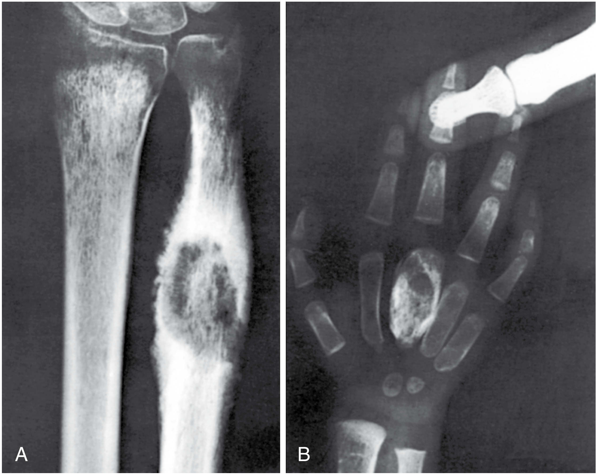
- Fusiform/spindle-shaped expansion of the diaphysis of a short tubular bone
- Cystic/lytic lesions within the expanded bone - the "balloon-like" appearance
- Thinned cortex stretched over the expanded cavity
- Little periosteal reaction (unlike pyogenic osteomyelitis)
- Sequestrum: uncommon but may occur in chronic cases
- Sclerosis in long-standing cases
- Diaphyseal predominance (vs. metaphyseal predominance in pyogenic osteomyelitis)
- Most sensitive for early marrow involvement and soft tissue extension
- T2-weighted: high signal marrow edema; periosseous soft tissue swelling
- Contrast: peripheral rim enhancement of cold abscess
- Useful for assessing neurovascular involvement and guiding biopsy
Histopathology (Diagnostic Confirmation)
- Biopsy is the definitive investigation
- Shows: epithelioid cell granuloma with Langhans giant cells, lymphocytes, plasma cells, and central caseating necrosis
- AFB may be demonstrable on Ziehl-Neelsen (ZN) staining
- Differentiates from other granulomatous conditions and tumors
Differential Diagnosis
| Condition | Distinguishing features |
|---|---|
| Pyogenic osteomyelitis | Acute onset, high fever, marked tenderness, no caseation on biopsy, periosteal reaction prominent |
| Syphilitic dactylitis | Painless, sclerotic on X-ray, positive VDRL/TPHA |
| Sarcoidosis | Systemic features, ACE elevated, non-caseating granuloma on biopsy |
| Enchondroma | Central chondroid matrix calcification on X-ray, lobulated; no constitutional symptoms |
| Giant cell tumor | Epiphyseal, soap-bubble appearance, older age group |
| Ewing's sarcoma | "Onion peel" periosteal reaction, very aggressive, soft tissue mass; biopsy: small round blue cells |
| Leprosy dactylitis | Concentric absorption ("sucked candy" pattern), sensory loss, skin patches |
Treatment
Anti-Tubercular Therapy (ATT) - Mainstay of Treatment
| Phase | Duration | Drugs |
|---|---|---|
| Intensive phase | 2 months | Isoniazid (H) + Rifampicin (R) + Pyrazinamide (Z) + Ethambutol (E) |
| Continuation phase | 6-10 months | Isoniazid (H) + Rifampicin (R) |
- Total duration: 9-12 months for bone TB (some authorities extend to 18 months for extensive disease)
- Pyridoxine (Vitamin B6) given alongside INH to prevent peripheral neuropathy
- Baseline and periodic LFT and visual acuity monitoring
- Compliance monitoring - DOTS (Directly Observed Treatment, Short-course)
Supportive Treatment
- Immobilization of the affected digit/extremity for pain relief and to protect bone integrity during early treatment (splint or POP cast)
- Nutritional supplementation
- Treatment of underlying HIV/immunosuppression if present
- Contact tracing and BCG vaccination of household contacts
Surgical Treatment (Reserved for select cases)
- Curettage of bone cavities in cystic tuberculosis to promote early healing (when cavities are large and healing is delayed on ATT alone)
- Drainage of cold abscess with secondary infection (pyogenic superinfection)
- Debridement + biopsy when diagnosis is uncertain pre-operatively
- Management of sinus tracts
- Bone grafting if significant structural defect persists after disease control
Prognosis
- Excellent with early diagnosis and complete ATT
- Most patients achieve full clinical and radiological resolution (as demonstrated in serial X-ray studies)
- The cystic expanded bone remodels completely in most children on ATT (remarkable regenerative capacity of paediatric bone)
- Neglected/late cases may develop:
- Pathological fracture
- Joint destruction and stiffness
- Permanent deformity
- Secondary pyogenic infection with sinus formation
Key Examination Points (Gold Medal Summary)
| Feature | Detail |
|---|---|
| Definition | TB dactylitis of short tubular bones - metacarpals, metatarsals, phalanges |
| Meaning of name | Spina = short bones; Ventosa = inflated/expanded |
| First described by | Carl von Linné (named); Rankin (1886, histology); Feilchenfeld (1896, radiology) |
| Age group | Children (< 6 years most common) |
| Route | Haematogenous from primary TB focus |
| Why diaphysis? | Active red marrow in paediatric short tubular bones - favorable for mycobacterial seeding |
| Pathology | Caseating granuloma → marrow destruction → periosteal expansion |
| Classic X-ray sign | Fusiform diaphyseal expansion with cystic lytic changes + thinned cortex |
| Little periosteal reaction | Key distinguisher from pyogenic osteomyelitis |
| Definitive diagnosis | Biopsy - caseating granuloma with Langhans giant cells |
| Treatment | ATT (2HRZE + 7-10HR); surgery rarely needed |
| Prognosis | Excellent; complete bone remodeling expected in children |
| Differential | Pyogenic OM, enchondroma, sarcoidosis, syphilitic dactylitis, Ewing's |