
Polyendicrine metabolic ovarian syndrome
"polycystic ovary syndrome" AND "metabolic syndrome"
polycystic ovary syndrome PCOS ovary morphology ultrasound diagram
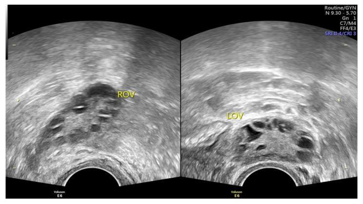
Two side-by-side grayscale pelvic ultrasound images displaying the right ovary (ROV) and left ovary (LOV) in a patient with polycystic ovary syndrome (PCOS). Both ovaries demonstrate classic polycystic ovarian morphology (PCOM). Each ovary contains numerous small, hypoechoic (dark), round-to-oval structures representing immature antral follicles, typically measuring 2–9 mm. The follicles are primarily arranged in a peripheral distribution, often referred to as the 'string of pearls' or necklace sign. The central ovarian stroma is notably hyperechoic (brighter) and appears increased in volume, a common feature of the androgen-induced stromal hyperplasia seen in PCOS. This diagnostic imaging is used in the Rotterdam criteria to identify ovarian dysfunction and chronic anovulation. The image is captured using a transvaginal probe, providing high-resolution visualization of the follicular arrangement and stromal density essential for gynecological and reproductive endocrinology assessment.
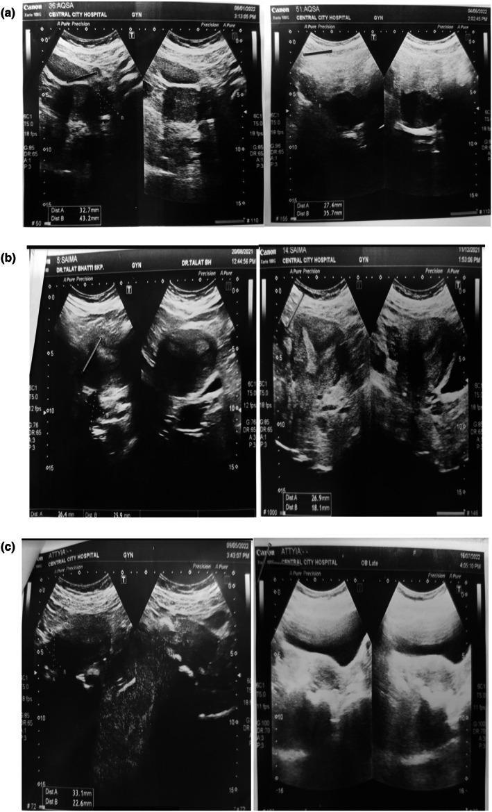
This composite of transvaginal pelvic ultrasound scans demonstrates the comparative morphology of ovaries in three patients (a, b, c) before and after a clinical intervention for polycystic ovary syndrome (PCOS). In the 'before' images (left columns), the ovaries exhibit classic polycystic ovarian morphology (PCOM): multiple small, hypoechoic, rounded cystic structures or follicles distributed throughout the ovarian stroma or arranged peripherally. Many of these ovaries appear bulky with heterogeneous internal echotexture. The 'after' images (right columns) show a notable reduction in the size and quantity of these cystic follicles. Quantitative measurements (Dist A and Dist B) across all panels indicate a decrease in overall ovarian dimensions post-treatment. The resulting ovarian parenchyma appears more uniform and homogeneous. These diagnostic images illustrate the transition from a multicystic state characteristic of PCOS to a more typical ovarian appearance, serving as a visual baseline for monitoring treatment efficacy in reproductive endocrinology and gynecology.
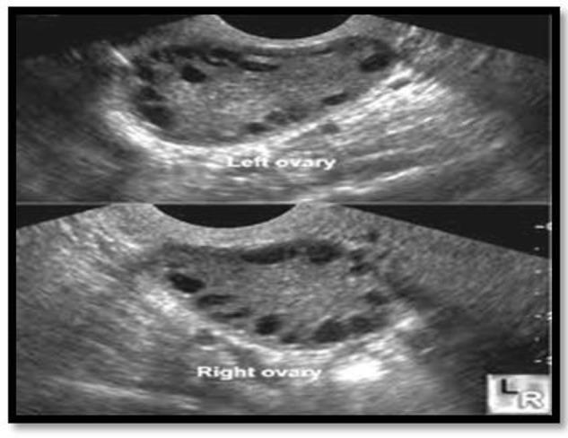
Diagnostic ultrasound imaging displaying a split-view of the left and right ovaries in a patient with Polycystic Ovary Syndrome (PCOS). Both ovaries exhibit classic sonographic features of polycystic morphology, characterized by an increased ovarian volume and a high count of small, peripherally arranged antral follicles. The follicles appear as multiple anechoic (dark), circular, or oval structures, typically measuring between 2 to 9 mm in diameter, creating a 'string of pearls' appearance. The central ovarian stroma is echogenic (brighter gray) and appears thickened or hypertrophied. Each ovary contains more than 12 visible follicles, meeting the Rotterdam diagnostic criteria for PCO. This clinical photograph is a primary tool in reproductive endocrinology and gynecology for evaluating menstrual irregularities, hyperandrogenism, and infertility. It serves as an educational reference for distinguishing normal follicular development from the pathological follicular arrest seen in PCOS.
Polycystic Ovary Syndrome (PCOS) - The Polyendocrine Metabolic Ovarian Syndrome
Epidemiology
- Affects 5-10% of women worldwide of reproductive age
- Most common cause of hyperandrogenism (HA), hirsutism, and anovulatory infertility in developed countries
- First described in 1935 by Stein and Leventhal (amenorrhea + bilateral polycystic ovaries + obesity)
- Genetic origin: likely polygenic and/or multifactorial; familial clustering is well recognized
The "Polyendocrine" Dimension - Pathophysiology
1. Hypothalamic-Pituitary Axis
- Elevated pulse frequency of GnRH leads to a disproportionately high LH:FSH ratio (elevated LH in ~two-thirds of cases)
- High LH stimulates ovarian theca cells to overproduce androgens
- Relatively low FSH fails to adequately stimulate follicular maturation
2. Ovarian Dysfunction
- Dysregulation of enzymes in androgen biosynthesis leads to excessive androgen production (testosterone, androstenedione, DHEAS)
- Follicular arrest: multiple small follicles (2-9 mm) are recruited but none reach dominance - the classical "string of pearls" morphology
- Chronic anovulation results
3. Pancreatic / Insulin Axis
- Insulin resistance (IR) with compensatory hyperinsulinemia is a central metabolic feature
- Insulin amplifies LH-driven androgen production in the ovary (theca cell stimulation)
- IR is present even in lean PCOS women, but is markedly worsened by obesity
4. Adrenal Contribution
- Elevated DHEAS from the adrenal cortex is seen in ~50% of PCOS patients
- This adrenal hyperandrogenism compounds ovarian androgen excess
Diagnostic Criteria
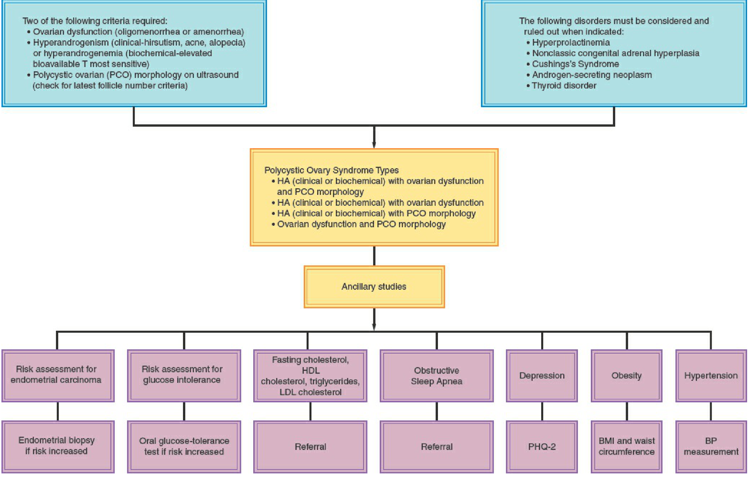
Rotterdam Criteria (2003) - 2 out of 3 required:
| Criterion | Details |
|---|---|
| 1. Oligo/anovulation | Irregular or absent menses |
| 2. Clinical or biochemical hyperandrogenism | Hirsutism, acne, male-pattern alopecia; or elevated free testosterone |
| 3. Polycystic ovarian morphology on ultrasound | ≥20 follicles (2-9 mm) in either ovary and/or ovarian volume >10 mL |
- HA + ovarian dysfunction + PCO morphology (classic full phenotype)
- HA + ovarian dysfunction (without PCO on US)
- HA + PCO morphology (with regular cycles)
- Ovarian dysfunction + PCO morphology (without HA)
Clinical Features
| System | Manifestations |
|---|---|
| Menstrual | Oligomenorrhea, amenorrhea, irregular cycles; primary amenorrhea rarely |
| Androgenic skin | Hirsutism (~70% in US), acne, male-pattern alopecia, acanthosis nigricans |
| Metabolic | Obesity (>50%), central adiposity, IR, dyslipidemia |
| Reproductive | Anovulatory infertility, recurrent pregnancy loss, increased miscarriage risk |
| Cardiovascular | Hypertension (40% by perimenopause), atherosclerosis, ~7-fold increased MI risk |
| Psychiatric | Depression, anxiety |
| Oncologic | Increased risk of endometrial hyperplasia/carcinoma (from unopposed estrogen) |
Ultrasound Appearance
Metabolic Consequences (The "Metabolic" Dimension)
- Impaired glucose tolerance (IGT): ~30% of obese PCOS; ~10% of non-obese PCOS
- Type 2 diabetes: 7.5-10% of obese PCOS; 1.5% of non-obese PCOS
- Dyslipidemia: elevated total cholesterol, LDL, triglycerides; low HDL (particularly HDL2α)
- Impaired fibrinolysis: elevated plasminogen activator inhibitor (PAI-1)
- Obstructive sleep apnea: increased prevalence
- All women with PCOS should be screened with fasting glucose + 75-g OGTT
Management
Lifestyle (all patients)
- Weight loss of even 5-10% can restore ovulation and reduce IR
- Exercise and dietary modification are first-line
Menstrual Regulation + Endometrial Protection
- Combined oral contraceptive pills (COCPs): first-line - regulate cycles, decrease androgens by increasing SHBG, protect endometrium
- Progestins (medroxyprogesterone 10 mg x 10-14 days every 3 months, or levonorgestrel IUD): for endometrial protection if COCPs contraindicated
Hyperandrogenism
- COCPs reduce androgens within 2-3 months, but hirsutism may take longer to respond
- Anti-androgens (spironolactone, flutamide): if inadequate COCP response at 6 months
- Eflornithine cream: for facial hirsutism
Metabolic / Insulin Resistance
- Metformin: for prevention of cardiometabolic risk in overweight/obese women; adjunct to diet/exercise; also reduces insulin and androgen levels
- Screen and manage: hypertension, dyslipidemia, sleep apnea, depression
Fertility / Ovulation Induction
- Letrozole (aromatase inhibitor): first-line for ovulation induction
- Clomiphene citrate ± metformin: second-line
- Injectable gonadotropins: by specialists (PCOS increases hyperstimulation risk)
- Metformin alone: NOT recommended for infertility treatment