Mri wrist joint anatomy images from textbook
Reading File
Finding Sources
Finding Sources
Finding Sources
Reading File
Reading File
Reading File
Reading File
Reading File
Reading File
Reading File
Reading File
Loading Image
Loading Image
Loading Image
Loading Image
Loading Image
Loading Image
Loading Image
Loading Image
I now have a comprehensive collection of high-quality MRI wrist anatomy images from the textbook. Let me present them all with proper context.
MRI Wrist Joint Anatomy - Textbook Images
All images are from Imaging Anatomy: Text and Atlas, Volume 3 - Bones, Joints, Vessels and Nerves (Chapter 17: Wrist).
1. TFCC - Coronal & Axial MRI (Fig. 17.35)
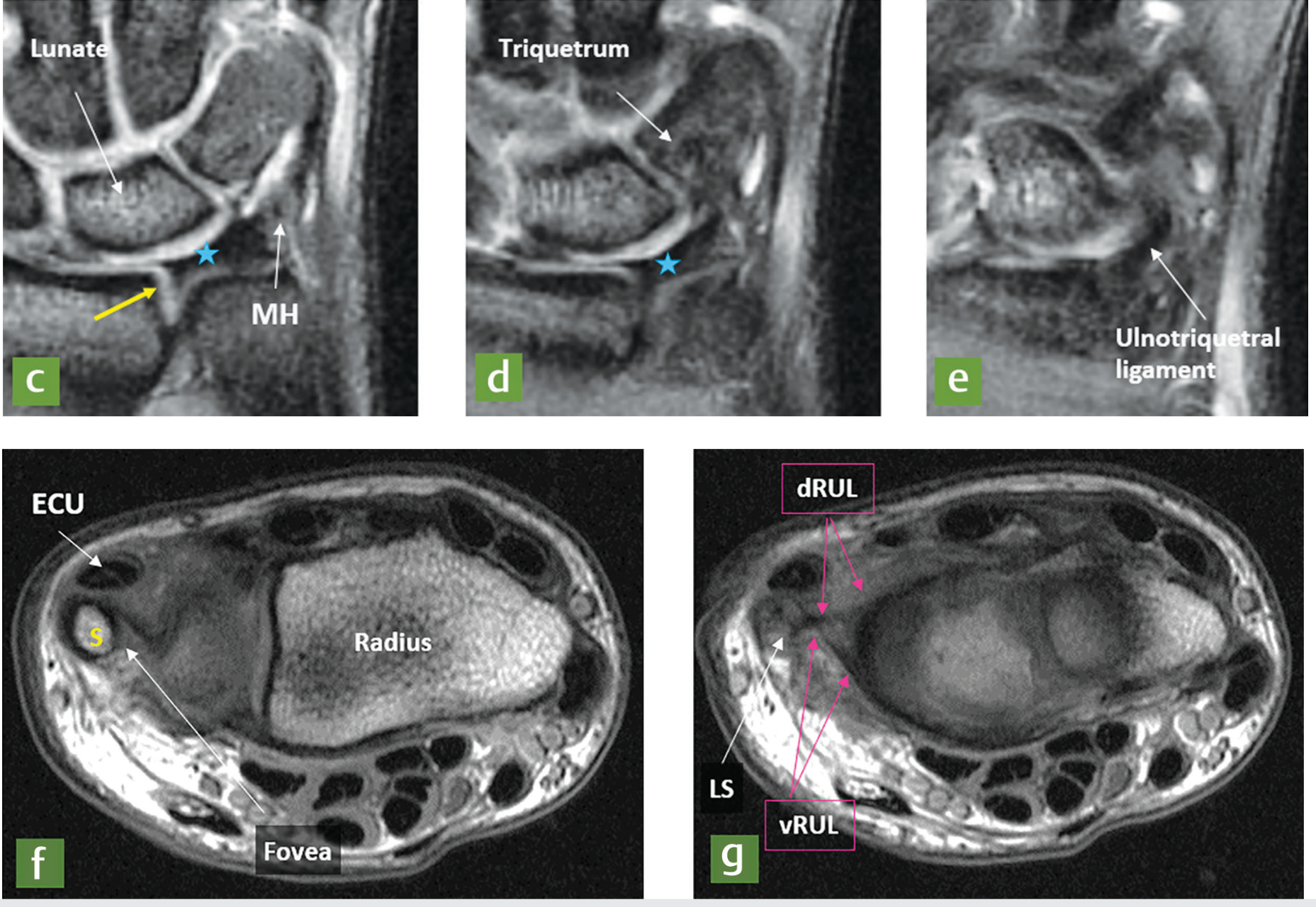
Fig. 17.35 - Triangular fibrocartilage (TFC) serial coronal images (a-e) from dorsal to palmar, and axial MRI (f, g). Key structures labeled:
- TFC proper (star) attaching to the sigmoid notch of the distal radius
- Proximal (arrowhead) and distal (thick arrow) laminae attaching at the fovea and ulnar styloid
- Lunate, Triquetrum, ulnotriquetral ligament (image e)
- Axial views: ECU tendon, fovea, ulnar styloid (S), dorsal radioulnar ligament (dRUL), volar radioulnar ligament (vRUL), ligamentum subcruentum (LS)
- Meniscus homologue (MH), ulnar collateral ligament (UCL)
2. TFCC - MRI Coronal Detail (Fig. 17.35a-b)
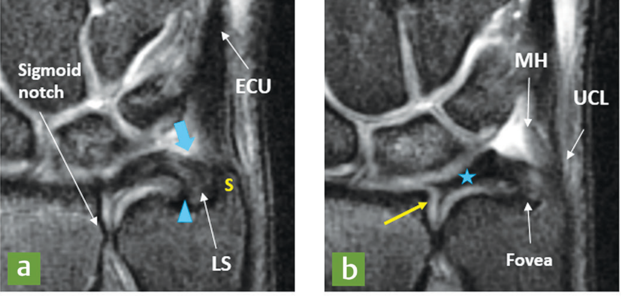
Fig. 17.35 (a, b) - Close-up coronal MRI views:
- (a): Sigmoid notch, ECU tendon, ligamentum subcruentum (LS), ulnar styloid (S), with the proximal lamina (arrowhead) and TFC proper (blue arrow)
- (b): Meniscus homologue (MH), ulnar collateral ligament (UCL), fovea - showing the precise distal insertion of the TFCC
3. TFCC - Coronal Section Anatomy Diagram (Fig. 17.34)
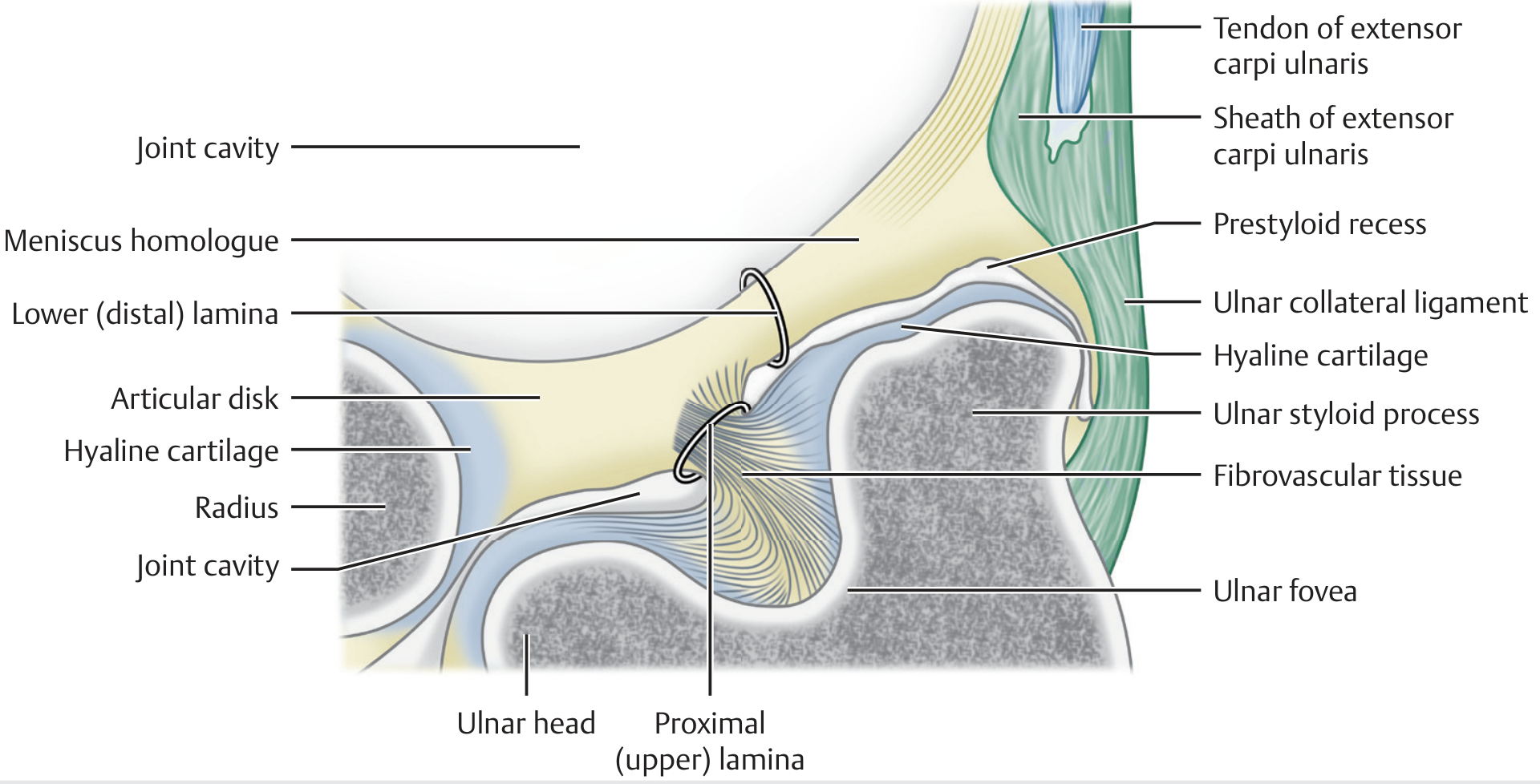
Fig. 17.34 - Labeled coronal section showing the full TFCC architecture: articular disk, proximal (upper) and distal (lower) laminae, meniscus homologue, ulnar collateral ligament, ECU tendon sheath, prestyloid recess, hyaline cartilage, ulnar fovea, and the ulnar head.
4. Wrist Cross-Section - Tendons, Nerves & Compartments (Fig. 17.50)
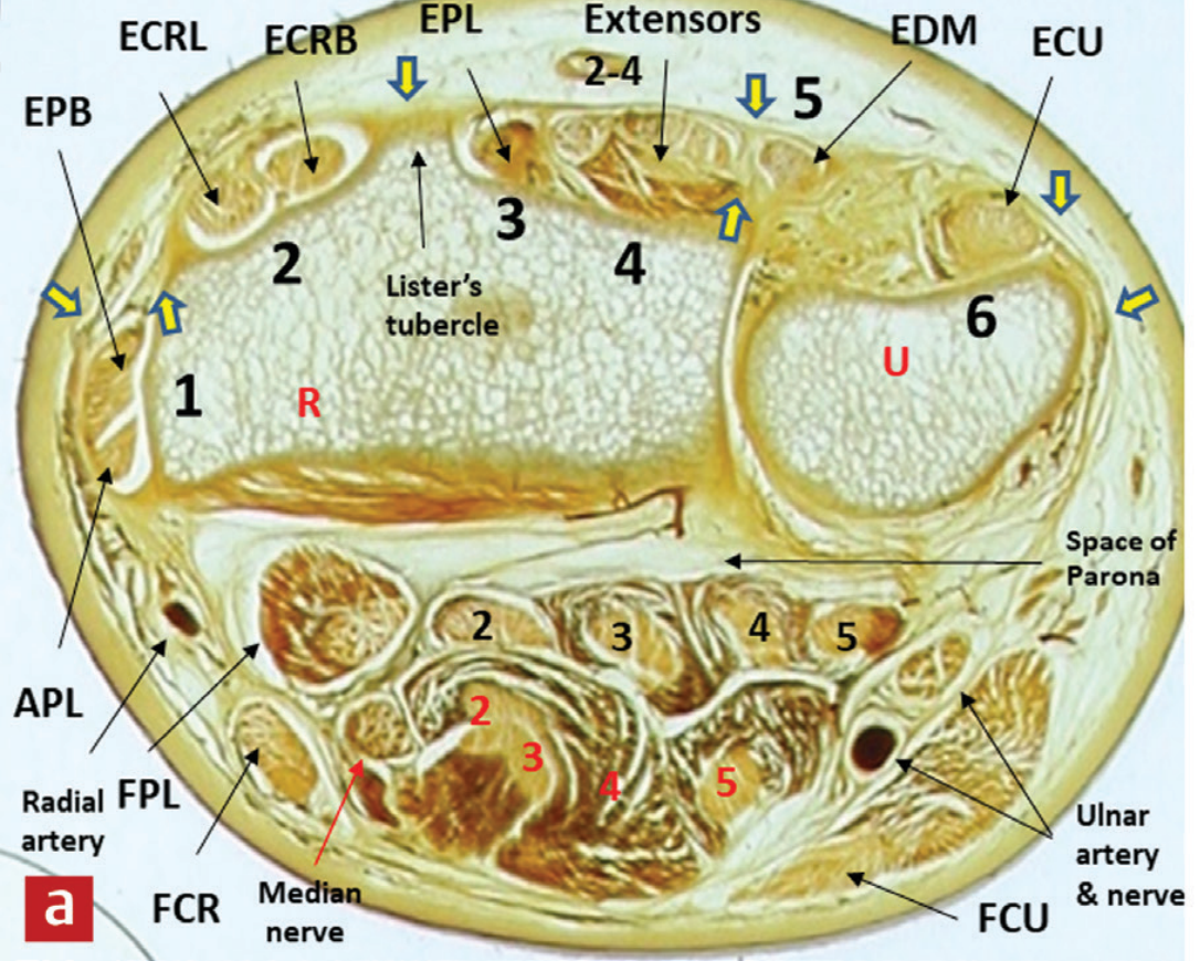
Fig. 17.50 - Labeled axial cross-section at the level of the distal radius (R) and ulna (U), showing:
- 6 extensor compartments (dorsal): EPB/APL (1), ECRL/ECRB (2), EPL (3), Extensors 2-4 (4), EDM (5), ECU (6)
- Lister's tubercle separating compartments 2 and 3
- Volar structures: FPL, FCR, Median nerve, FCU, Radial artery, Ulnar artery & nerve
- Space of Parona (deep palmar space)
5. Wrist MRI - Coronal Overview & ECU Tendinosis (Fig. 17.64)
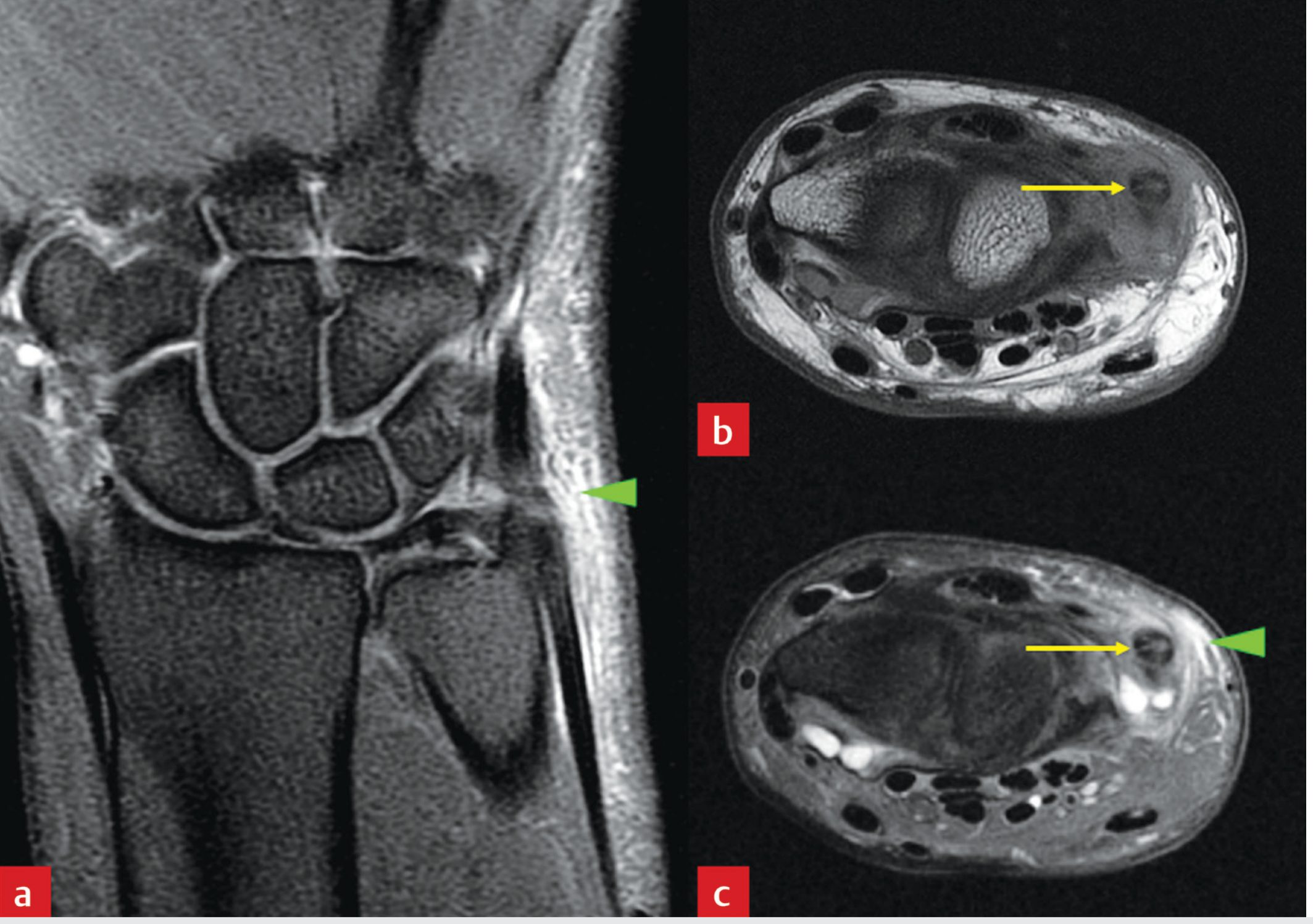
Fig. 17.64 - Coronal (a) and axial (b, c) proton density MRI views:
- (a) Coronal view: All 8 carpal bones clearly visible in their natural arrangement (scaphoid, lunate, triquetrum, pisiform in proximal row; trapezium, trapezoid, capitate, hamate in distal row); radiocarpal joint space well-defined
- (b, c) Axial views: ECU tendon at the ulnar groove with surrounding signal changes
6. De Quervain's Tenosynovitis - Axial MRI (Fig. 17.63)
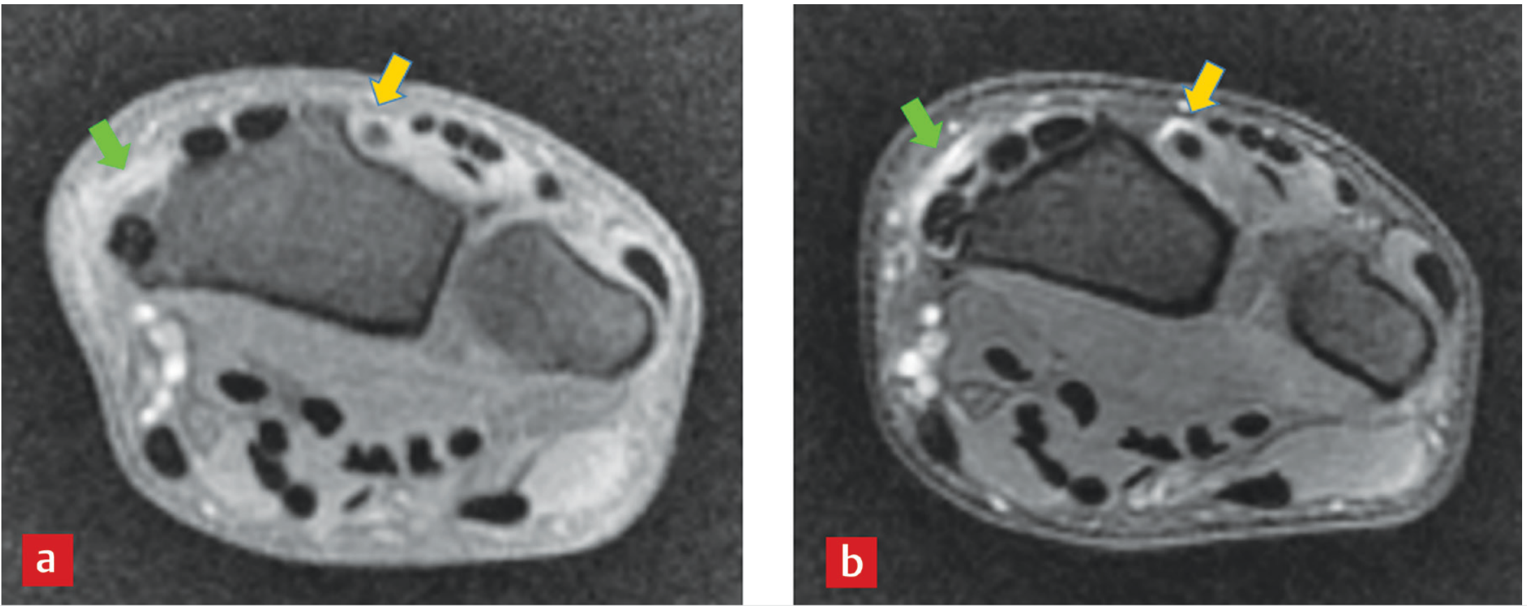
Fig. 17.63 - Axial fat-saturated T1W (a) and fat-saturated proton density (b) MRI demonstrating the first extensor compartment (EPB and APL tendons) and tenosynovitis appearance. Note: While this shows pathology, it clearly demonstrates normal axial MRI wrist anatomy including the round tendon cross-sections.
7. Guyon's Canal (Ulnar Tunnel) - Serial Axial MRI (Fig. 17.53a)
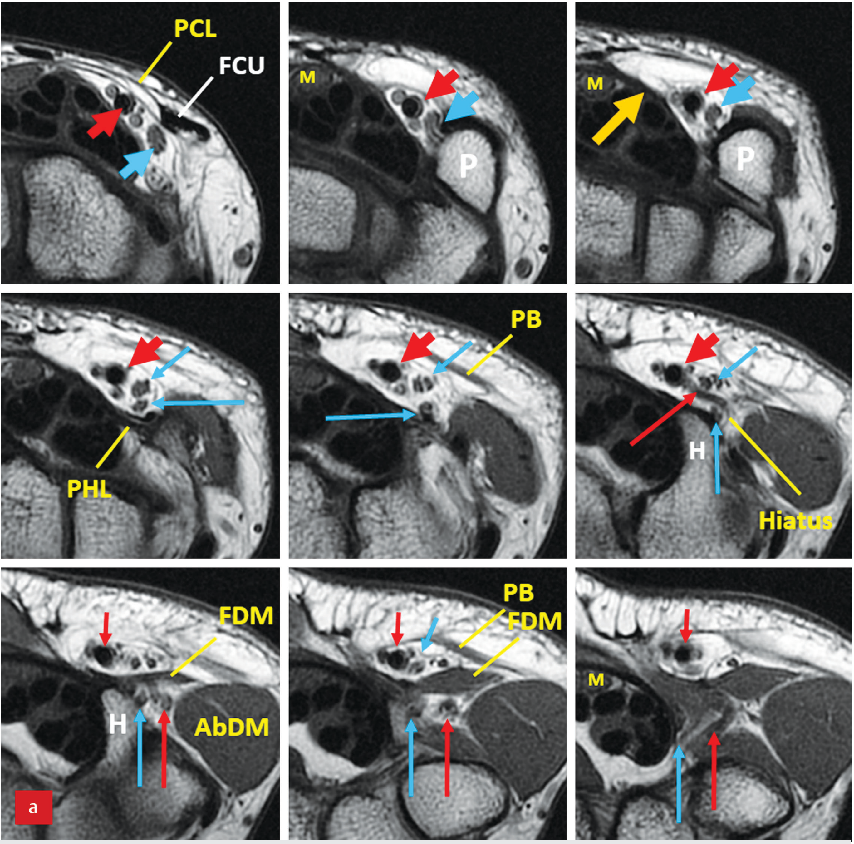
Fig. 17.53 (a) - Serial axial MRI from proximal to distal through Guyon's canal:
- Red arrows: Ulnar artery (thick = main; thin short = superficial branch; thin long = deep branch)
- Blue arrows: Ulnar nerve (thick = main; thin = superficial/motor branches)
- P = pisiform, H = hook of hamate, Hiatus = pisohamate hiatus
- PCL (palmar carpal ligament), FCU, FDM (flexor digiti minimi), AbDM (abductor digiti minimi), PB (palmaris brevis), PHL (pisohamate ligament)
8. Guyon's Canal - Serial Coronal MRI (Fig. 17.53b)
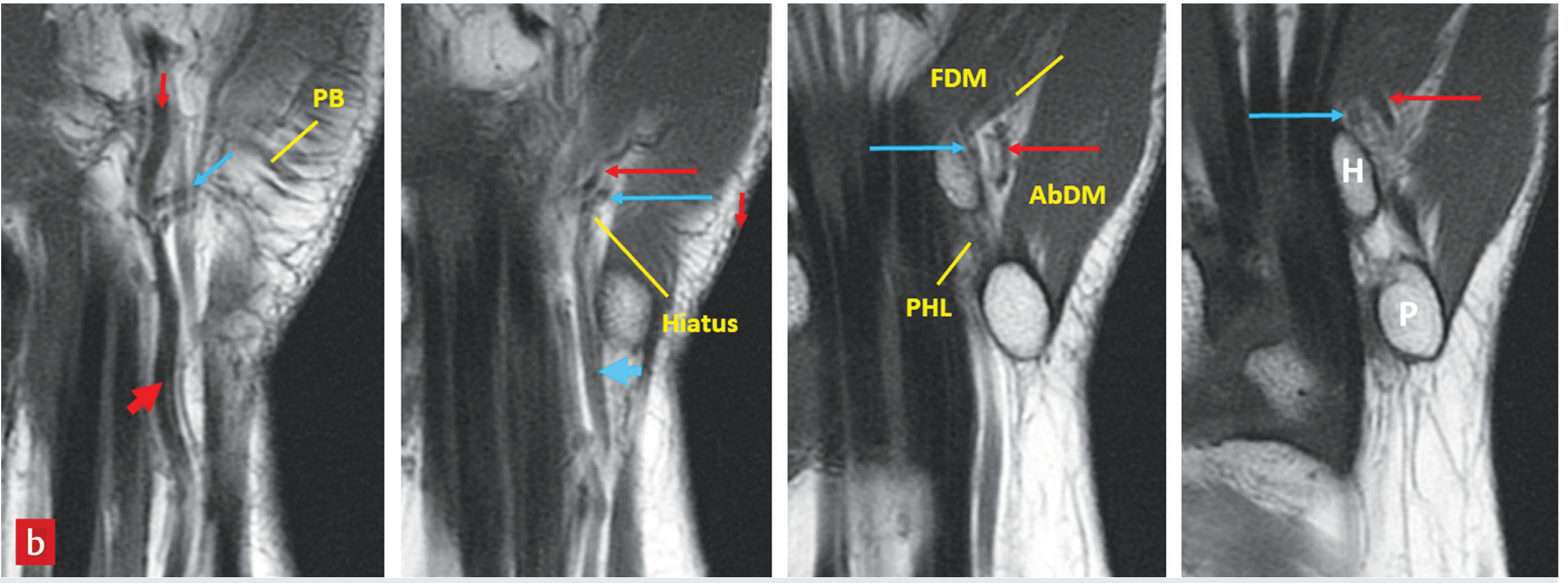
Fig. 17.53 (b) - Serial coronal MRI views showing the deep motor branch of the ulnar nerve (blue arrows) and superficial branch, the pisohamate hiatus (Hiatus), AbDM, FDM, PHL structures, with pisiform (P) and hamate (H) landmark bones clearly visible.
Summary of Key MRI Wrist Anatomy Structures
| Structure | Best MRI Plane | Sequence |
|---|---|---|
| TFCC / TFC proper | Coronal | PD or T2 fat-sat |
| Carpal ligaments (SLL, LTL) | Coronal | PD fat-sat |
| Extensor compartments | Axial | T1 or PD |
| Carpal tunnel (median nerve) | Axial | T1 or PD |
| Guyon's canal (ulnar nerve/artery) | Axial + Coronal | T1 |
| Radioulnar ligaments (DRUL/VRUL) | Axial | PD fat-sat |
| Carpal bones (osseous anatomy) | Coronal | T1 |
Source: Imaging Anatomy Text and Atlas, Vol. 3 - Bones, Joints, Vessels and Nerves, Chapter 17: Wrist (pp. 589-615)
Send pdf of this images
Reading File
Running Command
Writing File
Running Command
Your PDF is ready! Here's the download link:
MRI Wrist Joint Anatomy
PDF Document · PDF
What's inside (10 pages):
- Cover page with a contents table listing all 8 figures
- 8 image pages, each with the full-resolution MRI image + figure label + detailed anatomical caption covering:
- TFCC coronal & axial MRI (Fig. 17.35)
- TFCC coronal detail close-ups
- TFCC anatomy diagram (Fig. 17.34)
- Wrist axial cross-section showing all 6 extensor compartments (Fig. 17.50)
- Carpal bones coronal overview & ECU tendon (Fig. 17.64)
- First extensor compartment axial MRI (Fig. 17.63)
- Guyon's canal serial axial MRI (Fig. 17.53a)
- Guyon's canal serial coronal MRI (Fig. 17.53b)
- Quick Reference table - optimal MRI plane and sequence for each wrist structure
Source: Imaging Anatomy Text and Atlas, Vol. 3, Chapter 17: Wrist (pp. 589–615)
This is a shared conversation. Sign in to Orris to start your own chat.