
Chronic Lymph oedema pt treatment after axillary lymph node removal
"lymphedema" AND "axillary lymph node dissection" AND treatment
lymphedema arm breast cancer treatment compression bandaging
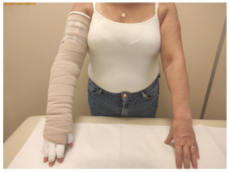
Clinical photograph of a patient undergoing treatment for upper limb lymphedema, likely following breast cancer-related interventions. The image demonstrates a side-by-side comparison between a treated left arm and an untreated right arm. The left arm is encased in a multi-layer compression bandaging system (CB), utilizing beige short-stretch bandages applied in a spiral, overlapping pattern. The bandaging extends from the axilla down to the hand, with individual white cotton gauze or padding applied to the fingers to manage digital swelling. The treated limb exhibits increased bulk due to the therapeutic padding and compression materials, which typically include a cotton stockinet and foam padding layers beneath the external bandages. The right arm serves as a baseline, showing natural morphology and skin tone. This visual illustrates the clinical application of Complete Decongestive Therapy (CDT) in its intensive phase, focusing on mechanical reduction of limb volume through graduated compression.
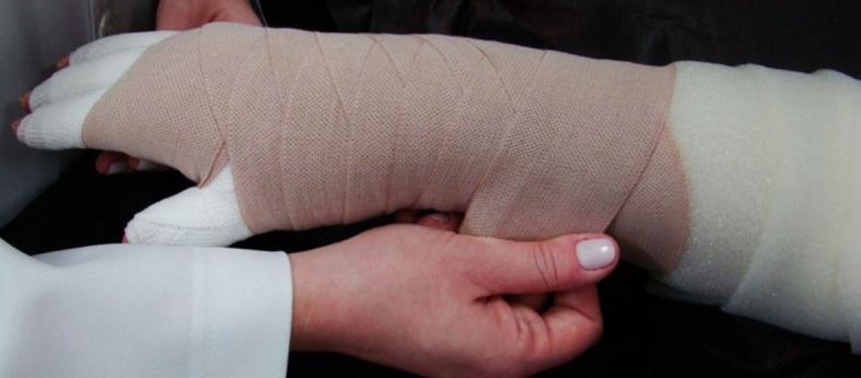
This clinical photograph illustrates the application of multi-layer bandaging (MLB) on the hand and forearm of a patient, a primary intervention for breast cancer-related lymphedema. The image shows a structured approach to compression therapy: an innermost layer of white, soft padding material is applied directly over the fingers, thumb, and arm to protect the skin and ensure even pressure distribution. Overlapping this is a secondary layer consisting of a light brown, short-stretch woven elastic bandage applied in a spiral and figure-eight pattern. This specific arrangement provides high working pressure and low resting pressure, essential for promoting lymphatic drainage and reducing limb volume. The anatomical focus is the upper extremity, demonstrating the complex distal-to-proximal wrapping required to manage interstitial fluid accumulation. This visual serves as an educational tool for physical therapy and oncology nursing regarding Complete Decongestive Therapy (CDT) techniques.
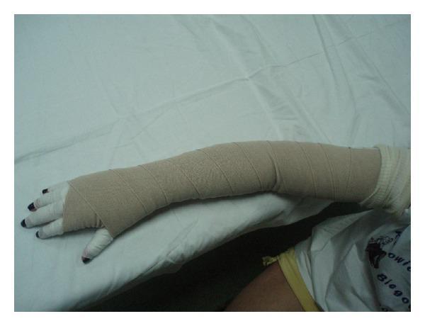
This clinical photograph displays a multilayered compression bandaging technique applied to a human upper extremity, commonly used in the management of breast cancer-related lymphedema. The bandage system consists of a white stockinette or padding under-layer, visible at the fingers and the proximal axillary region, followed by beige short-stretch bandages applied in a spiral, overlapping fashion. The wrapping extends from the metacarpals, through the wrist and elbow, up to the upper arm, conforming to the limb's anatomical contour while maintaining even pressure. Distal phalanges are left exposed to monitor neurovascular status. This visual demonstrates the standard application of Complex Decongestive Therapy (CDT) components designed to facilitate lymphatic drainage and reduce limb volume in chronic edematous states (Stages II and III). The image serves as an educational reference for correct bandaging morphology, showing the typical 'ribbed' texture created by precise overlapping layers to prevent garment slippage and ensure therapeutic compression levels of 50-60 mmHg.
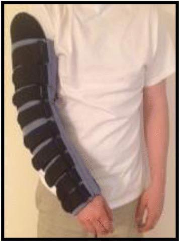
This clinical photograph displays a strapped Night-time Compression System Garment (NCSG) applied to the right upper extremity of a patient. The device is designed for the management of breast cancer-related lymphedema. The garment extends from the proximal upper arm to the wrist, featuring a dual-layer construction: a light-colored, padded foam liner that provides soft interface against the skin, and an overlying series of multiple, wide, dark-colored inelastic straps. These straps are arranged horizontally and use a hook-and-loop fastening mechanism (Velcro) to allow for precise adjustment of compression levels along the limb. The system facilitates the application of gentle gradient pressure, promoting lymphatic drainage while maintaining low resting pressure suitable for overnight use. This visual serves as an educational example of alternative self-management strategies to traditional multilayer compression bandaging (CB), emphasizing ease of application and customizable fit for patients with varying degrees of swelling.
Chronic Lymphoedema Treatment After Axillary Lymph Node Removal (BCRL)
ISL Staging (International Society of Lymphology)
| Stage | Description |
|---|---|
| 0 | Latent/subclinical - impaired transport, no visible oedema |
| 1 | Spontaneously reversible - pitting oedema, subsides with elevation |
| 2 | Spontaneously irreversible - pitting may or may not occur, fibrosis begins |
| 3 | Lymphostatic elephantiasis - non-pitting, skin changes (acanthosis, hyperkeratosis) |
Conservative (Non-Operative) Treatment
1. Complex Decongestive Therapy (CDT) - The Gold Standard
- Manual Lymphatic Drainage (MLD)
- Compression (bandages + garments)
- Skin care
- Exercise
- MLD performed by a certified therapist 5 days/week for 1-4 weeks until limb volume plateaus
- Techniques: Light massage starting on the contralateral normal side first, then the trunk quadrant adjacent to the affected limb, then the proximal zones of the swollen extremity, proceeding distally
- After each session, the arm is wrapped with a low-stretch compression bandage
- Volume reduction of 60-70% is achievable; compliant patients retain 90% of this reduction
- Patient continues compression garments during the day, bandages at night
- Maintenance MLD every 2-3 months
- Daily skin care and remedial exercises
- Lifelong adherence is imperative - fluid reaccumulates without it
2. Compression Therapy
- Compression garments: Custom-fitted, delivering 30-60 mmHg pressure; worn at all times except when the limb is elevated above heart level
- Multilayer inelastic short-stretch bandages: High working pressure (enhances lymphatic pumping during muscle contraction), low resting pressure (comfortable for continuous wear)
- Pneumatic compression pump therapy: Used especially in advanced cases; sequential external compressive devices
- These methods reduce oedema volume by approximately 30-45%
- Contraindications: Active arterial insufficiency, active infection, active malignancy in the limb
3. Limb Elevation
- Elevate the affected arm above heart level, particularly at night
- Adjunct to all other therapies; especially important in Stage 1
4. Skin Care and Infection Prevention
- Meticulous daily hygiene to prevent skin breakdown
- Prompt treatment of any infection (cellulitis/lymphangitis) with antibiotics targeted at gram-positive cocci (streptococcal/staphylococcal)
- Hydrocortisone cream for eczematous changes
- Protect the limb from injury (burns, cuts, insect bites) - compression garments help
- Avoid venepuncture/blood pressure cuffs on the affected arm
5. Exercise and Lifestyle
- Daily range-of-motion and remedial exercises complement compression and elevation
- Weight loss and aerobic exercise have been proven to decrease lymphoedema severity in large clinical studies
- Maintain ideal body weight (high BMI is a major risk factor)
6. Pharmacological Treatments
- No effective medications exist specifically for lymphoedema
- Antibiotics: Indicated for superimposed cellulitis/lymphangitis only
- Diuretics: NOT recommended - may worsen fibrosis and increase interstitial protein accumulation
- NSAIDs (Ketoprofen): Investigational; inhibits 5-lipoxygenase (upregulated in lymphoedema) - shown some benefit in histopathology but not volume reduction
- Supplements (sodium selenite, vitamin E + pentoxifylline): No proven benefit
Surgical (Operative) Treatment
A. Physiologic Procedures (Restore Lymphatic Flow)
1. Lymphovenous Anastomosis (LVA)
- Microsurgical connection between lymphatic channels and nearby venules
- Creates a bypass around the obstructed lymphatics
- Best results in early-stage disease (before fibrosis)
- Recent meta-analyses (PMID: 40403351) confirm LVA is effective in preventing BCRL when done immediately after ALND (Immediate LVA / LYMPHA procedure)
2. Vascularized Lymph Node Transplant (VLNT)
- Free flap transfer of lymph nodes from a donor site (cervical/neck, groin/inguinal, omentum) to the axilla or forearm
- Donor sites: contralateral neck (between omohyoid and anterior scalene), inguinal region (using superficial circumflex vessels), or omentum (via laparoscopic harvest)
- A systematic review of 18 studies (305 patients) showed 86-90% improved swelling, mean volume reduction of ~22%, and ~50% of patients no longer needing compression garments
- Reverse lymphatic mapping is used during harvest to avoid donor site lymphoedema
3. Immediate Lymphatic Reconstruction (LYMPHA)
- Performed at the time of ALND to prevent lymphoedema from developing
- Meta-analyses (PMID: 40862309, PMID: 39547134) confirm significant reduction in BCRL incidence post-ALND
B. Reductive Procedures (Debulk Tissue)
4. Suction-Assisted Lipectomy / Liposuction
- Removes fibroadipose tissue in advanced chronic lymphoedema where fluid accumulation is replaced by solid tissue
- Must be combined with lifelong compression therapy post-operatively to maintain results
- Does not address the underlying lymphatic dysfunction
5. Charles Procedure (Radical Excision)
- Excision of all subcutaneous tissue down to the deep fascia with skin grafting
- Reserved for severe, end-stage lymphoedema (Stage 3 / elephantiasis)
- Coverage preferably by full-thickness skin grafts from excised skin
Multidisciplinary Team
- Certified lymphoedema physiotherapist (MLD and CDT)
- Occupational therapist (ADL modification, garment fitting)
- Oncology nurse / breast care nurse
- Plastic/reconstructive surgeon (for surgical candidates)
- Social worker + psychosocial support (anxiety, depression from chronic disfigurement are common)
Key Complications to Watch For
| Complication | Notes |
|---|---|
| Cellulitis / lymphangitis | Treat aggressively with antibiotics; recurrent episodes accelerate fibrosis |
| Skin breakdown / ulceration | Rigorous hygiene and skin care |
| Stewart-Treves syndrome | Rare lymphangiosarcoma complicating long-standing (>10 years) lymphoedema; presents as subcutaneous nodules + worsening oedema; very poor prognosis; treatment: chemo-radiation + radical excision (often amputation) |
Recent Evidence Update (2024-2026)
- Immediate LVA meta-analysis (2025) confirms LVA at time of ALND significantly reduces BCRL incidence
- Immediate lymphatic reconstruction meta-analysis (2025) confirms LYMPHA procedure is effective preventively
- Risk factors meta-analysis (2024, PMID 39321505) found radiotherapy is a significant independent risk factor for arm symptoms alongside ALND