I now have comprehensive data to compile the full answer. Let me update the todos and write the complete structured response.Here is the complete, structured answer for all topics in both chapters.
CHAPTER 1 - VIRUSES CAUSING HEPATITIS
ENUMERATE THE VIRUSES CAUSING HEPATITIS
Hepatitis viruses are a taxonomically diverse (heterogeneous) group of viruses that share one common feature: they are hepatotropic (they infect and damage the liver). They are classified into six types - HAV through HGV (excluding "F" which was disproved):
| Virus | Family | Genome | Transmission | Chronicity |
|---|
| HAV | Picornaviridae (genus Hepatovirus) | ssRNA (+) | Fecal-oral | No |
| HBV | Hepadnaviridae | Partially ds-DNA | Parenteral, sexual, vertical | Yes (5-10%) |
| HCV | Flaviviridae (genus Hepacivirus) | ssRNA (+) | Parenteral | Yes (>70%) |
| HDV | Unclassified (deltavirus) | ssRNA (-), circular | Parenteral (requires HBV) | Yes (with HBV) |
| HEV | Hepeviridae | ssRNA (+) | Fecal-oral | No (rarely in immunosuppressed) |
| HGV | Flaviviridae | ssRNA (+) | Parenteral | Uncertain significance |
Other viruses that can cause sporadic hepatitis (not primarily hepatotropic):
- Cytomegalovirus (CMV)
- Epstein-Barr virus (EBV)
- Herpes simplex virus (HSV)
- Adenoviruses
- Rubella virus
- Hantaviruses
- Mumps virus
- Enteroviruses (Coxsackieviruses)
- Yellow fever virus - an arbovirus (Flaviviridae) causing hemorrhagic fever with hepatitis
HEPATITIS B VIRUS (HBV) - COMPREHENSIVE
1. MORPHOLOGY (Structure)
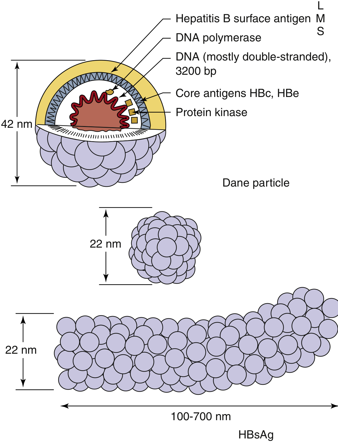
HBV belongs to the family Hepadnaviridae. The complete infectious virion is called the Dane particle.
Dane Particle:
- Size: 42 nm in diameter
- Enveloped virus
- Contains:
- Outer envelope: carries HBsAg (Hepatitis B surface antigen) in three forms: L (large/gp42), M (medium/gp36), S (small/gp27)
- Inner icosahedral nucleocapsid/core: composed of HBcAg (Hepatitis B core antigen)
- Genome: Partially double-stranded circular DNA, ~3200 base pairs (unique among DNA viruses - replicates via RNA intermediate using reverse transcriptase)
- DNA polymerase with reverse transcriptase and RNase H activity
- Protein kinase
- HBeAg: non-structural protein encoded by precore region; secreted into serum
Fig: Hepatitis B virus (Dane particle) and HBsAg particles - Medical Microbiology 9e
HBsAg particles in serum (non-infectious, outnumber virions):
- Spherical particles: 22 nm diameter, composed mainly of S-form HBsAg
- Tubular/Filamentous particles: 22 nm wide, 100-700 nm long, composed of S, M, and L forms
Genome Organization (4 major open reading frames):
- S gene: encodes HBsAg (S, M, L forms = surface antigen)
- C gene / Precore: encodes HBcAg (core) and HBeAg (precore)
- P gene: encodes DNA polymerase (reverse transcriptase)
- X gene: encodes HBxAg - a transactivating factor implicated in hepatocellular carcinoma (HCC)
Serotypes/Genotypes:
- HBV divided into 4 major serotypes (adr, adw, ayr, ayw) based on antigenic epitopes on HBsAg envelope protein
- 8-10 major genotypes (A through H/J) based on DNA sequence; immunity is NOT serotype-specific
- Genotype C: associated with higher risk of cirrhosis and HCC; low response to interferon treatment
2. MODES OF TRANSMISSION
HBV is present in blood, serum, saliva, semen, vaginal secretions, breast milk, and other body fluids.
- Sexual transmission - most common route in developed countries (particularly homosexual males at higher risk)
- Parenteral / Blood-borne transmission:
- Blood transfusions (residual risk ~1 in 600,000 with screening)
- Sharing IV drug use needles
- Needle-stick injuries (health care workers)
- Hemodialysis
- Tattooing, acupuncture with unsterilized equipment
- Vertical (Perinatal) transmission - from HBsAg-positive mother to neonate (usually at or around delivery, not in utero). Carrier rate if infected perinatally: ~90%; if infected as adults: ~5-10%
- Horizontal transmission in childhood - skin abrasions, contact with infected secretions (common in endemic areas)
- HBV is NOT transmitted by the fecal-oral route, breast milk (generally), casual contact (hugging, kissing), contaminated food, or water
Key risk factor for chronicity: Age at infection inversely correlates with chronicity - perinatal infection → 90% chronic; early childhood → 30%; adults → 5-10%
3. PATHOGENESIS
HBV pathogenesis is primarily immune-mediated, not directly cytopathic.
Steps:
- Entry: HBV attaches to hepatocytes via HBsAg glycoproteins binding to the sodium/bile acid cotransporter NTCP (sodium taurocholate cotransporting polypeptide)
- After entry, the nucleocapsid delivers the partially ds-DNA genome to the nucleus where it is completed into a covalently closed circular DNA (cccDNA) - a viral minichromosome that persists in infected hepatocytes
- The genome is transcribed, producing 4 types of mRNA including a 3500-base pregenomic RNA (pgRNA)
- Replication: pgRNA is packaged into core particles; reverse transcriptase synthesizes negative-sense DNA from the RNA template; RNA is degraded; positive-sense DNA is partially synthesized → new virions released by exocytosis
Immune-mediated liver damage:
- Hepatocytes carrying viral antigens are recognized by CD8+ cytotoxic T cells and NK cells - these kill infected hepatocytes → liver damage
- In adults with intact immunity: robust immune response → clearance (~95%)
- In neonates/infants: immature immune response → virus tolerated → carrier state (immune tolerance phase)
Consequences of chronic infection:
- Chronic active hepatitis → cirrhosis → hepatocellular carcinoma (HCC)
- HBV X gene product (HBxAg) transactivates cellular growth genes, promotes carcinogenesis
- Integration of HBV DNA into host genome triggers chromosomal rearrangements
- PHC (primary hepatocellular carcinoma) - HBV one of the three most common causes of cancer mortality worldwide
4. LABORATORY DIAGNOSIS OF HBV - SEROLOGICAL MARKERS AND SIGNIFICANCE
The serological picture describes the course and nature of HBV infection. There are three antigen-antibody systems:
A. Hepatitis B Surface Antigen/Antibody (HBsAg / Anti-HBs)
HBsAg (Australia antigen):
- First detectable marker; appears 1-10 weeks after exposure (before symptoms)
- Indicates active infection (acute or chronic)
- Presence for >6 months = chronic infection
- Detected by: ELISA, chemiluminescence, ICT
Anti-HBs (Antibody to HBsAg):
- Appears after clearance of HBsAg (after resolution)
- Indicates: recovery + immunity OR successful vaccination
- Only marker positive in vaccinated individuals
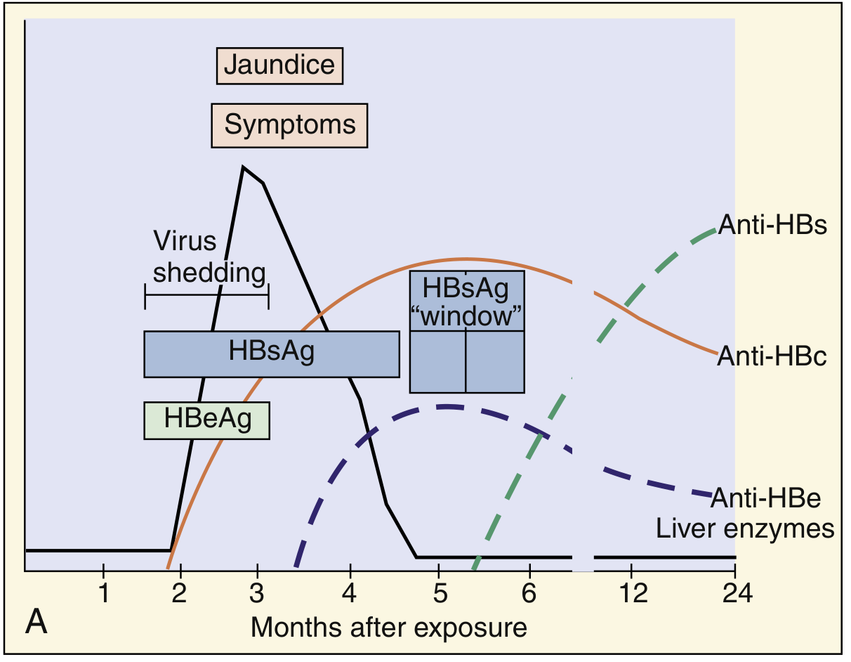
- HBsAg and anti-HBs cannot be detected simultaneously (immune complex obscures both) → "window period"
B. Hepatitis B Core Antigen/Antibody (HBcAg / Anti-HBc)
HBcAg:
- Hidden antigen; surrounds the viral core
- NOT detectable in serum (non-secretory, enclosed within HBsAg coat)
- Detectable only in hepatocytes by immunofluorescence
Anti-HBc:
- First antibody to appear; detected during the "window period" (after HBsAg disappears but before anti-HBs appears)
- IgM anti-HBc = marker of acute HBV infection (diagnostic during window period)
- IgG anti-HBc = marker of past infection; persists for decades
- Anti-HBc is present in both acute AND chronic infection; absent only in vaccinated persons
Window Period: The HBsAg window is the diagnostic gap when HBsAg has cleared but anti-HBs has not yet appeared. During this time, IgM anti-HBc is the only detectable marker - it is the diagnostic marker of acute HBV.
C. Hepatitis Be Antigen/Antibody (HBeAg / Anti-HBe)
HBeAg:
- Encoded by precore region; a soluble, secreted protein
- Present in serum when active viral replication is occurring
- Best correlate of viral infectivity - its detection indicates high viral load and high infectiousness
- Its persistence beyond 3 months indicates progression toward chronic hepatitis
- NOT detectable in "precore mutant" strains of HBV
Anti-HBe:
- Appears after clearance of HBeAg
- Signals reduction in infectivity and viral replication
- In chronic HBV, transition from HBeAg+ to anti-HBe+ (seroconversion) = good prognostic sign
Serological Marker Interpretation Table
| HBsAg | HBeAg | Anti-HBc (IgM) | Anti-HBc (IgG) | Anti-HBe | Anti-HBs | Interpretation |
|---|
| + | + | + | - | - | - | Acute HBV, high infectivity |
| + | - | - | + | + | - | Chronic HBV, low infectivity (precore mutant possible) |
| + | + | - | + | - | - | Chronic active HBV, high infectivity |
| - | - | + | - | - | - | Window period (acute) |
| - | - | - | + | + | + | Past infection, resolved |
| - | - | - | - | - | + | Vaccination (only anti-HBs) |
Additional Tests:
- HBV DNA (Viral Load): Detected by PCR; quantified by real-time PCR; most sensitive indicator of viral replication; used for monitoring treatment response and guiding antiviral therapy
- Liver enzymes (ALT, AST): Elevated in hepatitis; not specific but indicate hepatocyte injury
- Liver biopsy: "Gold standard" for staging HBV infection (degree of fibrosis); Fibroscan (elastography) is non-invasive alternative
- HBcAg: Detected in liver biopsy specimens by immunofluorescence
Fig: Serological events in acute HBV infection - Medical Microbiology 9e
Markers of ACUTE HBV infection (summary):
- HBsAg - earliest marker
- HBeAg - active replication marker
- IgM anti-HBc - the most specific marker of ACUTE infection (present even during window period)
- Elevated serum ALT/AST
5. PREVENTION OF HBV
A. Passive Immunization (Post-exposure prophylaxis)
- Hepatitis B Immunoglobulin (HBIG): Contains high-titer anti-HBs
- Given within 7 days of exposure (needle-stick) or within 24 hours of birth to neonate of HBsAg+ mother
- Provides immediate but temporary protection
B. Active Immunization - Hepatitis B Vaccines
First-generation vaccine (now obsolete):
- Plasma-derived vaccine: HBsAg purified from plasma of chronic carriers
- Safe but concerns about blood-borne pathogens
Second-generation vaccine (currently used):
- Recombinant DNA (yeast-derived) vaccine - most widely used globally
- HBsAg (S-protein) expressed in Saccharomyces cerevisiae by recombinant DNA technology
- Trade names: Recombivax HB, Engerix-B
- Highly immunogenic, safe (no blood products)
Third-generation vaccine:
- Contains pre-S1 and pre-S2 antigens in addition to S protein
- Better immune response in non-responders
Schedule (standard):
- 3 doses: 0, 1, and 6 months (IM in deltoid)
- Infants of HBsAg+ mothers: HBIG + vaccine within 24 hours of birth
- For newborns: 0, 1-2, and 6-18 months
Seroconversion rates:
- Healthy adults: ~95% respond
- Non-responders (~5-10%): do not develop anti-HBs even after 6 doses; HLA-linked non-response
- Immunocompromised, obese, smokers, elderly: lower response rates
Combined vaccines available:
- Twinrix = HAV + HBV vaccine
- Pentavalent vaccine (includes HBV, DTP, Hib)
Other prevention measures:
- Screening blood donations for HBsAg and anti-HBc
- Universal precautions for healthcare workers
- Needle exchange programs
- Safe sex (condoms)
- No sharing of razors, toothbrushes, needles
HEPATITIS A - LABORATORY DIAGNOSIS
HAV Morphology: 27-32 nm icosahedral particle, non-enveloped, linear ssRNA(+), family Picornaviridae (genus Hepatovirus). Single serotype; 7 genotypes.
Laboratory Diagnosis of HAV:
-
Anti-HAV IgM (Method of choice for acute diagnosis):
- Detected by ELISA
- Appears at onset of symptoms; persists 3-6 months
- Positive = acute HAV infection
-
Anti-HAV IgG:
- Appears 1 week after IgM
- Persists for decades (lifelong)
- Detection in absence of IgM = past infection or vaccination (immunity)
- Used in seroprevalence studies
-
HAV Antigen detection (rarely used clinically):
- ELISA or immunofluorescence on stool samples
- HAV shed in feces 2 weeks before to 2 weeks after onset of jaundice
-
PCR (RT-PCR for HAV RNA):
- Detects HAV RNA in blood and stool during viremic phase
- Used for genotyping and outbreak investigation
-
Liver function tests (ALT/AST): Elevated, non-specific
Key point: HAV does NOT cause chronic infection. No carrier state. ELISA is the method of choice; many rapid test formats available.
HEPATITIS C - LABORATORY DIAGNOSIS
HCV morphology: Spherical, 40 nm, enveloped, ssRNA(+). Family Flaviviridae, genus Hepacivirus. Has three structural proteins: nucleocapsid core protein C; two envelope glycoproteins E1 and E2. Six non-structural proteins (NS2, NS3, NS4A, NS4B, NS5A, NS5B). One p7 membrane protein.
HCV genotypes: 6 major genotypes (1-6); genotype 1 most common worldwide. Genotyping important for treatment decisions.
Laboratory Diagnosis:
-
Anti-HCV (ELISA / CIA):
- Screening test of choice
- First and second generation ELISA: now obsolete (less sensitive)
- Third-generation ELISA: uses antigens from NS5 region in addition to core, NS3, NS4 regions; sensitivity/specificity >99%; becomes positive ~5 weeks post-infection
- Limitation: does not distinguish acute from chronic or resolved infection
-
HCV RNA (RT-PCR/NAAT):
- Most sensitive test; detects viremia within 1-2 weeks of infection
- Qualitative PCR: for diagnosis and confirmation of anti-HCV positive results
- Quantitative RT-PCR (viral load): for monitoring antiviral treatment
- HCV RNA prevalence in general population is >0.1%
-
Recombinant Immunoblot Assay (RIBA) - formerly used as confirmatory test after ELISA; now obsolete as PCR is preferred
-
HCV Core Antigen:
- ELISA-based
- Detectable earlier than anti-HCV; correlates with viral load
- Useful when NAT unavailable
-
HCV Genotyping (PCR-based): Required before initiating antiviral therapy (direct-acting antivirals)
-
Liver biopsy / Fibroscan: Staging of liver fibrosis
HCV does NOT spread through breast milk, food, water, hugging, kissing, or casual contact. Vertical transmission rate is ~4% (much lower than HBV ~20%). Sexual transmission is rare.
YELLOW FEVER
Classification: Family Flaviviridae. Prototype flavivirus. Single serotype; 7 genotypes (5 in Africa, 2 in South America).
Vector: Aedes aegypti mosquito (urban cycle); Haemagogus species (jungle/sylvatic cycle)
Transmission cycles:
- Jungle (sylvatic) cycle: Between forest mosquitoes and monkeys
- Urban cycle: Aedes aegypti → human → Aedes aegypti
Pathogenesis:
- Mosquito inoculates virus into skin
- Virus multiplies in local lymph nodes → spreads to liver, spleen, kidney, bone marrow, heart
- Viremia during early phase (source of infection for mosquitoes)
- Liver: midzone necrosis of hepatocytes, Councilman bodies (eosinophilic inclusion bodies - necrotic hepatocytes), fatty change
- Kidneys: fatty degeneration and necrosis of tubular epithelium
Clinical Features:
- Incubation: 3-6 days
- Three phases:
- Acute phase (infection): fever, chills, headache, myalgia, backache, nausea, vomiting, bradycardia
- Remission: brief improvement
- Toxic phase (~15% of cases): jaundice, renal failure, hemorrhage (hematemesis - "black vomit"), myocarditis; mortality 20-50%
Laboratory Diagnosis:
- Virus isolation: From blood (first 4 days); intracerebral inoculation of mice; cell culture
- Antigen detection: ELISA antigen capture, immunohistochemistry in tissue
- PCR (RT-PCR): Detects viral RNA in blood/tissue
- Serology:
- Anti-YF IgM (ELISA): Appears after viremia; diagnostic for recent infection
- Plaque Reduction Neutralization Test (PRNT): Most specific; confirms diagnosis
- IgG antibodies persist for years
- Histopathology (postmortem): Midzone necrosis, Councilman bodies, fatty change in liver
Prevention:
- Live attenuated vaccine 17D strain: Highly effective, single dose provides lifelong immunity; required for travel to endemic areas
- Vector control: eradication of Aedes aegypti breeding sites
CHAPTER 2 - PARASITIC INFECTIONS OF HEPATOBILIARY SYSTEM
CESTODES (TAPEWORMS)
Classification of Cestodes
Cestodes (tapeworms) are flat, ribbon-like, hermaphroditic helminths of the class Cestoda.
Classification:
Order Cyclophyllidea (most medically important):
| Species | Common Name | Intermediate Host | Definitive Host |
|---|
| Taenia solium | Pork tapeworm | Pig (cysticercus) | Human |
| Taenia saginata | Beef tapeworm | Cattle | Human |
| Echinococcus granulosus | Dog tapeworm/Hydatid | Sheep/cattle/humans | Dog |
| Echinococcus multilocularis | Alveolar hydatid | Rodents/humans | Fox |
| Hymenolepis nana | Dwarf tapeworm | None (direct) | Human |
| Diphyllobothrium latum | Fish tapeworm | Copepods + fish | Human |
Order Pseudophyllidea:
General Features of Cestodes:
- No alimentary canal - absorb nutrients through tegument
- Hermaphroditic - both male and female reproductive organs in each segment
- Scolex (head): attachment organ with suckers (and hooks in some)
- Neck: zone of proliferation
- Strobila: chain of proglottids
- Proglottids: segments - immature, mature, gravid (contain eggs)
- Life cycle requires two hosts (definitive + intermediate), except H. nana
ECHINOCOCCUS GRANULOSUS (HYDATID DISEASE / CYSTIC ECHINOCOCCOSIS)
Morphology
Adult worm (in dog intestine):
- 3-6 mm long; smallest tapeworm of medical importance
- Scolex: with 4 suckers + double row of hooklets (rostellum)
- Neck: short
- Strobila: only 3-4 proglottids: 1 immature + 1 mature + 1-2 gravid
- Gravid proglottid: contains 500-800 eggs
Eggs:
- Spherical, thick-walled, 30-40 μm
- Contain oncosphere (hexacanth embryo) with 6 hooklets
- Indistinguishable from Taenia eggs
Hydatid Cyst (larval stage in intermediate host/human):
- Unilocular fluid-filled cyst
- Three layers:
- Pericyst (Host layer) - outermost; fibrous layer derived from host tissue (compressed liver parenchyma)
- Ectocyst (Laminated/Outer layer) - middle; white, laminated, acellular, non-nucleated membrane; produced by the parasite
- Endocyst (Germinal/Inner layer) - innermost; single layer of nucleated germinal epithelium; gives rise to all internal structures
Internal structures of the hydatid cyst:
- Brood capsules: daughter cysts budding inward from the germinal layer; each contains protoscoleces (immature tapeworm heads with hooklets)
- Daughter cysts: smaller cysts within the main cyst
- Hydatid fluid: clear, colorless, under pressure; contains oncospheres and protoscoleces; antigenic and anaphylactogenic
- Hydatid sand: sediment at bottom of cyst fluid; contains free protoscoleces + hooklets + brood capsule fragments
Neat Labeled Diagram of Hydatid Cyst
                 ┌──────────────────────────────┐
                 │  PERICYST (host fibrous wall) │
                 ├──────────────────────────────┤
                 │ ECTOCYST (laminated membrane) │
                 ├──────────────────────────────┤
                 │  GERMINAL LAYER (endocyst)    │◄── produces all structures
                 │    ┌──────────────┐           │
                 │    │ Brood Capsule│           │
                 │    │ ┌──────────┐ │           │
                 │    │ │Protoscoleces│           │
                 │    │ │(with hooks)│ │           │
                 │    │ └──────────┘ │           │
                 │    └──────────────┘           │
                 │   [ Daughter Cysts ]           │
                 │   [ Hydatid Fluid  ]           │
                 │   [ Hydatid Sand   ]           │
                 └──────────────────────────────┘
Life Cycle of E. granulosus
Dog (Definitive Host) ←────────────────────────────┐
    ↓ Adult worm in intestine                       │
    ↓ Gravid proglottids shed eggs in feces         │
    ↓                                               │
Eggs ingested by intermediate host                  │
(sheep, cattle, pig, or HUMAN - accidental host)    │
    ↓ Oncosphere hatches in small intestine         │
    ↓ Penetrates intestinal wall                    │
    ↓ Travels via portal circulation → liver        │
    ↓ (or lung, brain, bone, spleen rarely)         │
    ↓ Forms HYDATID CYST (grows slowly over years)  │
    ↓                                               │
Dog ingests raw viscera of infected sheep/cattle ───┘
(containing protoscoleces which develop into adult worms in dog intestine)
Humans are accidental intermediate hosts - infected by ingesting eggs from dog feces (hand-to-mouth, contaminated vegetables, water). The cycle does NOT continue because humans are not eaten by dogs (in most settings).
Pathogenesis
- Eggs ingested by human → oncosphere hatches in duodenum → penetrates gut wall → enters portal circulation
- Arrested in liver first (most common site, 60-70%)
- If passes liver → lungs (20-30%); rarely brain, bone, spleen
- Develops into hydatid cyst over months to years (slow-growing)
- Clinical manifestations depend on site, size, complications:
- Liver cyst: hepatomegaly, abdominal pain, obstructive jaundice (bile duct compression)
- Lung cyst: cough, chest pain, hemoptysis
- Cyst rupture (most dangerous):
- Spillage of antigenic cyst fluid → anaphylactic shock (life-threatening)
- Secondary seeding: daughter cysts implant and grow in peritoneum/other organs
- Hydatid fluid is extremely antigenic
Laboratory Diagnosis
1. Imaging:
- Ultrasound (USG): First-line; shows cystic lesion with daughter cysts, calcification, hydatid sand; WHO classification of cysts (CL, CE1-CE5)
- CT scan / MRI: Better anatomical detail, shows daughter cysts within mother cyst
- X-ray: May show calcified cyst (eggshell calcification)
2. Serology (Immunological tests):
- ELISA: Most sensitive; detects anti-Echinococcus antibodies (IgG); screening test
- Indirect Hemagglutination (IHA): Positive in ~80% of liver cysts
- Latex Agglutination Test (LAT)
- Casoni's Intradermal Test (Casoni's test): Injection of sterile hydatid cyst fluid intradermally
- Positive: wheal >5 mm at 20 minutes = hypersensitivity to hydatid antigens
- Sensitivity: 80%; many false positives and negatives; now largely replaced by serology
- Historically important but no longer widely used
3. Aspiration (PAIR procedure):
- Puncture, Aspiration, Injection, Re-aspiration - guided by ultrasound
- Aspirated fluid examined microscopically for protoscoleces and hooklets (diagnostic)
- Hydatid sand: scolex + hooks + brood capsule debris seen under microscope
- Risk of spillage limits its use to specialist centers
4. Indirect methods:
- Blood count: Eosinophilia (present in ~25% of patients; higher if cyst has leaked)
- Elevated ALP/GGT if biliary obstruction
5. Casoni's Test (detail):
- 0.2 mL sterile hydatid cyst fluid injected intradermally
- Read at 15-30 minutes: Immediate reaction (wheal + flare >5mm = positive)
- Read at 24-48 hours: Delayed reaction (induration = positive for cellular immunity)
- Sensitivity ~70-80% but lacks specificity (cross-reacts with other cestode infections)
Two key diagnostic tests to name:
- ELISA (most sensitive serological test)
- Casoni's intradermal test (classic, though now obsolete clinically)
INVASIVE (EXTRAINTESTINAL) AMOEBIASIS
Extraintestinal Manifestations of Amoebiasis
Entamoeba histolytica can spread beyond the intestine to cause:
- Amoebic Liver Abscess (ALA) - most common (accounts for ~50% of liver abscess cases)
- Route: portal bloodstream from large intestine → right lobe of liver preferentially
- "Anchovy sauce" pus (chocolate-brown fluid = liquefied hepatic tissue)
- Pleuropulmonary amoebiasis: direct extension from liver abscess through diaphragm
- Amoebic brain abscess: rare, hematogenous spread
- Cutaneous amoebiasis: perianal skin; rare
- Amoebic pericarditis: extension from left lobe liver abscess (rare, life-threatening)
Laboratory Diagnosis of Extraintestinal Amoebiasis / Amoebic Liver Abscess
1. Microscopy:
- Aspiration of liver abscess fluid: Examine for trophozoites at the wall of the abscess (not in the necrotic pus itself)
- Trophozoites with ingested red blood cells (erythrophagocytosis) seen on wet prep
- Yield is low (~10-40%); better from the last aspirated material
2. Serology (most useful for extraintestinal amoebiasis):
- ELISA / IFA (Immunofluorescence Assay): Most sensitive (~90-99%); detects anti-amoeba IgG antibodies
- Countercurrent Immunoelectrophoresis (CIE): Rapid, highly specific
- Indirect Hemagglutination Assay (IHA): Positive in >90% of ALA
- Latex Agglutination: Rapid, useful in field settings
- Note: Serology may be negative early in illness; repeat if initially negative
3. Antigen detection:
- ELISA for Gal/GalNAc lectin antigen in serum (highly specific for E. histolytica)
- Fecal antigen ELISA: for intestinal amoebiasis; not useful for ALA
4. Molecular diagnosis:
- PCR (stool, biopsy): Differentiates E. histolytica from non-pathogenic E. dispar and E. moshkovskii (morphologically identical)
- Most specific available test
5. Imaging:
- Ultrasound: First-line; ALA appears as hypoechoic lesion in right lobe of liver
- CT/MRI: Better characterization; round/oval lesion with peripheral enhancement ("ring sign")
6. Stool examination (for intestinal amoebiasis):
- Direct wet mount: Trophozoites with ingested RBCs (diagnostic), or cysts
- Iodine preparation: Better visualization of cysts (4 nuclei in E. histolytica)
- Culture (Jones' medium / Robinson's medium): rarely done clinically
- Biopsy of colonic ulcer: shows "flask-shaped" ulcers with trophozoites
7. Complete blood count:
- Leukocytosis (neutrophilia)
- Elevated ESR, CRP
- Elevated serum ALP (liver involvement)
LIVER FLUKES - ENUMERATION
Liver flukes are trematodes (flukes) that infect the biliary system:
| Species | Common Name | Intermediate Hosts | Final Host |
|---|
| Fasciola hepatica | Liver fluke (sheep liver rot) | Snail (Lymnaea) + water vegetation | Sheep, cattle, humans |
| Fasciola gigantica | Giant liver fluke | Snail + aquatic plants | Cattle, buffaloes, humans |
| Clonorchis sinensis | Chinese/Oriental liver fluke | Snail + freshwater fish | Humans, dogs, cats |
| Opisthorchis viverrini | Southeast Asian liver fluke | Snail + freshwater fish | Humans, cats |
| Opisthorchis felineus | Cat liver fluke | Snail + freshwater fish | Cats, humans |
| Dicrocoelium dendriticum | Lancet fluke | Land snail + ants | Sheep, cattle, humans (rare) |
LARVA MIGRANS
Larva migrans refers to the prolonged migration of larval helminths through human tissues, as humans are dead-end/aberrant hosts.
1. Cutaneous Larva Migrans (CLM) - "Creeping Eruption"
Causative agents: Ancylostoma braziliense (most common), A. caninum, Uncinaria stenocephala
- Larvae from cat/dog hookworms penetrate human skin
- Unable to complete life cycle in humans → migrate in dermis creating serpiginous (snake-like), intensely itchy tracks
- Diagnosis: Clinical; skin biopsy; serology
- Treatment: Albendazole or ivermectin
2. Visceral Larva Migrans (VLM)
Causative agents: Toxocara canis (dog roundworm) most common; T. cati (cat roundworm)
Route: Ingestion of Toxocara eggs from contaminated soil (geophagia, contact with dogs)
Pathogenesis: Larvae hatch in intestine → penetrate gut wall → migrate hematogenously → arrested in tissues (liver, lungs, eye, brain) → eosinophilic granulomas form around larvae
Clinical features:
- Systemic VLM: Hepatomegaly (most common), fever, cough, wheeze (pulmonary infiltrates), urticaria
- Ocular Larva Migrans (OLM): Larvae in eye → endophthalmitis, granuloma, retinal damage, can mimic retinoblastoma
- Covert/Common toxocariasis: Mild symptoms in older children; abdominal pain, neurological symptoms
Diagnosis:
- Eosinophilia (prominent, often >30%)
- Serology: ELISA for anti-Toxocara antibodies (larval excretory-secretory [ES] antigens; highly sensitive and specific)
- Imaging: Liver USG/CT - multiple small hypodense lesions
- Ophthalmic examination (for OLM)
- Liver biopsy (rarely needed): eosinophilic granulomas with larvae
Treatment: Albendazole or mebendazole; corticosteroids for severe disease/OLM
3. Neural Larva Migrans
- Baylisascaris procyonis (raccoon roundworm): causes fatal eosinophilic meningoencephalitis
SOURCES
- Medical Microbiology 9e (Murray, Rosenthal & Pfaller)
- Tietz Textbook of Laboratory Medicine, 7th Edition
- Jawetz, Melnick & Adelberg's Medical Microbiology, 28th Edition
- Robbins & Kumar Basic Pathology