
Atherosclerosis in detail for 5 marks
I'll read the medical library skill first, then search for comprehensive atherosclerosis content.
Reading File
Now let me search the library in parallel for atherosclerosis content.
Finding Sources
Finding Sources
Excellent - Robbins Pathology has the best dedicated section. Let me read it along with the Robbins Cotran and Guyton content in parallel.
Reading File
Reading File
Reading File
Now let me read the Robbins pathogenesis section and morphology content in detail, plus fetch key images.
Reading File
Reading File
I now have rich content from Robbins Basic Pathology and Robbins Cotran. Let me fetch the key images and read the morphology/consequences section.
Reading File
Loading Image
Loading Image
I now have all the content needed for a comprehensive 5-mark answer. Let me compile the full response.
Atherosclerosis
Definition
Atherosclerosis is a chronic inflammatory disease of large and medium-sized arteries characterized by intimal lesions called atheromas (atherosclerotic plaques) - raised lesions composed of a soft, friable lipid core (mainly cholesterol and cholesterol esters with necrotic debris) covered by a fibrous cap. It is the single leading cause of morbidity and mortality in the Western world, responsible for roughly half of all deaths.
- Robbins & Kumar Basic Pathology, p. 314
Epidemiology & Risk Factors
Risk factors are categorized as:
| Constitutional (Non-modifiable) | Modifiable |
|---|---|
| Genetic variation / family history | Hyperlipidemia (high LDL, low HDL) |
| Increasing age (incidence of MI rises 5x between ages 40-60) | Hypertension |
| Male sex (premenopausal women relatively protected) | Cigarette smoking |
| - | Diabetes mellitus |
| - | Inflammation (elevated CRP, homocysteine) |
| - | Obesity / sedentary lifestyle |
Risk factors are multiplicative: hyperlipidemia + hypertension + smoking increases MI risk ~7-fold.
Pathogenesis: "Response-to-Injury" Hypothesis
The central concept is that chronic endothelial injury initiates a cascade of events. The sequence involves:
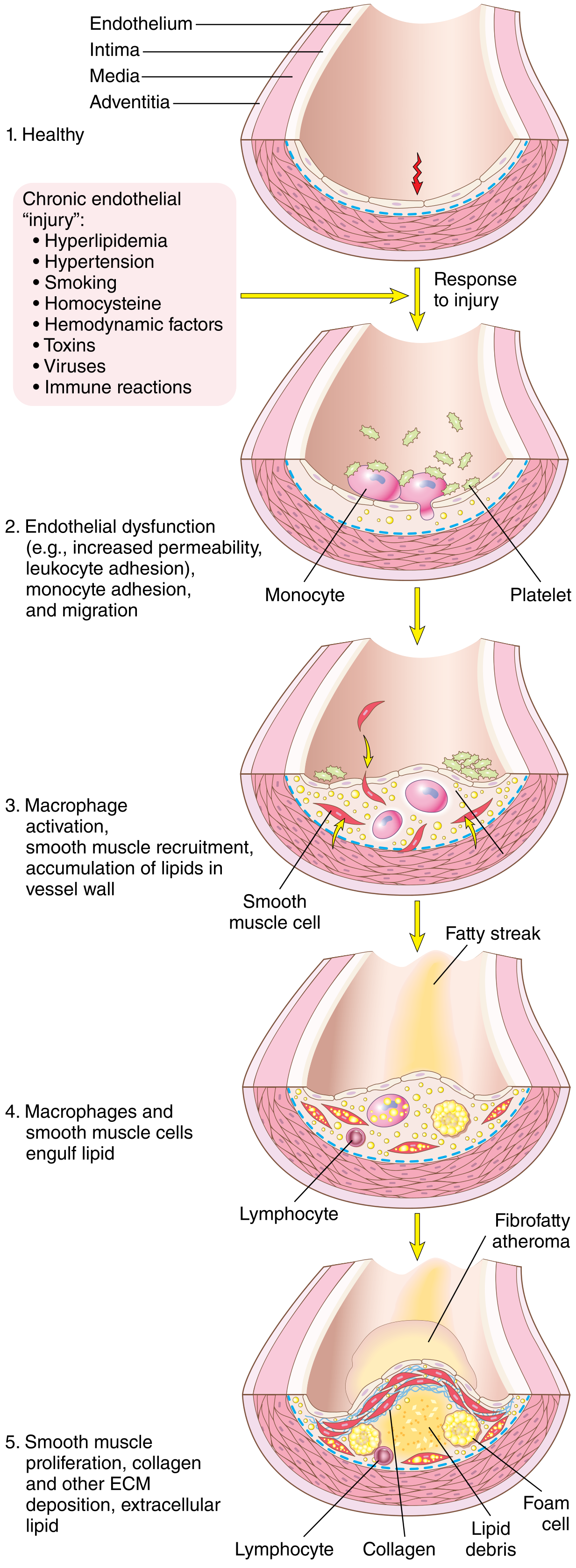
Step 1 - Endothelial Injury & Dysfunction
- Causes: hemodynamic turbulence (at branch points, ostia), hypercholesterolemia, smoking, hypertension, toxins, homocysteine
- Results in: increased permeability, upregulation of adhesion molecules (VCAM-1, ICAM-1), decreased nitric oxide production, altered gene expression
- Laminar flow normally induces the transcription factor KLF-2 which suppresses pro-inflammatory genes ("atheroprotective"); turbulent flow abolishes this effect
Step 2 - Lipoprotein Accumulation & Oxidation
- LDL enters the intima through the dysfunctional endothelium and is oxidized (ox-LDL)
- Oxidized LDL is pro-inflammatory: it stimulates endothelial cells to express more adhesion molecules and chemokines
- HDL has a protective role by reverse cholesterol transport
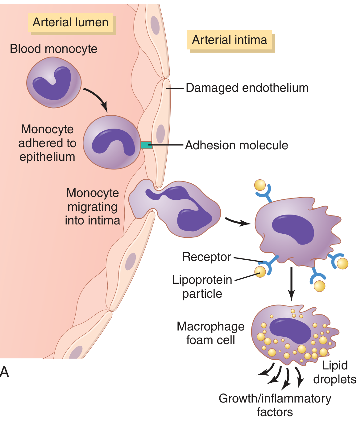
Step 3 - Monocyte Recruitment & Foam Cell Formation
- Monocytes adhere to the activated endothelium via adhesion molecules, then migrate into the intima
- In the intima, monocytes differentiate into macrophages, which ingest oxidized LDL via scavenger receptors (not downregulated like normal LDL receptors) → become foam cells (lipid-laden macrophages)
- Foam cells aggregate to form visible fatty streaks - the earliest grossly visible lesion (reversible, seen even in aortas of children)
Step 4 - Smooth Muscle Cell (SMC) Migration & Proliferation
- Activated macrophages, platelets, and endothelial cells release growth factors (PDGF, FGF, TGF-β) and cytokines
- SMCs migrate from the media into the intima, proliferate, and synthesize extracellular matrix (collagen, proteoglycans) → forming a neointima
- T lymphocytes also accumulate and sustain local inflammation
Step 5 - Advanced Plaque Formation
- Progressive accumulation of lipid, foam cells, SMCs, ECM, and T cells forms a full fibrous atheromatous plaque
- Central lipid core (necrotic debris from dead foam cells) is covered by a fibrous cap (collagen-rich, SMC-derived)
- Late stages: calcification of ECM and necrotic debris ("hardening of arteries")
Morphology of Atherosclerotic Plaques
Gross:
- Yellow-white raised intimal lesions
- Sites: aorta (esp. posterior abdominal), coronary arteries, popliteal arteries, carotid bifurcation, circle of Willis
Microscopic components:
- Fibrous cap - SMCs, collagen, proteoglycans, T cells
- Lipid core - cholesterol crystals, necrotic debris, foam cells, extracellular lipid
- Shoulder region - more cellular (macrophages, foam cells, T cells) - most vulnerable to rupture
- Neovascularization and calcification in advanced lesions
Consequences of Atherosclerosis
1. Atherosclerotic Stenosis
- Gradual plaque growth occludes the lumen
- Initially compensated by Glagov phenomenon (outward remodeling of the media)
- Critical stenosis occurs when the lumen cross-section is reduced by 70-75% - causes tissue ischemia and angina
- Result: chronic ischemic heart disease, peripheral vascular disease
2. Acute Plaque Change (Plaque Rupture / Erosion)
- The most dangerous complication
- Rupture of the fibrous cap (especially at the thin "shoulder" region) exposes the thrombogenic lipid core
- Triggers acute thrombosis → sudden occlusion → acute MI, stroke, sudden cardiac death
- Erosion (endothelial loss without rupture) can also trigger thrombosis
3. Aneurysm Formation
- Plaque thickens the intima, impeding diffusion from lumen to media
- Ischemia of the media weakens the arterial wall → aortic aneurysm (especially abdominal aorta)
4. Embolism
- Thrombus or atheromatous debris can embolize distally → embolic stroke, renal infarction, limb ischemia
Summary Table
| Feature | Details |
|---|---|
| Primary lesion | Atheroma (fibrous plaque) |
| Earliest lesion | Fatty streak (reversible) |
| Key cell | Macrophage foam cell |
| Central mechanism | Endothelial injury → lipid accumulation → inflammation → fibrosis |
| Key arteries affected | Aorta, coronary, carotid, popliteal |
| Main complications | MI, stroke, aneurysm, peripheral vascular disease |
| Critical stenosis threshold | 70-75% luminal cross-sectional area reduction |
Sources: Robbins & Kumar Basic Pathology (10e), p. 313-321; Robbins, Cotran & Kumar Pathologic Basis of Disease, p. 466-475; Guyton and Hall Textbook of Medical Physiology (14e), p. 851
This is a shared conversation. Sign in to Orris to start your own chat.