
Leprosy
Reading File
Now let me search the library for leprosy content in parallel.
Finding Sources
Searching PubMed
leprosy Hansen disease
Good hits. Now let me read the most comprehensive sources in parallel - Robbins Pathology and the Family Medicine textbook for broad clinical coverage.
Reading File
Reading File
Finding Sources
Excellent content from Robbins Pathology. Now let me read the Robbins morphology continuation and Harrison's leprosy section in parallel.
Reading File
Reading File
Reading File
Excellent content from Harrison's and Red Book. Let me now get the Harrison's treatment section and fetch the key pathology image.
Reading File
Loading Image
I now have comprehensive content. Let me compile the full answer.
Leprosy (Hansen's Disease)
Definition and Overview
Leprosy is a chronic infectious disease caused by Mycobacterium leprae (and, rarely, the more recently described M. lepromatosis, implicated mainly in Mexico and Central America). It primarily affects the skin, peripheral nerves, upper respiratory tract, and eyes. Despite being one of the oldest recorded diseases, it remains a neglected tropical disease - 174,087 new cases from 182 countries were reported in 2022, with the highest burden in India, Brazil, and parts of Africa.
- Harrison's Principles of Internal Medicine 22E, Ch. 184
- Robbins, Cotran & Kumar Pathologic Basis of Disease
Causative Organism
- Obligate intracellular, acid-fast, rod-shaped bacterium (1-8 µm long)
- Cannot be cultured in vitro - best grows in the footpads of mice or nine-banded armadillos
- Proliferates optimally at 27-34°C - explaining its predilection for cooler body surfaces (skin, peripheral nerves, testes, upper airways)
- Only bacterium known to infect Schwann cells of peripheral nerves - demonstration of AFB in peripheral nerves is pathognomonic
- Virulence is partly mediated by the cell wall lipid PGL-1, which facilitates host cell invasion
- Inhibits mitochondrial energy metabolism to evade host immune response
- Remains viable in the environment for up to 9 days
- Robbins Pathologic Basis of Disease, p. 356
Transmission and Epidemiology
- Transmission is primarily via respiratory secretions (nasal droplets) from untreated multibacillary patients
- Zoonotic transmission from 9-banded armadillos (Dasypus novemcinctus) occurs in the southern US; red squirrels in the British Isles can harbor both M. leprae and M. lepromatosis
- Not highly infectious - spouses of leprosy patients are at low risk, but biological relatives (parents, children, siblings) of untreated multibacillary patients have some increased risk, suggesting a genetic susceptibility component
- HIV co-infection does not increase risk of acquiring leprosy, but HIV treatment can precipitate immune reconstitution inflammatory syndrome (IRIS) in co-infected patients
- Incubation period: typically 3-10 years (range 6 months to 40 years)
- Red Book 2021, p. 761; Harrison's 22E
Immunopathogenesis and Disease Spectrum
The key determinant of clinical presentation is the host T-helper lymphocyte response to M. leprae. Disease exists on a continuous spectrum:
| Feature | Tuberculoid (TT) | Borderline (BB) | Lepromatous (LL) |
|---|---|---|---|
| Immune response | Strong Th1, IL-2, IFN-γ, Th17 | Intermediate | Weak Th1; Th2 predominance |
| Bacterial load | Very low (paucibacillary) | Intermediate | Very high (multibacillary) |
| Skin lesions | Few, well-defined, anaesthetic | Multiple, variable | Innumerable, symmetric |
| Nerve involvement | Asymmetric, severe | Moderate | Symmetric, late |
| Lepromin test | Strongly positive | Variable | Negative |
| Antibody production | Low | Moderate | High (but non-protective) |
In tuberculoid leprosy, IFN-γ mobilizes effective macrophage killing, granulomas form, and bacterial burden is low. In lepromatous leprosy, the poor Th1 response means bacteria replicate unchecked within macrophages (forming characteristic "lepra cells" stuffed with organisms).
In lepromatous leprosy, non-protective antibodies form immune complexes that can cause erythema nodosum, vasculitis, and glomerulonephritis.
- Robbins Pathologic Basis of Disease, p. 356-357
Histopathology
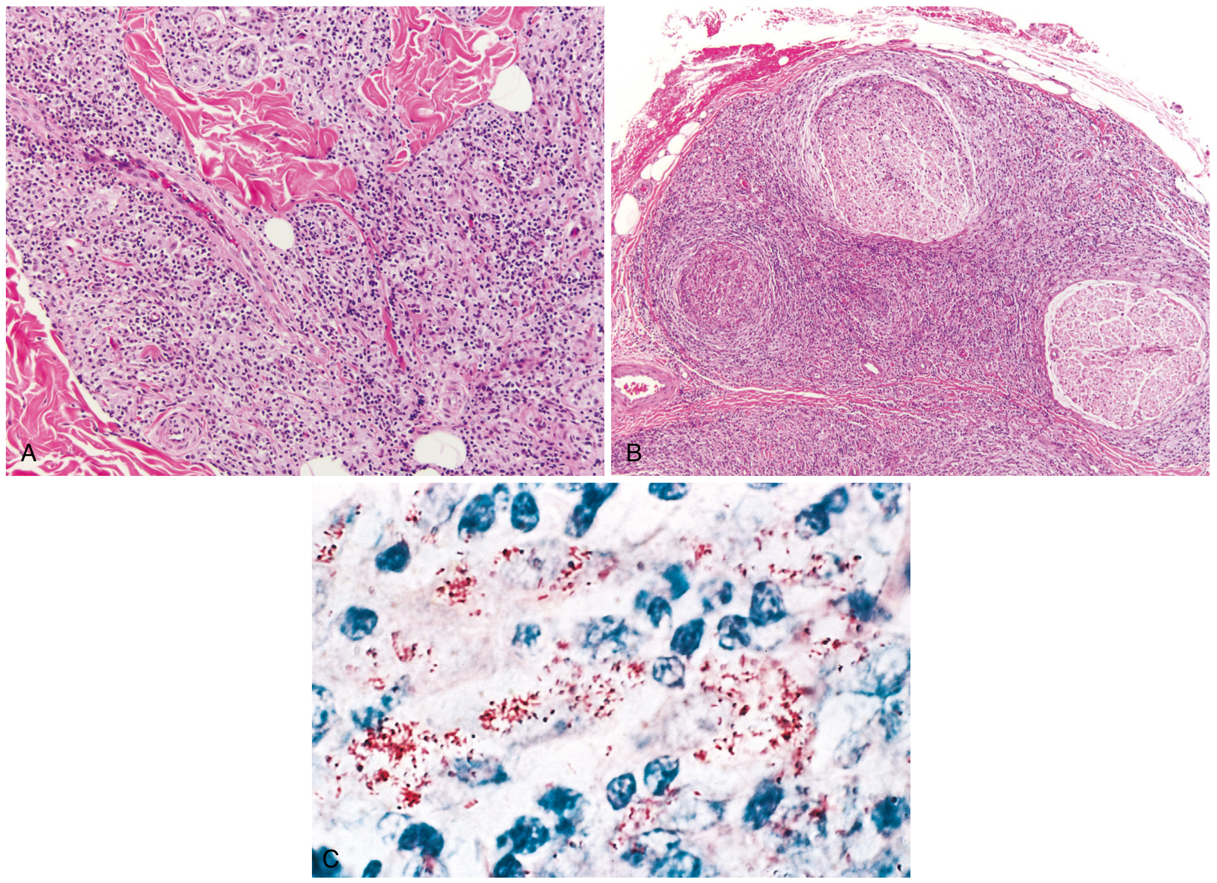
Fig. 8.33 Leprosy: (A) Tuberculoid leprosy - dense dermal macrophage infiltration surrounding adnexa, vessels, and nerves. (B) Lepromatous leprosy - dense lymphocytic and macrophage infiltration into large nerve bundles (mononeuropathy). (C) Acid-fast bacilli within macrophages in the lepromatous form (Fite stain). - Robbins Pathologic Basis of Disease
Tuberculoid leprosy:
- Well-formed epithelioid granulomas resembling tuberculosis
- Minimal or no bacteria visible (paucibacillary)
- Nerves enclosed within granulomatous reactions and destroyed if small
- Reflects strong T-cell immunity
Lepromatous leprosy:
- Large aggregates of lipid-laden macrophages ("lepra cells") filled with masses ("globi") of AFB
- No true granuloma formation - reflects weak cell-mediated immunity
- Multibacillary; bacteriologic index 4+ to 6+
- Stained best with Fite stain (not standard Ziehl-Neelsen)
- Robbins Pathologic Basis of Disease, p. 357
Clinical Features
Tuberculoid Leprosy (TT)
- Anesthetic plaque - focally hypopigmented skin lesion with loss of touch and pain sensation (hallmark)
- Few, well-defined lesions with indurated, elevated, hyperpigmented margins and depressed pale centers (central healing)
- Asymmetric peripheral nerve involvement - nerve trunks become enlarged and palpable
- Nerve degeneration leads to anesthesia, skin and muscle atrophy, susceptibility to trauma, chronic ulcers, contractures, paralyses, and autoamputation of digits
- Facial nerve involvement can cause lagophthalmos, keratitis, corneal ulceration
Lepromatous Leprosy (LL)
- Innumerable bilateral symmetrically distributed, erythematous, copper-colored patches, plaques, and nodules
- Spreads over face, earlobes, ears, extensor extremities, back, buttocks
- Progressive coarsening of skin folds leads to "leonine facies" (lion face)
- Bilateral earlobe thickening and loss of eyebrows (madarosis)
- Saddle-nose deformity from septal cartilage destruction
- Systemic involvement: lymph nodes, spleen, liver, bone marrow, testes (sterility, gynecomastia), eyes
- Eye complications: corneal anesthesia, episcleritis, iridocyclitis, iris atrophy, cataract, glaucoma, blindness
- Symmetric peripheral nerve involvement with sensory loss in "glove-and-stocking" distribution (late)
Rare Forms
- Histoid leprosy - waxy, shiny firm nodules on normal-looking skin; high bacteriologic index; spindle-cell granulomas on histology
- Lucio's phenomenon / Diffuse leprosy of Lucio and Latapi - nonnodular LL leprosy with diffuse shiny infiltration; occurs in Mexico and Central America; "lepra bonita" (beautiful leprosy)
- Primary neuritic leprosy - only peripheral nerve involvement, no skin lesions (2-10% of cases in India and Nepal)
- Harrison's 22E, Ch. 184
Leprosy Reactions (Acute Exacerbations)
These are immunologically mediated acute-on-chronic episodes; common in early treatment but can occur without therapy.
Type 1 - Reversal Reaction (LR-1)
- Seen in borderline tuberculoid and borderline lepromatous leprosy
- Due to sudden increase in effective cell-mediated immunity
- Acute tenderness, swelling, and erythema at existing skin and neural lesions; new lesions may appear
- Fever and systemic toxicity uncommon
- No PMNs in lesion on histology
- Treatment: corticosteroids
Type 2 - Erythema Nodosum Leprosum (ENL / LR-2)
- Occurs in borderline and lepromatous leprosy
- Systemic inflammatory reaction (immune complex mediated)
- Tender red subcutaneous papules/nodules resembling erythema nodosum
- High fever, migrating polyarthralgia, painful lymph node/spleen swelling, iridocyclitis, and rarely nephritis
- Treatment: thalidomide (drug of choice) or corticosteroids
- Red Book 2021, p. 761; Harrison's 22E
Diagnosis
| Method | Notes |
|---|---|
| Clinical | Skin patch + loss of sensation + thickened peripheral nerve = classic triad |
| Slit-skin smear | AFB seen in lepromatous; absent/rare in tuberculoid |
| Skin biopsy (Fite stain) | Gold standard for confirmation |
| Lepromin test (Mitsuda) | Positive in tuberculoid (strong immunity), negative in lepromatous |
| PCR | Useful where smear/biopsy inconclusive |
| Nerve biopsy | AFB in peripheral nerves = pathognomonic |
WHO Classification for Treatment
| Type | Criteria | Regimen |
|---|---|---|
| Paucibacillary (PB) | 1-5 skin lesions, smear-negative | 6-month MDT |
| Multibacillary (MB) | >5 lesions, smear-positive | 12-month MDT |
WHO Multidrug Therapy (MDT) - Standard Regimens
Paucibacillary (6 months):
- Rifampicin 600 mg once monthly (supervised)
- Dapsone 100 mg daily (self-administered)
Multibacillary (12 months):
- Rifampicin 600 mg once monthly (supervised)
- Clofazimine 300 mg once monthly + 50 mg daily (self-administered)
- Dapsone 100 mg daily (self-administered)
MDT cures the disease and prevents most chronic complications. The bacteriologic index falls approximately 1 log unit per year with MDT.
Complications and Disability
- Peripheral neuropathy leading to anesthesia - most serious long-term complication
- Repeated unrecognized trauma from insensate limbs leads to: ulcers, fractures, bone resorption, and autoamputation
- Lagophthalmos and corneal ulceration from facial nerve involvement
- Leprosy is a leading cause of permanent physical disability among communicable diseases worldwide
- Stigma and social exclusion remain major psychosocial burdens
Key Points Summary
- M. leprae - obligate intracellular, acid-fast, cannot be cultured in vitro, infects Schwann cells
- Disease spectrum determined by host Th1 immunity: strong = tuberculoid (paucibacillary); weak = lepromatous (multibacillary)
- Hallmark of tuberculoid: anesthetic hypopigmented skin patch
- Hallmark of lepromatous: leonine facies, madarosis, symmetric involvement, high AFB load
- Two reaction types: Type 1 (reversal) and Type 2 (ENL) - can occur during or without treatment
- Treatment: WHO MDT (rifampicin + dapsone ± clofazimine)
- BCG vaccination confers partial cross-protection against leprosy
Recent evidence note: A 2025 systematic review and meta-analysis (PMID 41082563) analyzed leprosy relapse after MDT - relevant for monitoring post-treatment outcomes. A 2024 systematic review on leprosy immunopathogenesis (PMID 39308868) provides updated insights consistent with the classical Th1/Th2 paradigm described above.
This is a shared conversation. Sign in to Orris to start your own chat.