
Reason the list for the sudden swellingon the face near eyes
periorbital swelling causes face near eyes
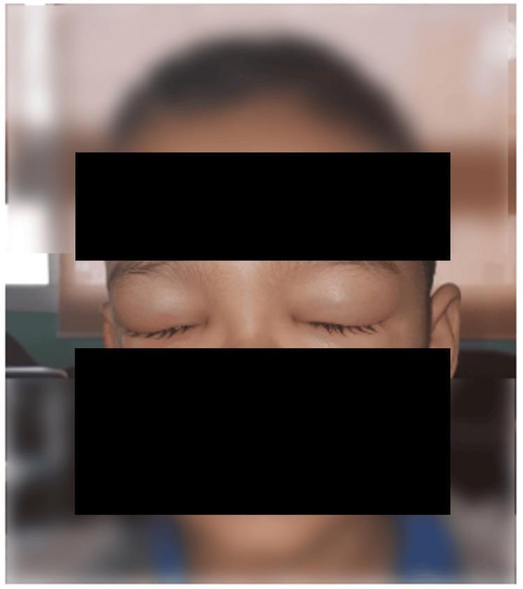
Clinical photograph of a pediatric patient's face, specifically focusing on the periorbital region. The image demonstrates significant bilateral eyelid edema, characterized by puffy and distended upper and lower eyelids. The swelling is severe enough to cause the involuntary closure of both eyes. The overlying skin appears tense and smooth due to the underlying fluid accumulation. This finding is presented in the context of Henoch-Schönlein purpura (HSP) and subsequent IgA nephropathy, illustrating systemic manifestations of small-vessel vasculitis. The educational focus is on identifying periorbital swelling as a clinical sign that may be associated with pediatric renal complications, such as proteinuria or hypoalbuminemia. Black bars are placed across the forehead and mid-face for patient privacy.
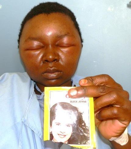
This clinical photograph displays a frontal view of a 29-year-old female patient with severe, generalized facial angioedema. The primary pathological finding is marked periorbital edema, resulting in complete involuntary closure of both eyes due to eyelid swelling. The edema extends across the forehead, malar regions, and lower face, causing a loss of normal anatomical contours and a tense, shiny appearance to the skin. The patient is holding a commercial hair dye packet labeled 'BLACK HENNA', identifying the likely allergen as paraphenylenediamine (PPD). This image illustrates a severe case of allergic contact dermatitis with secondary systemic manifestations following the use of topical hair dye. The clinical presentation is critical for differential diagnosis against orbital cellulitis or idiopathic angioedema. This visual serves as an educational tool for dermatology and emergency medicine regarding type IV hypersensitivity reactions and the risks associated with chemical additives in cosmetic products like PPD.
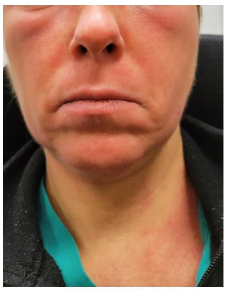
Clinical photograph of a patient's face and neck demonstrating significant cutaneous manifestations of a systemic inflammatory or allergic process. The image shows diffuse, confluent erythema (flushing) most prominent on the cheeks, bridge of the nose, and forehead. Notable periorbital edema is present, manifesting as puffiness and swelling around the eyes. The erythema extends inferiorly to the neck and upper chest area in a patchy, macular distribution. While the inflammatory process is diffuse, the perioral region appears relatively spared compared to the malar and periorbital areas. The skin surface exhibits a slight sheen, suggestive of underlying edema or acute inflammation. This presentation is clinically significant for the evaluation of Mast Cell Activation Syndrome (MCAS), chronic urticaria, angioedema, or autoimmune connective tissue diseases like dermatomyositis (heliotrope rash/malar rash). The image serves as a teaching aid for identifying facial flushing and soft tissue swelling in the context of systemic hypersensitivity or autonomic dysfunction.
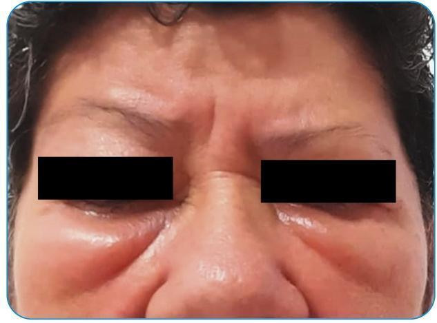
Clinical photograph of a 63-year-old female's mid and upper face, demonstrating significant bilateral periocular angioedema. The primary pathology is localized in the infraorbital and periorbital regions, presenting as pronounced soft-tissue swelling (edema) of the lower eyelids and malar areas. The affected skin appears taut, shiny, and erythematous, with visible skin wrinkling beneath the eyes indicative of acute fluid accumulation and tissue distension. The forehead and glabella regions exhibit horizontal and vertical rhytides (wrinkles), which appear accentuated by the surrounding facial edema. The eyes are partially obscured by black bars for patient privacy, yet the surrounding inflammatory reaction is clearly visible. This image serves as a clinical example of a localized Type I hypersensitivity-like reaction, specifically angioedema, involving the subcutaneous tissues and conjunctiva. Key educational concepts include the recognition of facial angioedema, distinguishing it from periorbital cellulitis, and understanding it as a potential, though rare, adverse reaction following immunization.
Causes of Sudden Periorbital (Peri-eye) Swelling
1. Allergic Reactions (Most Common Acute Cause)
- Triggers: Foods (nuts, shellfish, eggs), medications (NSAIDs, ACE inhibitors, penicillins), insect stings, latex, hair dyes (paraphenylenediamine/PPD)
- The swelling is typically soft, non-pitting, not tender, and resolves within hours to days
- May accompany urticaria, itching, or systemic anaphylaxis
2. Preseptal (Periorbital) Cellulitis - Infection
- Causes: Skin trauma (insect bites, lacerations), sinusitis (especially ethmoid), dacryocystitis, or spread from a stye/hordeolum
- Associated with fever, pain, redness, warmth
- Must be distinguished from the more dangerous orbital (postseptal) cellulitis, which also causes proptosis, restricted eye movement, and vision changes
- Spectrum of severity: Preseptal cellulitis → Orbital cellulitis → Subperiosteal abscess → Orbital abscess → Cavernous sinus thrombosis (most severe)
3. Insect Bite or Sting
4. Contact Dermatitis
- Allergic contact dermatitis: Type IV (delayed) hypersensitivity to cosmetics, eye drops, metals (nickel), nail polish (transferred by touching)
- Irritant contact dermatitis: Direct irritation from soaps, shampoos, topical medications
- Presents with itching, redness, scaling, and swelling
5. Nephrotic Syndrome (Systemic Cause)
"In the nephrotic patient, edema is often periorbital in the morning, whereas the face is not affected overnight in heart failure."
- Associated with foamy urine (proteinuria), generalized edema, hypoalbuminemia
- Causes include: glomerulonephritis, diabetic nephropathy, minimal change disease
6. Trauma / Injury
- Blunt facial trauma → rapid periorbital hematoma ("black eye") and edema
- Midfacial (Le Fort) fractures → periorbital bruising and swelling
- Septal hematoma after nasal trauma
7. Dermatomyositis
- Accompanied by proximal muscle weakness, Gottron's papules
- Considered when periorbital swelling has a purple hue
8. Thyroid Eye Disease (Graves' Orbitopathy)
- Autoimmune inflammation of orbital tissues
- Causes bilateral periorbital and eyelid swelling, proptosis (bulging eyes), lid retraction
- Associated with hyperthyroidism symptoms
9. Sinusitis Complications
10. Other Causes
| Cause | Key Feature |
|---|---|
| Stye (Hordeolum) | Localized, tender nodule on eyelid margin |
| Chalazion | Non-tender, chronic eyelid lump |
| Dacryocystitis | Swelling near inner corner of eye (lacrimal sac) |
| Superior vena cava (SVC) syndrome | Bilateral facial/neck swelling, venous distension |
| Hypothyroidism (myxedema) | Puffy, non-pitting facial edema |
| Melkersson-Rosenthal syndrome | Recurring facial edema, facial nerve palsy |
| Cluster headache | Unilateral periorbital swelling with severe pain, tearing |
| Rosacea | Chronic facial edema, especially ocular subtype |
Red Flag Features (Seek Urgent Care)
- Proptosis (eye pushing forward) - suggests orbital cellulitis or abscess
- Pain with eye movement - orbital involvement
- Restricted eye movement or double vision - orbital/cavernous sinus emergency
- Vision changes - urgent
- Systemic symptoms (high fever, altered consciousness) - sepsis risk
- Difficulty breathing - severe angioedema/anaphylaxis