
Mechanism of action of ashwagandha,and which pathway it act
ashwagandha Withania somnifera mechanism action
ashwagandha mechanism of action signaling pathways withanolides 2024
ashwagandha withanolides mechanism of action signaling pathways NF-kB
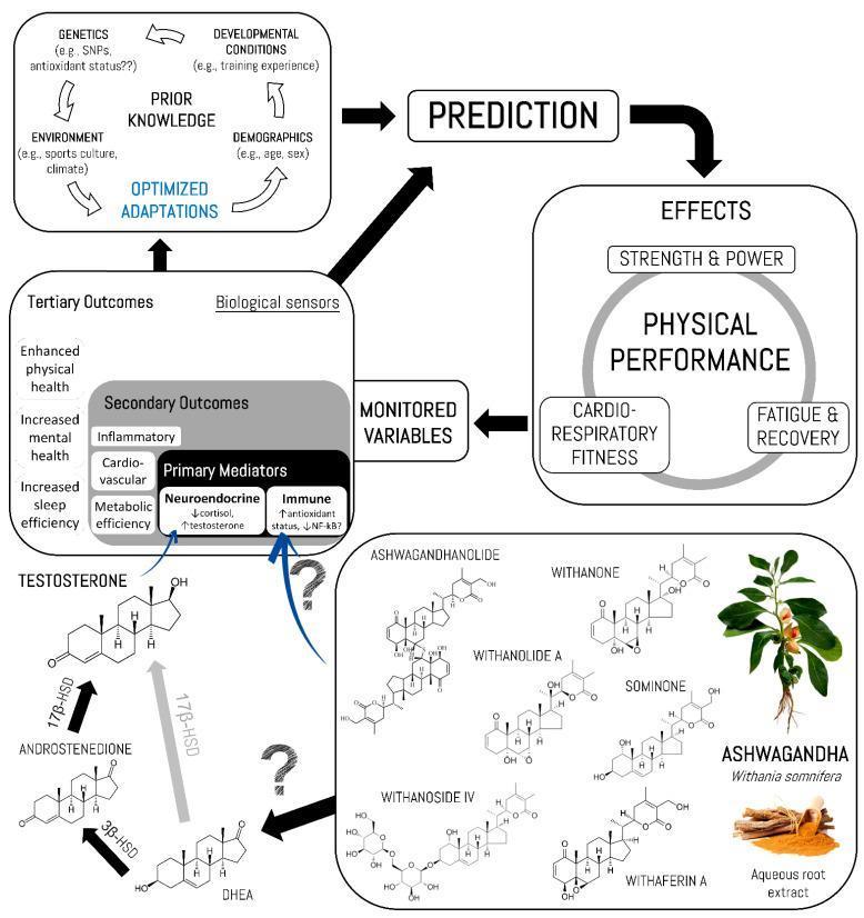
A comprehensive pathophysiology and pharmacology diagram illustrating the mechanisms by which Ashwagandha (Withania somnifera) influences human physical performance. The flowchart integrates predictive regulation models with biochemical pathways. At the top, 'Prior Knowledge' (comprising genetics, environment, developmental conditions, and demographics) leads to 'Optimized Adaptations,' which inform a 'Prediction' loop affecting 'Physical Performance' outcomes, including Strength & Power, Cardio-Respiratory Fitness, and Fatigue & Recovery. The biochemical section details the conversion of DHEA to Androstenedione via 3̢β-HSD, and the reversible conversion between Androstenedione and Testosterone via 17β-HSD. Ashwagandha components, specifically withanolides (Withaferin A, Withanoside IV, Withanolide A, Ashwagandhanolide, Withanone, and Sominone), are shown as primary mediators. These compounds influence neuroendocrine systems (decreasing cortisol, increasing testosterone) and immune responses (increasing antioxidant status, decreasing NF-κκB). These primary mediators cascade into secondary outcomes (inflammatory, cardiovascular, and metabolic efficiency) and tertiary outcomes (enhanced physical and mental health, sleep efficiency), which are monitored as biological sensors to provide feedback for predictive physical performance regulation.
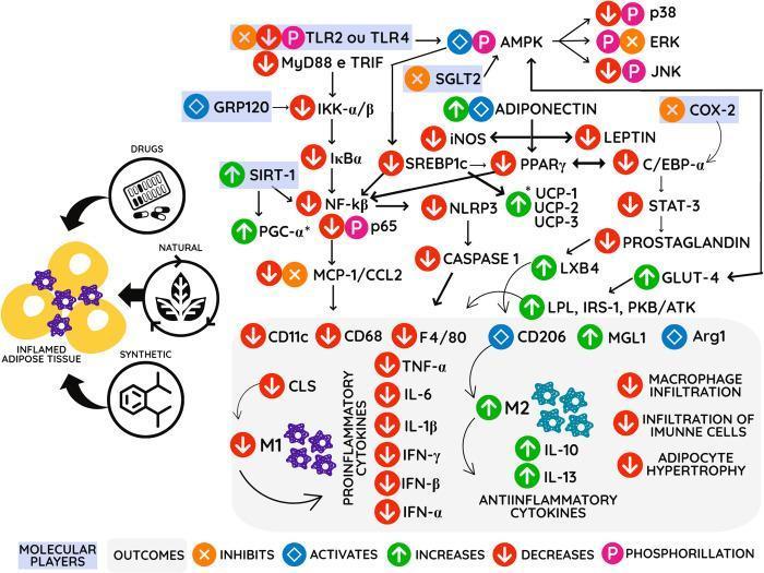
A complex pathophysiology diagram and signaling pathway illustrating the mechanism of action for anti-inflammatory agents (drugs, natural, and synthetic compounds) on inflamed adipose tissue. The visual maps a cascade of intracellular and extracellular molecular players within a hypothetical adipocyte and surrounding immune environment. Key signaling nodes include the inhibition of TLR2/4, MyD88, and TRIF, alongside the activation of AMPK, GRP120, and SIRT-1. The diagram details the downregulation of pro-inflammatory pathways, specifically the NF-kβ complex and its downstream chemokines like MCP-1/CCL2. It also highlights the modulation of metabolic regulators such as PPAR-γ, SREBP1c, and UCP-1/2/3. In the lower section, the diagram shows the transition from a pro-inflammatory M1 macrophage phenotype (characterized by TNF-α, IL-6, and IL-1β) toward an anti-inflammatory M2 phenotype (indicated by IL-10, IL-13, and Arg1). The overall clinical outcomes depicted include reduced macrophage infiltration, decreased adipocyte hypertrophy, and improved insulin sensitivity markers such as GLUT-4 and IRS-1. Symbol legends indicate activation, inhibition, and phosphorylation states for each molecular player.
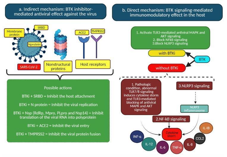
A medical infographic and signaling pathway diagram illustrating the dual mechanisms of Bruton’s tyrosine kinase inhibitors (BTKi) in the treatment of SARS-CoV-2. Section (a) details the 'Indirect mechanism' focused on antiviral effects. It depicts a SARS-CoV-2 virion with structural components (Membrane protein, SRBD, N protein) and nonstructural proteins (Nsp). It also shows host cell receptors ACE2 and TMPRSS2. A summary table outlines how BTKi may inhibit host attachment, viral replication, RNA translation, viral entry, and protein fusion by targeting these specific viral and host components. Section (b) illustrates the 'Direct mechanism' focusing on immunomodulatory effects via BTK signaling. It compares two states: 'with BTKi' and 'without BTKi'. Without inhibition, a pathologic state involves abnormal TLR7/8 and TLR3 signaling, leading to NF-kB and NLRP3 activation, resulting in a 'cytokine storm' (TNF-a, IL-6, IL-12, etc.). With BTKi, the pathway shifts to activate TLR3-mediated antiviral MAPK/AKT signaling while blocking NF-kB and NLRP3 pro-inflammatory pathways. This educational content demonstrates pharmacological strategies for drug repurposing in infectious disease and immunology.
https://pmc.ncbi.nlm.nih.gov/articles/PMC12423730
Ashwagandha (Withania somnifera) - Mechanism of Action
Active Compounds
| Compound Class | Key Members | Primary Role |
|---|---|---|
| Withanolides (steroidal lactones) | Withaferin A (WA), Withanolide A, Withanone, Withanoside IV, Sominone | Anti-inflammatory, anticancer, adaptogenic |
| Alkaloids | Isopelletierine, Anaferine | CNS modulation |
| Sitoindosides | Sitoindoside VII, VIII | Immunomodulation, neuroprotection |
| Saponins | Multiple glycosides | Antioxidant, adaptogenic |
Key Pathways
1. NF-kB Pathway (Anti-inflammatory, Anticancer)
- Withanolides inhibit NF-kB (Nuclear Factor kappa B) activation
- This suppresses downstream pro-inflammatory cytokines: TNF-alpha, IL-6, IL-1-beta
- Simultaneously upregulates IL-10 (anti-inflammatory)
- Mechanism: blocks protein kinase signaling cascades upstream of NF-kB; also blocks Hsp90-Cdc37 co-chaperone interaction (a critical step for kinase activation)
- Clinical relevance: reduces chronic inflammation, joint inflammation (rheumatoid arthritis), and tumor growth
2. HPA Axis (Adaptogenic / Stress Response)
- Modulates the hypothalamic-pituitary-adrenal (HPA) axis
- Reduces circulating cortisol levels - confirmed by systematic review (PMID: 38140274)
- Reduces DHEA-to-androstenedione conversion (via 3-beta-HSD) while supporting testosterone levels
- Net effect: blunts chronic stress response without suppressing acute adaptive stress reactivity
3. GABAergic Pathway (Anxiolytic / Sedative)
- Acts as a GABA-A receptor agonist (direct or indirect)
- Enhances GABAergic signaling in the CNS
- This is the primary basis for ashwagandha's anxiolytic and sedative effects
- Also partly explains its use in generalized anxiety disorder (GAD) - referenced in Kaplan & Sadock's Comprehensive Textbook of Psychiatry
4. PI3K/Akt/mTOR Pathway (Anticancer, Metabolic)
- Withanolides target Akt and its downstream effectors
- Inhibits the PI3K/Akt/mTOR axis, leading to:
- Reduced tumor cell proliferation
- Induction of apoptosis
- Improved insulin sensitivity and metabolic regulation
- Also inactivates via thiol-mediated oxidative stress in cancer cells
5. Nrf2 Pathway (Antioxidant)
- Activates Nrf2 (Nuclear factor erythroid 2-related factor 2)
- Nrf2 upregulates antioxidant response element (ARE) genes
- Results in increased levels of endogenous antioxidant enzymes (SOD, catalase, glutathione peroxidase)
- Reduces reactive oxygen species (ROS) and lipid peroxidation
- This protects neural integrity and reduces oxidative stress in the CNS
6. MAPK Pathway (Inflammation, Stress Response)
- Modulates mitogen-activated protein kinase (MAPK) signaling
- Works in coordination with NF-kB inhibition
- Regulates cell survival, differentiation, and inflammatory gene expression
7. JAK/STAT Pathway (Immunomodulation)
- Withanolides inhibit JAK/STAT signaling
- This pathway is involved in cytokine receptor signaling
- Results in immunomodulation - enhances NK cells and macrophage activity while dampening pathological inflammation
8. PPAR-gamma Pathway (Metabolic)
- Activates PPARgamma (Peroxisome proliferator-activated receptor gamma)
- Relevant to insulin sensitivity, adipogenesis, and anti-inflammatory effects
Neuroprotective Mechanisms (Summary)
| Action | Target | Effect |
|---|---|---|
| Antioxidant | Nrf2/ROS | Protects neurons |
| Anti-inflammatory | NF-kB, TNF-alpha | Reduces neuroinflammation |
| Neurogenesis support | BDNF upregulation | Promotes brain plasticity |
| Stress axis control | HPA axis / cortisol | Reduces chronic stress damage |
| Receptor modulation | GABA-A, NMDA | Anxiolytic, cognitive benefits |
Diagram - Ashwagandha Mechanism of Action
Summary Table
| Effect | Primary Pathway | Key Compound |
|---|---|---|
| Anti-stress / adaptogenic | HPA axis, cortisol reduction | Withanolides, sitoindosides |
| Anxiolytic / sedative | GABAergic (GABA-A) | Alkaloids, withanolides |
| Anti-inflammatory | NF-kB inhibition, MAPK | Withaferin A |
| Antioxidant | Nrf2 activation | Withanolides |
| Anticancer | PI3K/Akt/mTOR, NF-kB, JAK/STAT | Withaferin A |
| Immunomodulation | JAK/STAT, NK cell/macrophage activation | Sitoindosides |
| Cognitive/neuroprotective | BDNF, Nrf2, NMDA modulation | Withanolide A, Withanone |
| Metabolic | PPARgamma, PI3K/Akt | Withanolides |
Curcumin MOA and which pathway it act on to show anti-inflammatory action
curcumin anti-inflammatory mechanism NF-kB
curcumin mechanism of action anti-inflammatory signaling pathways NF-kB COX-2 2024
curcumin anti-inflammatory NF-kB signaling pathway mechanism diagram
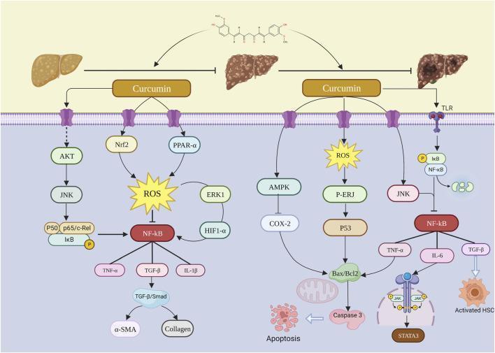
A pathophysiology diagram illustrating the molecular mechanisms of curcumin in inhibiting the progression of Non-Alcoholic Fatty Liver Disease (NAFLD) to Liver Fibrosis (LF) and Hepatocellular Carcinoma (HCC). The top section shows three liver states: fatty liver, fibrotic liver, and cancerous liver, with arrows representing disease progression and inhibitory T-bars indicating curcumin's intervention. Below, a cellular cross-section details curcumin's interaction with transmembrane receptors and intracellular signaling pathways. Key regulatory cascades shown include the inhibition of NF-̄kB and its downstream inflammatory cytokines (TNF-́, IL-6, IL-1̂), and the TGF-̂/Smad pathway leading to ́-SMA and collagen production in activated Hepatic Stellate Cells (HSC). On the right, pathways involving ROS, AMPK, p53, and Bax/Bcl2 demonstrate the modulation of oxidative stress and the induction of apoptosis via Caspase 3. Additional signaled molecules include AKT, JNK, Nrf2, PPAR-́, and STAT3, collectively highlighting curcumin's multifaceted role in anti-inflammatory, anti-fibrotic, and anti-tumorigenic processes within hepatocytes.
A complex pathophysiology diagram and signaling pathway illustrating the mechanism of action for anti-inflammatory agents (drugs, natural, and synthetic compounds) on inflamed adipose tissue. The visual maps a cascade of intracellular and extracellular molecular players within a hypothetical adipocyte and surrounding immune environment. Key signaling nodes include the inhibition of TLR2/4, MyD88, and TRIF, alongside the activation of AMPK, GRP120, and SIRT-1. The diagram details the downregulation of pro-inflammatory pathways, specifically the NF-kβ complex and its downstream chemokines like MCP-1/CCL2. It also highlights the modulation of metabolic regulators such as PPAR-γ, SREBP1c, and UCP-1/2/3. In the lower section, the diagram shows the transition from a pro-inflammatory M1 macrophage phenotype (characterized by TNF-α, IL-6, and IL-1β) toward an anti-inflammatory M2 phenotype (indicated by IL-10, IL-13, and Arg1). The overall clinical outcomes depicted include reduced macrophage infiltration, decreased adipocyte hypertrophy, and improved insulin sensitivity markers such as GLUT-4 and IRS-1. Symbol legends indicate activation, inhibition, and phosphorylation states for each molecular player.
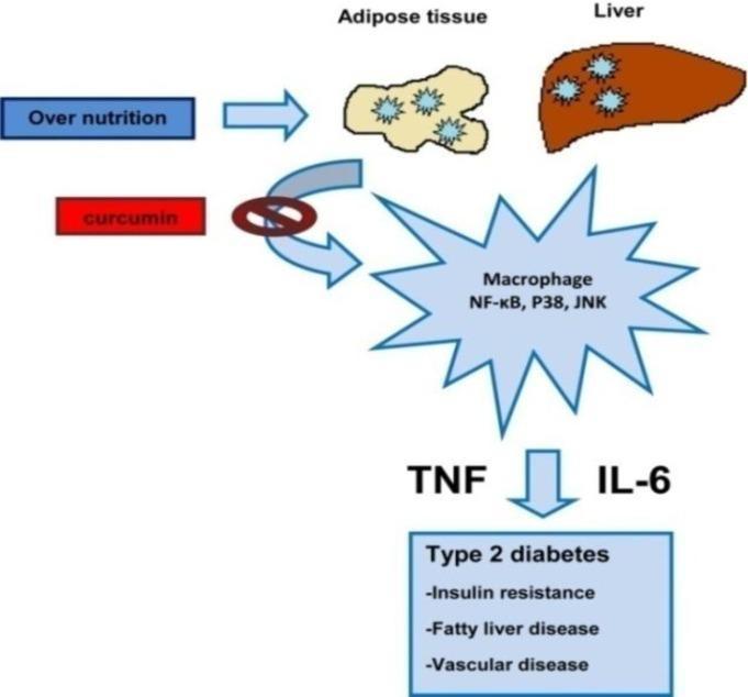
This pathophysiology diagram illustrates the inflammatory signaling pathway linking over-nutrition to metabolic disorders. The process begins with 'Over nutrition' (blue box), which acts upon adipose tissue (yellow lobular illustration) and the liver (brown anatomical representation). These tissues trigger macrophage activation, characterized by the signaling molecules NF-kB, P38, and JNK. The activated macrophages release pro-inflammatory cytokines TNF and IL-6. This inflammatory cascade leads to a clinical outcome box labeled 'Type 2 diabetes,' encompassing insulin resistance, fatty liver disease, and vascular disease. Additionally, the diagram features a therapeutic intervention point where 'curcumin' (red box) is shown with an inhibitory symbol (crossed circle) blocking the signaling pathway from the tissues to the macrophages. This content is relevant for endocrinology and metabolic health education, specifically demonstrating how nutritional excess promotes chronic systemic inflammation and secondary chronic diseases.
https://www.frontiersin.org/journals/pharmacology/articles/1…
Curcumin - Mechanism of Action & Anti-Inflammatory Pathways
What is Curcumin?
- Two aromatic rings (each with hydroxyl and methoxy groups)
- A seven-carbon linker with double bonds and a central ketone (beta-diketone moiety)
Core Anti-Inflammatory Pathways
1. NF-kB Pathway (PRIMARY - most important)
TLR stimulus → MyD88 → IRAK1/4 → TRAF6 → TAK1 → IKK complex → IkB phosphorylation → IkB degradation (26S proteasome) → NF-kB nuclear translocation → pro-inflammatory gene transcription (TNF-alpha, IL-1beta, IL-6, COX-2, iNOS)
- Inhibits TLR4 homodimerization (upstream block - prevents initial signal)
- Suppresses MyD88 adaptor protein interaction
- Inhibits IRAK1 and IRAK4 kinases
- Blocks TAK1-mediated IKK activation
- Directly inhibits IKK-beta (possibly by occupying its ATP binding site)
- Prevents IkB phosphorylation - so IkB stays bound to NF-kB, keeping it inactive in the cytoplasm
- Blocks p65 nuclear translocation
- Upregulates miR-146a, a microRNA that further suppresses NF-kB signaling
2. MAPK Pathway (Three Branches)
| Branch | Role | Curcumin Effect |
|---|---|---|
| ERK (Extracellular signal-regulated kinase) | Cell proliferation, survival | Inhibits ERK phosphorylation |
| JNK (c-Jun N-terminal kinase) | Stress response, apoptosis | Inhibits JNK activation |
| p38 MAPK | Inflammatory cytokine production | Inhibits p38 phosphorylation |
- Pro-inflammatory gene expression
- Cell proliferation in inflammation
- Cytokine storm amplification
3. JAK-STAT Pathway
- Curcumin reduces JAK1/2 phosphorylation
- Inhibits STAT1 and STAT3 phosphorylation
- STAT3 is particularly relevant - it drives chronic inflammation and promotes tumor survival
- By blocking JAK-STAT: reduces IFN-gamma-mediated inflammation, IL-6 signaling, and oncogenic signaling in cancer-associated inflammation
4. NLRP3 Inflammasome Inhibition
- The NLRP3 inflammasome activates caspase-1, which cleaves pro-IL-1beta into active IL-1beta
- Curcumin directly inhibits NLRP3 assembly and activation
- This blocks the downstream release of IL-1beta and IL-18 (key drivers of acute and chronic inflammation)
- Relevant in gout, metabolic syndrome, atherosclerosis, and neuroinflammation
5. COX-2 and LOX Pathway (Arachidonic Acid Cascade)
- Curcumin directly inhibits COX-2 (cyclooxygenase-2) expression - both by blocking NF-kB-driven COX-2 transcription AND by direct enzyme inhibition
- Inhibits 5-LOX (5-lipoxygenase) - reduces leukotriene production (LTB4, LTC4)
- Inhibits iNOS (inducible nitric oxide synthase) - reduces NO-mediated tissue damage
6. Nrf2/ARE Pathway (Antioxidant - Supports Anti-inflammation)
- Curcumin activates Nrf2 (Nuclear factor erythroid 2-related factor 2)
- Nrf2 binds the ARE (Antioxidant Response Element) in gene promoters
- Induces: HO-1 (heme oxygenase-1), NQO-1, GSH (glutathione), SOD, catalase
- Reduces ROS → indirectly reduces oxidative activation of NF-kB
- HO-1 itself has direct anti-inflammatory effects by generating CO and biliverdin
7. AP-1 Pathway
- AP-1 (Activator Protein-1) is a transcription factor complex (c-Fos/c-Jun) that drives inflammatory and pro-apoptotic gene expression
- Curcumin downregulates AP-1 activity
- This reduces expression of anti-apoptotic proteins and contributes to its anti-cancer and anti-inflammatory effects
8. TGF-beta / Smad Pathway (Fibrosis)
- In chronic inflammation leading to fibrosis, TGF-beta activates Smad proteins
- Curcumin inhibits TGF-beta/Smad signaling
- Reduces alpha-SMA and collagen production in activated fibroblasts/stellate cells
- Clinically relevant in liver fibrosis, IBD, and pulmonary fibrosis
Visual: Curcumin Signaling Pathways
Summary Table: All Anti-Inflammatory Pathways
| Pathway | Curcumin Action | Downstream Effect |
|---|---|---|
| NF-kB | Blocks IKK, IkB phosphorylation, p65 translocation | ↓ TNF-alpha, IL-1beta, IL-6, COX-2 |
| MAPK (ERK/JNK/p38) | Inhibits all three branches | ↓ Cytokines, cell proliferation |
| JAK-STAT | Inhibits JAK1/2, STAT1/3 phosphorylation | ↓ IFN-gamma signaling, IL-6 cascade |
| NLRP3 Inflammasome | Blocks NLRP3 assembly | ↓ Caspase-1, IL-1beta, IL-18 |
| COX-2 / LOX | Direct enzyme inhibition + transcriptional repression | ↓ Prostaglandins, leukotrienes |
| iNOS | Transcriptional suppression | ↓ Nitric oxide-mediated tissue damage |
| Nrf2/ARE | Activates Nrf2 | ↑ HO-1, GSH, NQO-1; ↓ ROS |
| AP-1 | Downregulates AP-1 | ↓ Inflammatory + anti-apoptotic genes |
| TGF-beta/Smad | Inhibits Smad phosphorylation | ↓ Fibrosis, collagen deposition |
Key Textbook Reference
Why Curcumin Has Poor Bioavailability (and What's Done About It)
- Poor aqueous solubility
- Rapid metabolism and conjugation
- Fast elimination