
Describe etiology, clinical features and management of Sub-Conjunctival hemmorage 5 marks
Reading File
Now let me search the medical library for subconjunctival hemorrhage content.
Finding Sources
Finding Sources
Good results. Let me read the most relevant sections from Wills Eye Manual and Rosen's Emergency Medicine.
Reading File
Reading File
Let me read the management section from Wills Eye Manual and also get the Tintinalli content.
Reading File
Reading File
I now have comprehensive content. Let me fetch the clinical image for a visual reference.
Loading Image
I now have all the information needed for a complete 5-mark answer. Here is the full structured response:
Subconjunctival Hemorrhage
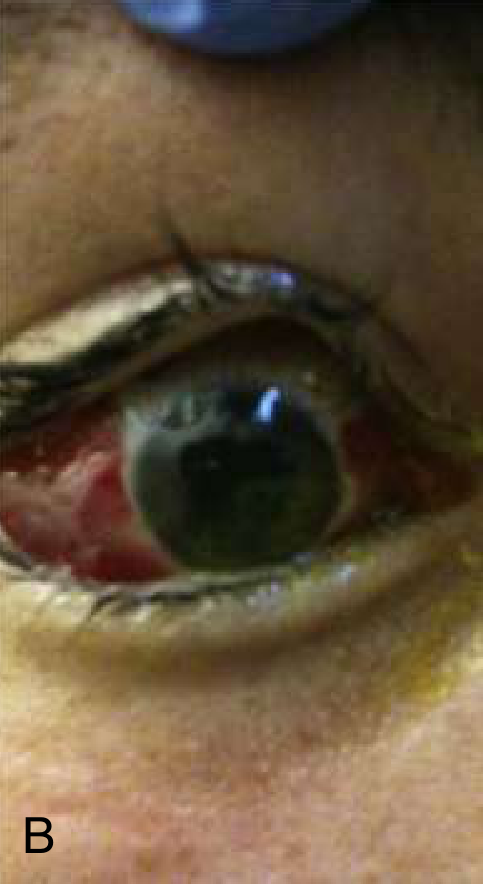
Fig. Subconjunctival hemorrhage. (Rosen's Emergency Medicine)
Definition
Subconjunctival hemorrhage (SCH) is bleeding beneath the bulbar conjunctiva due to rupture of small subconjunctival blood vessels, resulting in a bright red patch on the white of the eye.
Etiology
Causes are grouped as follows:
1. Valsalva/Increased Venous Pressure
- Forceful coughing, sneezing, vomiting
- Bearing down (constipation, childbirth, heavy lifting)
- Vigorous eye rubbing
2. Traumatic
- Blunt or penetrating eye injury - the most common cause in emergency settings
- May be isolated or associated with retrobulbar hemorrhage or ruptured globe
- Rule out globe perforation when there is 360-degree involvement, chemosis, pain, or reduced vision
3. Systemic Conditions
- Hypertension (check BP in all patients)
- Diabetes mellitus
- Bleeding disorders (thrombocytopenia, coagulopathy, leukemia)
4. Medications
- Antiplatelet agents: aspirin, clopidogrel, ticagrelor
- Anticoagulants: warfarin, dabigatran, rivaroxaban, apixaban, edoxaban
- Topical steroids (prolonged use)
5. Others
- Orbital mass (rare - hemorrhage due to underlying lesion)
- Idiopathic - most spontaneous cases have no identifiable cause
Clinical Features
Symptoms:
- Usually asymptomatic - patient typically notices a red eye while looking in the mirror
- Mild diffuse foreign body sensation may occur depending on size and location
- No change in visual acuity (if vision is affected, suspect associated injury)
- No pain in spontaneous cases
Signs:
- Bright red, well-demarcated patch of blood beneath the bulbar conjunctiva
- Blood appears smooth and flat; no vessels visible behind the hemorrhage (distinguishes it from conjunctivitis)
- May be localized to one sector or involve the entire visible sclera
- Overlying conjunctiva is minimally and smoothly raised
- No discharge, no corneal involvement
Warning signs suggesting serious underlying injury (especially in trauma):
- 360-degree involvement with chemosis
- Pain or decreased vision
- Abnormally deep or shallow anterior chamber
- Hyphema or uveal prolapse - raise suspicion for globe rupture
Management
Workup
- History: Trauma, medications, bleeding history, Valsalva events, recurrent episodes, cough
- Blood pressure measurement in all patients
- Ocular examination: Slit-lamp exam; check visual acuity, IOP, extraocular movements; slit-lamp with fluorescein to rule out corneal abrasion, globe perforation (Seidel's test)
- Lab investigations (if recurrent or bleeding history): PT, aPTT, CBC with peripheral smear, liver function tests, protein C and S
- Imaging (CT/MRI orbits with contrast): only if orbital signs are present (proptosis, restricted motility, elevated IOP) to rule out orbital mass
Treatment
- No treatment required for uncomplicated spontaneous SCH
- Artificial tears (lubricant drops q.i.d.) if mild irritation is present
- Discourage elective use of aspirin and NSAIDs - do not stop prescribed anticoagulants without clearance from the primary physician
- Treat underlying cause (control hypertension, manage systemic illness)
Follow-Up
- Resolves spontaneously within 2 to 4 weeks as blood reabsorbs; may turn yellow-green as it resolves (similar to a bruise)
- Advise patient to return if blood does not fully resolve or recurrence occurs
- Refer to internist/GP if hypertension or bleeding diathesis is suspected
Sources: The Wills Eye Manual, 7th ed. (Section 5.5); Rosen's Emergency Medicine, 9th ed. (Chapter 57)
This is a shared conversation. Sign in to Orris to start your own chat.