
Tb
Reading File
The query "Tb" is quite brief. Before I dive into a full search, let me clarify what you need:
Asking for Preferences
What aspect of Tuberculosis (TB) are you asking about?
Searching sources...
Finding Sources
Finding Sources
Good - I have the best sources. Now let me read the most comprehensive sections from Harrison's (gold standard reference) and Murray & Nadel's (respiratory textbook).
Reading File
Reading File
Finding Sources
Reading File
Reading File
Reading File
Reading File
Finding Sources
Finding Sources
Reading File
Excellent - I now have rich content from both Harrison's and Murray & Nadel's. Let me fetch the key chest X-ray images and compile the full answer.
Loading Image
Loading Image
I now have comprehensive material. Here is a detailed, well-structured answer on the diagnosis and investigations of TB.
Tuberculosis (TB) - Diagnosis & Investigations
1. Clinical Suspicion - When to Investigate
Clinical suspicion is the first step. Key symptoms include:
- Cough - persistent, productive, often with mucus or blood (note: 10-25% of bacteriologically confirmed TB patients do not report cough)
- Systemic symptoms - fever, night sweats, weight loss
- Hemoptysis - seen with more extensive disease
- Lymphadenopathy - especially in HIV-positive patients
Risk factors that heighten suspicion include: HIV infection, recent exposure to a known case, immunosuppression, residence in high-incidence areas, and certain occupational exposures. Using a broader symptom screen (any one of: cough of any duration, fever, night sweats, or weight loss) in high-risk groups gives a negative predictive value of ~97.7% for TB.
- Murray & Nadel's Textbook of Respiratory Medicine, p. 1187-1188
2. Chest Radiograph
The CXR is usually the first investigation and is highly informative, though not diagnostic on its own.
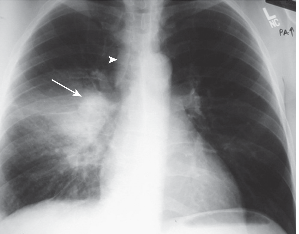
Primary TB:
- Middle or lower lung zone consolidation with ipsilateral hilar/paratracheal lymphadenopathy
- Atelectasis from airway compression
- May show pleural effusion
Primary TB (right lower lobe consolidation + right hilar lymphadenopathy):
Reactivation (Post-primary) TB:
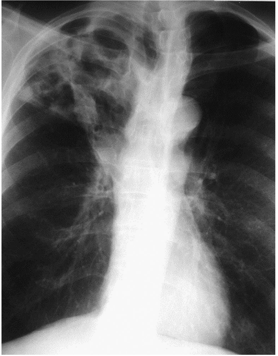
- Upper lobe involvement (apical and posterior segments of right upper lobe; apical-posterior segment of left upper lobe)
- Cavitation is common
- Fibrotic scarring with lung shrinkage and calcification on healing
- Bronchogenic spread can cause lower lobe opacities
- Miliary pattern if hematogenous dissemination occurs
Cavitary TB (right upper lobe):
In HIV:
- Early HIV - typical upper lobe pattern
- Advanced HIV (low CD4) - atypical pattern: lower zone / diffuse opacities, intrathoracic adenopathy, less cavitation
Limitation: CXR cannot determine activity from a single film alone, and cannot give a definitive diagnosis. Microbiologic evaluation is always needed.
- Murray & Nadel's Textbook of Respiratory Medicine, p. 1188-1189
3. Microbiological Tests
A. AFB Smear Microscopy (Ziehl-Neelsen / fluorescence stain)
- Collect at least 2 sputum specimens (US: 3 specimens are routine)
- Specimens include: early morning sputum, spot sputum, induced sputum, BAL (if unable to produce)
- Sensitivity: up to 90% with 3 specimens; however, often lower in paucibacillary disease
- AFB smear cannot distinguish M. tuberculosis from non-tuberculous mycobacteria (NTM)
- Remains the most widely available test globally
B. Mycobacterial Culture
-
More sensitive than smear (~100% when adequate sputum is provided)
-
Results take 2-8 weeks (liquid culture faster than solid media)
-
Definitive identification of M. tuberculosis complex
-
Also allows drug susceptibility testing (DST)
-
Requires specialized lab infrastructure; often unavailable in resource-limited settings
-
Textbook of Family Medicine, 9e, p. 763
C. Nucleic Acid Amplification Testing (NAAT) - Xpert MTB/RIF
This is the most significant advance in rapid TB diagnosis:
-
Xpert MTB/RIF (GeneXpert): Self-contained, automated, results in ~90 minutes
-
Simultaneously detects M. tuberculosis complex AND rifampicin resistance (as a proxy for MDR-TB)
-
Distinguishes M. tuberculosis from NTM (unlike smear)
-
Interpretation:
- AFB smear-positive + NAAT-positive = rapid confirmation of TB
- AFB smear-negative + NAAT-positive = presumptive evidence in intermediate/high-probability cases
- NAAT-negative does NOT exclude active TB (insufficient sensitivity to replace culture)
-
WHO recommendation: Preferred initial test for all adults/children with suspected TB where feasible, especially for HIV-positive patients and those at risk of MDR-TB
-
US recommendation: Use as adjunct to smear + culture
-
Murray & Nadel's Textbook of Respiratory Medicine, p. 1189-1193
D. Line Probe Assays (LPAs)
- Molecular test detecting resistance to isoniazid, rifampin, fluoroquinolones, and injectables
- Useful for rapid MDR-TB and XDR-TB screening
- Applied to culture isolates or directly to smear-positive specimens
4. Tests for TB Infection (Latent TB)
These tests measure the host immune response and cannot distinguish latent from active TB. They are used for screening/infection detection.
A. Tuberculin Skin Test (TST / Mantoux)
- Intradermal injection of PPD (purified protein derivative)
- Read at 48-72 hours; induration measured in mm
- Cutoffs:
- ≥5 mm: HIV-infected, close contacts, immunosuppressed, CXR with fibrotic changes
- ≥10 mm: Recent immigrants, IV drug users, high-risk occupational groups, children <4 yrs
- ≥15 mm: No known risk factors
- Limitations:
- False-negative: immunosuppression, overwhelming TB, very early infection
- False-positive: prior BCG vaccination, NTM infection
- Low specificity due to cross-reactivity with conserved mycobacterial proteins
- Operator-dependent; batch variations
B. Interferon-Gamma Release Assays (IGRAs)
- Measure T-cell IFN-γ release in response to TB-specific antigens ESAT-6 and CFP-10 (RD1-encoded, absent from BCG and most NTMs)
- Types:
- QuantiFERON-TB Gold Plus (QFT-Plus): Whole-blood ELISA; measures both CD4+ and CD8+ T-cell responses
- T-SPOT.TB (Oxford Immunotec): ELISPOT assay
- Advantages over TST: Higher specificity; not affected by BCG vaccination; no booster effect; single visit
- Limitations: False-negatives in immunosuppressed; higher cost; need lab processing
C. New Antigen-Based Skin Tests (TBSTs)
-
Use ESAT-6 and CFP-10 antigens (same as IGRAs) but in skin-test format
-
WHO assessment: accuracy similar to IGRAs and greater than TST
-
Useful also for HIV-positive individuals, children, and BCG-vaccinated persons
-
Harrison's Principles of Internal Medicine 22E (2025), p. 1437
5. Non-Sputum-Based Tests
Urine LAM (Lipoarabinomannan) Antigen Test
-
Detects LAM - a mycobacterial cell-wall component - in urine
-
Particularly useful when sputum cannot be produced
-
Best performance in: HIV-positive patients with low CD4 counts (<200 cells/µL)
- Pooled sensitivity 42-62% (higher in hospitalized, symptomatic, low-CD4 patients)
- Pooled specificity ~91%
-
WHO recommendation: Recommended for all HIV-positive inpatients with TB symptoms, regardless of CD4 count; and for outpatients with CD4 <100 cells/µL
-
Not currently available in the US
-
Murray & Nadel's Textbook of Respiratory Medicine, p. 1193
6. Additional Investigations
| Investigation | Role |
|---|---|
| CT Chest | Better than CXR for cavitation, adenopathy, miliary disease, pleural disease; helpful when CXR is ambiguous |
| Bronchoscopy + BAL | When sputum cannot be obtained; allows smear, culture, NAAT of BAL fluid |
| Pleural fluid analysis | For TB pleuritis: exudate, lymphocyte-predominant; ADA elevated; pleural biopsy (granulomas + culture) has higher yield than fluid alone |
| Tissue biopsy | For extrapulmonary TB (lymph node, bone, pleura) - histology shows caseating granulomas; AFB stain + culture |
| PCR on non-sputum specimens | High specificity when positive; useful for pleural biopsy material; sensitivity too low (~30%) to exclude diagnosis |
| FNA (lymph node) | Useful in scrofula / extrapulmonary TB |
7. Summary Algorithm
Clinical suspicion (symptoms + risk factors)
        ↓
Chest X-ray
        ↓
Sputum × 2-3 for: AFB smear + mycobacterial culture + NAAT (Xpert MTB/RIF)
        ↓
[If smear-negative + high suspicion] → CT chest, bronchoscopy/BAL, induced sputum
        ↓
[Infection screening (no active disease suspected)] → TST or IGRA
        ↓
[HIV-positive / cannot produce sputum] → Urine LAM + Xpert
        ↓
Definitive diagnosis = positive culture OR positive NAAT (with clinical correlation)
Key points to remember:
- Smear alone cannot confirm TB species - NAAT distinguishes M. tuberculosis from NTM
- A single negative NAAT does not rule out active TB - culture remains the gold standard
- TST and IGRAs detect infection, not active disease
- Radiographs are suggestive but never diagnostic; microbiologic confirmation is always needed
- In HIV, expect atypical presentations and use urine LAM + Xpert as initial tests
Sources: Harrison's Principles of Internal Medicine 22E (2025), p. 1437-1438 | Murray & Nadel's Textbook of Respiratory Medicine, p. 1187-1193
This is a shared conversation. Sign in to Orris to start your own chat.