
ATT drugs induced aki
antituberculosis drugs acute kidney injury rifampicin
tuberculosis treatment nephrotoxicity kidney
PMID: 40916634
acute interstitial nephritis drug-induced histology kidney biopsy
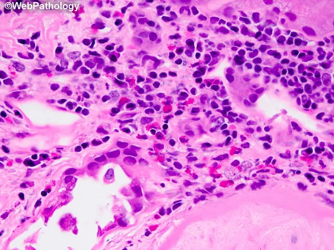
Renal biopsy tissue examined by light microscopy after Hematoxylin and Eosin staining reveals tubulointerstitial changes characteristic of diabetic nephropathy with superimposed inflammatory activity. The interstitial compartment shows a chronic inflammatory infiltrate dominated by T lymphocytes and macrophages, with scattered eosinophils forming small aggregates within fibrosed stroma. Tubules within the affected area display varying degrees of injury, including tubular atrophy and mild tubulitis, consistent with chronic kidney disease in diabetes. The surrounding interstitium appears fibrotic, with collagen deposition and loss of peritubular capillaries, creating a scar-like background that can obscure normal architecture. Notably, the image emphasizes the potential for neutrophilic infiltration and neutrophil casts, findings that may signal superimposed acute pyelonephritis, a known complication in diabetic patients. When infection is present, papillary necrosis and further destruction of renal parenchyma may ensue. Overall, the depicted histology aligns with a diabetic nephropathy spectrum where chronic inflammation coexists with fibrosis and occasional acute inflammatory components, raising clinical concerns for infection, rapid decline in renal function, and the need for targeted antimicrobial therapy and glycemic optimization. Clinically relevant differential considerations include tubulointerstitial nephritis of non-diabetic etiology and superimposed bacterial nephritis; correlation with serum creatinine and imaging improves diagnostic precision.
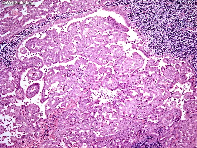
This is a light microscopy histopathology image of a human kidney biopsy stained with Hematoxylin and Eosin. The specimen comprises renal cortex with glomeruli and surrounding tubulointerstitial elements. The glomeruli appear variably cellular with focal expansion of the mesangial matrix, while the surrounding interstitium shows a mixed inflammatory infiltrate concentrated around tubular segments. Tubules exhibit epithelial flattening and mild tubular dilation with rare proteinaceous casts, and there is edema in the interstitial stroma. Vascular structures are not prominently featured in this field. Overall, there is evidence of tubulointerstitial inflammation with relative sparing of the glomerular capillary loops, a pattern that can be seen in acute interstitial nephritis or immune-mediated tubulointerstitial disease. No overt crescent formation or global glomerular necrosis is readily evident in this field. The density and distribution of inflammatory cells vary between areas, suggesting patchy involvement. The image highlights the interface between inflamed interstitium and nephron units, with preserved glomerular architecture in some regions and mild glomerular hypercellularity in others. Clinically, such histology supports inflammatory renal disease, guides differential diagnosis toward tubulointerstitial processes, and would prompt correlation with exposure history, complement levels, serologies, and urinalysis for diagnostic refinement and therapeutic decisions.
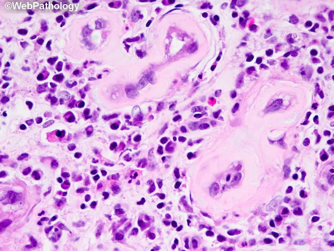
This histopathology image shows a renal biopsy section examined by light microscopy after Hematoxylin and Eosin staining, captured at high power to emphasize tubulointerstitial pathology associated with diabetic nephropathy. The cortical interstitium contains chronic inflammatory infiltrates composed predominantly of T lymphocytes and macrophages within areas of established interstitial fibrosis, reflecting long-standing injury. Eosinophil aggregates are conspicuous in the interstitium and can resemble tubulitis or tubulointerstitial nephritis. The tubular compartment demonstrates varying degrees of tubular atrophy and epithelial cell changes, with relative sparing of some glomeruli; when present, glomerular lesions of diabetic nephropathy such as mesangial expansion or nodular sclerosis may accompany the interstitial process but are not the defining feature in this field. Occasional neutrophils and, in places, neutrophil casts indicate a superimposed acute bacterial process and raise suspicion for acute pyelonephritis, a complication more common and often more severe in diabetes. Papillary necrosis is a theoretical progression in diabetics with infection and ischemic injury. Clinically, these findings correlate with chronic kidney disease from diabetes and heightened infection risk, underscoring the need for glycemic control, infection screening, and careful nephrotoxic exposure assessment. Differential diagnoses include chronic tubulointerstitial nephritis and drug-induced interstitial nephritis; clinicopathologic correlation is essential for definitive attribution.
ATT Drug-Induced Acute Kidney Injury (AKI)
1. Rifampicin (Rifampin) - Most Important Cause
Mechanisms
| Mechanism | Details |
|---|---|
| Acute Interstitial Nephritis (AIN) | Most common; immune-mediated (Type IV T-cell hypersensitivity, occasionally IgE-mediated Type I) |
| Acute Tubular Necrosis (ATN) | Direct tubular toxicity, particularly with high-dose or intermittent therapy |
| Immune-mediated hemolysis + pigment nephropathy | Anti-rifampicin IgG/IgM antibodies cause hemolysis; free hemoglobin is nephrotoxic |
| Thrombotic microangiopathy | Rare; immune-mediated platelet destruction |
Key Clinical Features
- Onset: typically 2-40 days after starting therapy
- More common with intermittent (twice or thrice weekly) dosing than daily regimens - this is a classic exam point
- Classic triad: fever + eosinophilia + rash (rash seen in ~25% of cases)
- Urinalysis: hematuria, mild proteinuria, leukocyturia (eosinophiluria)
- Serum creatinine rise or oliguric AKI in ~50% of cases
- Re-challenge is absolutely contraindicated - the drug must be permanently stopped
Park's Textbook of Preventive Medicine explicitly lists "Renal failure, shock, or thrombocytopenia" as reactions after which Rifampicin must never be given again.
Pathology
- Interstitial edema with lymphocytic and eosinophilic infiltration
- Tubulitis (lymphocytes invading tubular epithelium)
- Occasionally necrotizing granulomas
2. Streptomycin (and other injectable second-line drugs)
Mechanism: Direct Proximal Tubular Toxicity
- Cationic aminoglycoside binds to the brush border of proximal tubular cells via megalin (receptor-mediated endocytosis)
- Accumulates in lysosomes → lysosomal disruption → mitochondrial dysfunction → reactive oxygen species → proximal tubular cell death → ATN
- Streptomycin is the least nephrotoxic aminoglycoside (does not concentrate as much in the renal cortex compared to gentamicin or neomycin)
- Neomycin > Gentamicin > Tobramycin > Amikacin > Streptomycin (nephrotoxicity hierarchy)
Risk Factors
- High doses, prolonged therapy (>7-10 days)
- Pre-existing CKD, volume depletion
- Concomitant nephrotoxins (NSAIDs, amphotericin B, vancomycin, cyclosporine, ACE inhibitors)
Management
- Dose adjustment in renal impairment
- Therapeutic drug monitoring
- Avoid prolonged courses; switch to oral alternatives when culture sensitivities allow
3. Pyrazinamide - Hyperuricemia and Urate Nephropathy
Mechanism
- Pyrazinamide (and to a lesser extent ethambutol) inhibit renal tubular secretion of uric acid (via the URAT1 transporter)
- Result: hyperuricemia → urate crystal deposition in renal tubules → urate nephropathy
- Can also precipitate acute gouty attacks
Clinical Features
- Usually subclinical hyperuricemia during standard 2-month intensive phase
- Symptomatic gout in predisposed patients
- Frank urate nephropathy is uncommon but possible in prolonged MDR-TB regimens
Management
- Antigout agents (allopurinol - xanthine oxidase inhibitor) for symptomatic hyperuricemia
- Monitor uric acid and joint symptoms closely, especially in patients with pre-existing gout
- Pyrazinamide-induced hyperuricemia is largely not an indication to stop the drug unless symptomatic gout or severe hyperuricemia occurs
4. Ethambutol
- Primarily causes optic neuritis (dose-dependent, visual field defects)
- Also inhibits uric acid secretion like pyrazinamide (mild hyperuricemia)
- Dose-dependent renal excretion: requires dose reduction in renal impairment (GFR-guided dosing)
- In renal failure, reduced clearance of ethambutol markedly raises the risk of optic toxicity - so AKI from any cause while on ethambutol requires urgent dose review
5. Isoniazid (INH)
- Primarily hepatotoxic, not directly nephrotoxic at standard doses
- In overdose/poisoning: causes anion gap metabolic acidosis, seizures, and can cause rhabdomyolysis → secondary myoglobinuric AKI
- Oxidative stress and mitochondrial dysfunction contribute to organ damage (per the 2025 review on INH/RIF toxicity)
- Pyridoxine is the antidote for INH toxicity; hemodialysis has been used in severe poisoning
6. Second-Line ATT Drugs
| Drug | Renal Effect |
|---|---|
| Kanamycin / Amikacin | ATN (aminoglycosides - higher nephrotoxicity than streptomycin) |
| Capreomycin | Aminoglycoside-like ATN + hypokalemia, hypomagnesemia (tubular wasting) |
| Cycloserine | Renally excreted; accumulates in CKD causing CNS toxicity (not primary nephrotoxin) |
| Linezolid | Mitochondrial toxicity; rare lactic acidosis can contribute to AKI |
| Fluoroquinolones (moxifloxacin, levofloxacin) | Generally safe; rarely crystalline nephropathy |
| Bedaquiline / Delamanid | Limited renal data; use with caution in severe renal impairment |
Summary Table: ATT Drugs and Renal Effects
| Drug | Renal Lesion | Mechanism | Key Point |
|---|---|---|---|
| Rifampicin | AIN, ATN, TMA, hemolysis | Immune (T-cell / antibody) | Intermittent dosing risk; never re-challenge |
| Streptomycin | ATN (proximal tubule) | Direct aminoglycoside toxicity | Least nephrotoxic aminoglycoside |
| Pyrazinamide | Urate nephropathy | ↓ Uric acid excretion | Hyperuricemia; worsens gout |
| Ethambutol | Indirect (urate, drug accumulation) | ↓ Uric acid excretion | Dose-reduce in CKD; risk of optic toxicity |
| Isoniazid | Rhabdomyolysis-AKI (OD) | Mitochondrial/oxidative | Overdose; not at therapeutic doses |
| Kanamycin/Amikacin | ATN | Aminoglycoside | Higher nephrotoxicity than streptomycin |
| Capreomycin | ATN + tubular wasting | Aminoglycoside-like | Electrolyte wasting (K+, Mg2+) |
Clinical Approach When ATT-Induced AKI is Suspected
- Identify the offending drug - timing, pattern, urinalysis (eosinophiluria suggests AIN)
- Stop the most likely culprit - rifampicin if AIN pattern; aminoglycoside if ATN
- Renal biopsy if diagnosis uncertain or oliguria is severe
- Steroids may help for rifampicin-induced AIN (controversial, no RCTs)
- Restart ATT sequentially without the offending drug; redesign regimen
- Rifampicin must not be restarted if it caused AKI, shock, thrombocytopenia, or hemolysis
- Dose-adjust all renally-cleared ATT drugs (ethambutol, cycloserine, aminoglycosides) to GFR