
Regulation & mechanism of secretion of HCl in stomach
Reading File
Now let me search the medical library for HCl secretion in the stomach.
Finding Sources
Finding Sources
Excellent results. Let me read the most comprehensive sources in parallel.
Reading File
Reading File
Reading File
Good. Let me get the detailed mechanistic content from Yamada's and also fetch the diagram image, plus search for parietal cell mechanism details.
Reading File
Loading Image
Finding Sources
Excellent! Let me now read the Sabiston parietal cell activation section and also more of the Yamada content on intracellular signaling.
Reading File
Reading File
Let me fetch the parietal cell intracellular signaling diagram and also get more content on the phases and inhibition.
Loading Image
Reading File
Reading File
I now have comprehensive content. Let me also fetch the parietal cell intracellular signaling diagram from Schwartz's and the H+K+-ATPase pump diagram from Yamada's.
Loading Image
Reading File
I now have all the information needed to write a thorough, well-illustrated answer. Let me compile this.
Regulation & Mechanism of Secretion of HCl in the Stomach
The Parietal Cell - The Source of HCl
HCl is produced exclusively by parietal cells (oxyntic cells), which reside in the neck and pits of the oxyntic glands throughout the gastric corpus and fundus. The normal human stomach contains approximately 1 billion parietal cells, capable of secreting up to 30 mEq/hour of HCl in response to a meal. Parietal cells are metabolically intense - because H+ must be secreted against a gradient of more than a millionfold, nearly one-third of the parietal cell volume is occupied by mitochondria.
Cellular Mechanism of HCl Secretion
The H+/K+-ATPase proton pump is the final common pathway of HCl secretion. Here is how it works step by step:
The Proton Pump (H+/K+-ATPase)
- The H+/K+-ATPase is a heterodimer consisting of a catalytic α-subunit and a glycoprotein β-subunit, assembled in the endoplasmic reticulum.
- In the resting state, the pump is stored in intracellular tubulovesicles; no significant acid secretion occurs.
- Upon stimulation, cytoskeletal rearrangements drive translocation of tubulovesicles, which fuse with the apical secretory canaliculi, enlarging the secretory surface area 6-10 times. The H+/K+-ATPase is then inserted into the canalicular membrane.
- The pump performs electroneutral ion exchange: cytosolic H+ is actively pumped into the lumen in exchange for extracellular K+ (requires ATP since H+ is secreted against a massive gradient).
- Simultaneously, apical K+ and Cl- channels supply luminal K+ for exchange and Cl- for HCl formation.
- Basolateral bicarbonate-chloride exchangers export one HCO3- for every H+ secreted ("alkaline tide"), and import Cl- to replenish the apical Cl- supply.
- The net result: H+ + Cl- = HCl, secreted at pH ~0.8 (160 mM).
- Cessation of acid secretion involves endocytosis of H+/K+-ATPase back into tubulovesicles, triggered by a tyrosine-based signal on the cytoplasmic tail of the β-subunit.
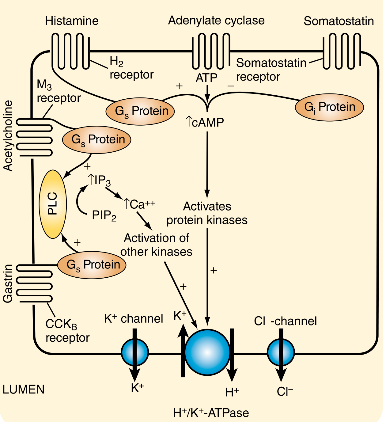
Three Stimulatory Pathways to the Parietal Cell
Three major agonists converge on the parietal cell, each through a distinct receptor and second-messenger system:
| Agonist | Receptor | G-protein | Second Messenger | Effect |
|---|---|---|---|---|
| Histamine | H2 receptor | Gs | ↑cAMP → PKA | Activates proton pump |
| Acetylcholine | M3 (muscarinic) | Gq | ↑IP3 → ↑Ca2+ | Activates proton pump |
| Gastrin | CCK-B (CCK2) | Gq | ↑IP3 → ↑Ca2+ | Activates proton pump |
| Somatostatin | SSTR2 | Gi | ↓cAMP, ↓Ca2+ | Inhibits proton pump |
Histamine is considered the most important stimulator because it acts via the H2 receptor as the final paracrine mediator. This is why H2-receptor antagonists and proton pump inhibitors (PPIs) are so effective clinically.
Regulatory Cell Types
| Cell | Location | Substance Released | Effect |
|---|---|---|---|
| Parietal cell | Oxyntic glands (corpus/fundus) | HCl, intrinsic factor | Acid secretion |
| ECL cell (enterochromaffin-like) | Oxyntic glands | Histamine | Stimulates parietal cell via H2 |
| G cell | Antral pyloric glands | Gastrin | Stimulates ECL and parietal cells |
| D cell | Antrum + corpus | Somatostatin | Inhibits G cells, ECL cells, and parietal cells |
| Chief cell | Oxyntic glands | Pepsinogen | Activated by HCl to pepsin |
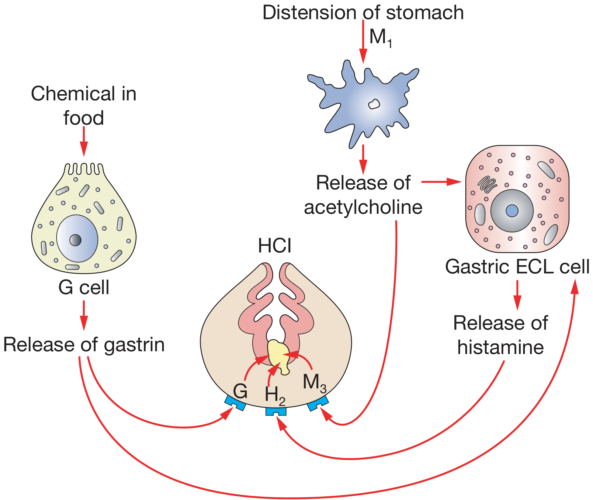
Phases of Gastric Acid Secretion (Physiological Regulation)
1. Cephalic Phase (~30% of meal-stimulated output)
- Triggered by the thought, sight, smell, and taste of food.
- Cortical/hypothalamic stimuli activate the vagal dorsal motor nucleus (DMN) and nucleus tractus solitarius (NTS).
- Efferent vagal fibers synapse with submucosal enteric neurons → release acetylcholine (ACh).
- ACh acts directly on parietal cells (M3) AND stimulates antral G cells (via GRP) to release gastrin.
- Vagal stimulation also sensitizes ECL cells to gastrin.
- Sham feeding (chewing and spitting) stimulates acid to ~50% of maximum - purely via this phase.
2. Gastric Phase (~60% of meal-stimulated output)
- Begins when food enters the stomach; lasts until the stomach empties.
- Mechanical component: Gastric distension activates vagovagal reflex arcs and local enteric reflexes → ACh release → parietal cell stimulation.
- Chemical component: Amino acids and small peptides directly stimulate antral G cells → gastrin release into blood → stimulates ECL cells (histamine) and parietal cells directly.
- The buffering effect of food (raising luminal pH) removes the tonic acid-inhibition of G cells, thus amplifying gastrin release.
3. Intestinal Phase (~10% or less of meal-stimulated output)
- As chyme enters the duodenum and small bowel.
- Initially mildly stimulatory (enterooxyntin from small intestinal mucosa).
- Rapidly becomes predominantly inhibitory once duodenal acidification occurs:
- Acidification → secretin release → inhibits gastric acid secretion.
- Additional enterogastrones (GIP, CCK, glucagon-like peptide-1) inhibit acid.
- Hyperosmolar/fatty chyme also triggers inhibitory reflexes.
Inhibitory Regulation (Negative Feedback)
| Mechanism | Stimulus | Mediator | Target | Effect |
|---|---|---|---|---|
| Acid feedback (antrum) | ↓Luminal pH (<3) | Somatostatin from D cells | G cells | ↓Gastrin release |
| Acid feedback (corpus) | ↓Luminal pH | Somatostatin from D cells | ECL + parietal cells | ↓Histamine + direct inhibition |
| CGRP pathway | ↑Luminal acid | CGRP from extrinsic sensory neurons | D cells (antrum + corpus) | ↑Somatostatin |
| Duodenal inhibition | Acid, fat, hyperosmolarity in duodenum | Secretin, CCK, GIP, PYY, GLP-1 | G cells, ECL cells, parietal cells | ↓Acid secretion |
| VIP on D cells | Vagal peptidergic neurons | VIP | D cells | ↑Somatostatin → inhibition |
| Prostaglandins | Various | PGE2, PGI2 | Parietal + ECL cells | ↓cAMP, ↓acid |
The somatostatin pathway is the principal "off switch": D cells act both locally (via cytoplasmic processes directly onto adjacent G, ECL, and parietal cells) and via the local circulation.
Summary Diagram
Stimulatory axis:
Vagus → ACh → M3 on parietal cell + G cell (via GRP) → Gastrin → CCK-2 on ECL cell → Histamine → H2 on parietal cell → cAMP → PKA → H+/K+-ATPase → HCl
Inhibitory axis:
Luminal acid → D cell → Somatostatin → SSTR2 on G cell + ECL cell + parietal cell → ↓Gastrin, ↓Histamine, ↓H+/K+-ATPase activity
Clinical Relevance
- PPIs (omeprazole, pantoprazole) - irreversibly bind the activated H+/K+-ATPase at the cysteine residues of the α-subunit; most effective when taken before meals when parietal cells are stimulated. Acid secretion recovers in days as new pump is synthesized.
- H2 blockers (famotidine) - block the H2 receptor; effective but limited since they block only one of three pathways; tachyphylaxis can occur due to CCK-2 receptor upregulation.
- Potassium-competitive acid blockers (PCABs) (vonoprazan) - bind ionically to H+/K+-ATPase, blocking K+ access; work even in the resting state unlike PPIs.
- Helicobacter pylori: antrum-predominant infection → ↑gastrin → hypersecretion; corpus-predominant infection → ↓acid (parietal cell mass lost).
- Zollinger-Ellison syndrome: gastrinoma → unregulated gastrin → massive acid hypersecretion → refractory peptic ulcers.
Sources: Yamada's Textbook of Gastroenterology 7th ed. | Bailey & Love's Short Practice of Surgery 28th ed. | Sabiston Textbook of Surgery | Schwartz's Principles of Surgery 11th ed.
Peptic ulcer (CASE)
Asking for Preferences
What kind of peptic ulcer case content do you need?
I'll provide a comprehensive clinical case-based discussion covering the full workup. Let me pull from the medical library first.
Finding Sources
Finding Sources
Reading File
Reading File
Reading File
Reading File
Finding Sources
Reading File
Finding Sources
Reading File
Finding Sources
Reading File
Finding Sources
Reading File
Reading File
I have comprehensive content. Let me get the Forrest classification table and the complications section fully, then compile the answer.
Reading File
I have all the material needed for a comprehensive case-based discussion. Now I'll write the full answer.
Peptic Ulcer Disease - Clinical Case Discussion
THE CASE
Mr. A, 45-year-old male, presents with:
- 3-month history of burning epigastric pain, worse 2-4 hours after meals and at night, relieved by eating food or antacids
- History of taking diclofenac (NSAID) regularly for knee pain for 6 months
- Smokes 10 cigarettes/day; occasional alcohol
- No weight loss, no vomiting of blood, no black stools
Examination:
- Mild epigastric tenderness on deep palpation
- No guarding or rigidity
- No palpable mass
- Vital signs: normal
STEP 1 - DIFFERENTIAL DIAGNOSIS
| Diagnosis | For | Against |
|---|---|---|
| Duodenal ulcer | Night pain, hunger pain, relief with food, NSAID use | - |
| Gastric ulcer | NSAID use, epigastric pain | Pain usually worsened by food in GU |
| GERD | Epigastric burning | No heartburn/regurgitation |
| Gastric carcinoma | Age, male | No weight loss, no dysphagia, short history |
| Functional dyspepsia | Epigastric symptoms | Organic cause likely here (NSAIDs) |
Working diagnosis: Peptic ulcer disease (likely duodenal) - NSAID-related +/- H. pylori
STEP 2 - PATHOPHYSIOLOGY (What's happening in this patient?)
Peptic ulceration results from an imbalance between mucosal defensive factors and aggressive/damaging factors.
Defensive Factors (reduced in PUD):
- Mucus-bicarbonate barrier (gel layer trapping HCO3-)
- Mucosal blood flow
- Prostaglandin E2 (maintains mucus/HCO3- secretion)
- Cell renewal and growth factors
Aggressive Factors (increased in PUD):
- HCl + pepsin
- H. pylori infection (present in ~90% of duodenal ulcers)
- NSAIDs - inhibit COX-1 → ↓prostaglandin synthesis → ↓mucus and HCO3- → mucosal injury; also direct topical injury
- Smoking (↓mucosal blood flow, ↓prostaglandins)
- Alcohol
- Stress (Curling's ulcer post-burn; Cushing's ulcer in CNS injury)
How H. pylori causes duodenal ulcer:
- H. pylori (gram-negative, microaerophilic, flagellated spiral bacillus) colonizes the gastric antrum
- Its urease enzyme converts urea → NH3 + HCO3-, creating an alkaline microenvironment allowing survival
- Colonization → inhibits somatostatin secretion by antral D cells
- ↓Somatostatin → removes tonic inhibition of G cells → ↑gastrin secretion
- ↑Gastrin → ↑parietal cell HCl output → excess H+ delivered to duodenum
- H. pylori also spreads to duodenum → inhibits duodenal HCO3- secretion
- Net result: ↑acid load + ↓buffering → duodenal mucosal erosion
STEP 3 - GASTRIC vs. DUODENAL ULCER: KEY DIFFERENCES
| Feature | Duodenal Ulcer | Gastric Ulcer |
|---|---|---|
| Site | 1st part duodenum (D1), anterior wall | Lesser curvature, antrum (Type I most common) |
| Age | Younger (30-50s) | Older (55-65s) |
| Sex | Male > Female | Male > Female |
| H. pylori | ~90% associated | ~70-90% associated |
| Acid secretion | Increased (hyperacidity) | Normal or low |
| Pain timing | Hunger pain (2-4h after meal), night pain | Shortly after eating (food worsens it) |
| Pain relief | Food, antacids relieve | Food may worsen |
| Malignancy risk | Extremely rare | Always exclude (biopsy mandatory) |
| H. pylori mechanism | Indirect - ↑acid via ↓somatostatin | Direct - cytotoxins break mucosal barrier |
| Gastrin levels | Elevated post-meal | Elevated (due to ↓net H+ from mucosal leak) |
Johnson Classification of Gastric Ulcer Types (surgical relevance):
| Type | Location | Acid Level | Notes |
|---|---|---|---|
| I | Lesser curve at incisura angularis | Low to normal | Most common (50-60%) |
| II | Gastric body + concurrent duodenal ulcer | Increased | 15-20% |
| III | Prepyloric | Increased | Behaves like DU |
| IV | High lesser curve, near GEJ | Normal | <10%, technically difficult |
| V | Anywhere | Normal | NSAID-induced |
STEP 4 - INVESTIGATIONS
For Diagnosis:
-
Upper GI Endoscopy (OGD) - gold standard
- Confirms location, depth, appearance of ulcer
- Biopsy of gastric ulcers: mandatory in all cases (4 quadrants - first biopsy alone has 70% sensitivity; 4 specimens = 95%; 7 specimens = 98%)
- Duodenal ulcers: biopsy not routinely needed for malignancy (rarely malignant)
- Can be therapeutic (bleeding control)
-
Barium meal (upper GI series) - largely replaced by endoscopy
- Single contrast: misses 50% of ulcers; double contrast: detects 80-90%
For H. pylori:
| Test | Type | Notes |
|---|---|---|
| Urea breath test (UBT) | Non-invasive | Patient drinks 13C-urea → H. pylori urease → 13CO2 exhaled and measured; best for confirmation of eradication |
| Stool antigen test | Non-invasive | Good for initial diagnosis and post-treatment confirmation |
| Rapid urease test (CLO test) | Invasive (endoscopy biopsy) | Quick, from antral biopsy |
| Histology | Invasive | Gold standard for tissue diagnosis |
| Culture | Invasive | Used for antibiotic sensitivity testing |
| Serology (IgG) | Non-invasive | Cannot distinguish active from past infection; not used for eradication testing |
Other labs:
- FBC (anemia in bleeding)
- Fasting serum gastrin (if ZES suspected - multiple/refractory ulcers, diarrhea, steatorrhea)
- LFTs, renal function
STEP 5 - TREATMENT
A. Lifestyle Modifications
- Stop NSAIDs/aspirin if possible
- Smoking cessation
- Avoid alcohol
- Small frequent meals (reduces acid peaks)
B. Pharmacological Treatment
1. Proton Pump Inhibitors (PPIs) - First-line acid suppression
- Omeprazole, pantoprazole, lansoprazole, rabeprazole, esomeprazole
- Mechanism: prodrug → activated in parietal cell canaliculus → covalent disulfide bond to cysteine on H+/K+-ATPase α-subunit → irreversible inhibition
- Healing rate: 85% at 4 weeks, 96% at 8 weeks
- Take before meals (parietal cell must be stimulated for drug activation)
- Maintenance PPI: for large ulcers (>2 cm), refractory disease, failed H. pylori eradication, or continued NSAID/aspirin requirement
2. H2 Receptor Antagonists (H2RAs)
- Famotidine, cimetidine, ranitidine (ranitidine withdrawn globally April 2020 - NDMA contamination risk)
- Second-line; may cause tachyphylaxis
- Do NOT combine with PPIs (H2RAs create alkaline environment, preventing PPI activation)
3. Antacids - Symptomatic relief only
4. Misoprostol (PGE1 analogue) - Used for NSAID-related ulcer prevention (stimulates mucus/HCO3-, ↑mucosal blood flow)
5. Sucralfate - Mucosal protective agent; binds to ulcer base
C. H. pylori Eradication (when H. pylori positive)
Eradication reduces recurrence to only ~2% (vs. 80% without eradication).
First-line regimens (14-day courses):
Standard Triple Therapy (no macrolide resistance risk, no penicillin allergy):
PPI + Clarithromycin + Amoxicillin (x 14 days)
Bismuth Quadruple Therapy (macrolide resistance/prior macrolide exposure/penicillin allergy):
PPI + Bismuth subcitrate + Tetracycline + Metronidazole (10-14 days)
Levofloxacin Triple Therapy (alternative):
PPI + Levofloxacin + Amoxicillin (10-14 days)
Confirm eradication at 4-6 weeks post-treatment with urea breath test or stool antigen (not serology).
~20-30% of patients fail initial therapy (increasing antibiotic resistance). These need second-line regimens guided by susceptibility.
STEP 6 - COMPLICATIONS OF PUD (The "BOOP" of PUD)
| Complication | Features | Management |
|---|---|---|
| Bleeding | Most common complication (15-20% of PUD) | See below |
| Obstruction | Pyloric/duodenal narrowing | Endoscopic dilation; surgery (gastrojejunostomy) |
| Perforation | Acute abdomen, pneumoperitoneum | Emergency surgery (patch/omental closure) |
| Intractability | Unresponsive to medical therapy | Surgical: truncal vagotomy ± antrectomy |
Bleeding PUD - Emergency Management:
- ABC resuscitation - large-bore IV access, fluids, blood products
- IV PPI immediately on admission
- Prokinetic (IV erythromycin) before endoscopy to improve visualization
- Urgent endoscopy within 24 hours (diagnostic + therapeutic)
- Endoscopic hemostasis: hemostatic clips, electrocoagulation, argon plasma coagulation, injection (epinephrine NOT as monotherapy - combine with another modality)
- Post-endoscopy: IV PPI for 72 hours, then oral PPI ≥2 weeks
- Rebleed → repeat endoscopy → if fails: interventional radiology (transarterial embolization) → if fails: surgery
Forrest Classification (endoscopic risk stratification):
| Forrest Class | Finding | Rebleed Risk |
|---|---|---|
| IA | Active spurting | 90% |
| IB | Active oozing | 10-20% |
| IIA | Non-bleeding visible vessel | 50% |
| IIB | Adherent clot | 25-30% |
| IIC | Flat pigmented spot | 5-10% |
| III | Clean-based ulcer | 3-5% |
Forrest IA, IB, IIA require endoscopic intervention. IIB - controversial. IIC/III - no intervention needed.
Perforation - Emergency:
- Sudden severe "board-like" epigastric rigidity
- Upright CXR: free gas under diaphragm (pneumoperitoneum)
- CT abdomen confirms
- Initial: IV PPI, NBM, IV antibiotics, NG tube decompression
- Surgical: omental patch repair (Graham patch) + H. pylori eradication
- Mortality 30-50% in elderly/debilitated patients
STEP 7 - SPECIAL SITUATIONS
Zollinger-Ellison Syndrome (ZES / Gastrinoma)
- Gastrin-secreting tumor (usually pancreatic islet cell; 25% in MEN-1)
- Multiple, refractory, atypical ulcers (jejunal ulcers)
- Diarrhea and steatorrhea (low duodenal pH inactivates pancreatic lipases)
- Diagnosis: fasting serum gastrin >1000 pg/mL; secretin stimulation test
- Gastrin is NOT inhibited by H+ (unlike physiological G cells)
- Treatment: High-dose PPI + surgical resection of tumor
NSAID-related ulcers
- COX-1 inhibition → ↓prostaglandins → ↓mucus/HCO3- + ↓mucosal blood flow
- Prevention: use COX-2 selective NSAIDs (celecoxib) OR add misoprostol/PPI
- Type V Johnson classification
- Often painless (NSAIDs also suppress pain perception) → may present with occult bleeding
Stress Ulcers (in ICU)
- Curling's ulcer: major burns (ischemia of mucosa)
- Cushing's ulcer: CNS injury (vagal hyperactivity → ↑acid)
- Prophylaxis with PPI or sucralfate in ventilated/coagulopathic ICU patients
SUMMARY TABLE: Gastric vs. Duodenal Ulcer at a Glance
| Gastric Ulcer | Duodenal Ulcer | |
|---|---|---|
| Incidence | Less common | More common (4:1) |
| Age | 55-65 | 30-50 |
| H. pylori | 70-90% | ~90% |
| NSAIDs | Major cause | Less so |
| Acid | Normal/low | High |
| Pain | Worsened by food | Relieved by food; hunger pain |
| Night pain | Less common | Common |
| Malignancy | Must exclude (biopsy all) | Extremely rare |
| Surgery type | Partial gastrectomy / oversew + H. pylori Rx | Oversew + H. pylori Rx |
Sources: Sabiston Textbook of Surgery | Costanzo Physiology 7th ed. | Robbins & Kumar Basic Pathology | Yamada's Textbook of Gastroenterology 7th ed. | Schwartz's Principles of Surgery 11th ed.
This is a shared conversation. Sign in to Orris to start your own chat.